Jump to content
In this section we have spot diagnoses posted on a daily basis since June 2010, now over 4000! You can review the archived cases and read the suggested diagnoses by users and the final comment by the contributors.
Case are uploaded each week day by 10 am UK time with the correct diagnosis will generally be posted at 8 pm UK time. Why not view the most recent spot diagnosis and proffer a diagnosis?

Case Number : Case 1880 - 11 August - Dr Arti Bakshi Posted By: Guest

Please read the clinical history and view the images by clicking on them before you proffer your diagnosis.
Submitted Date :
   (0 reviews)

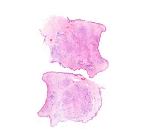
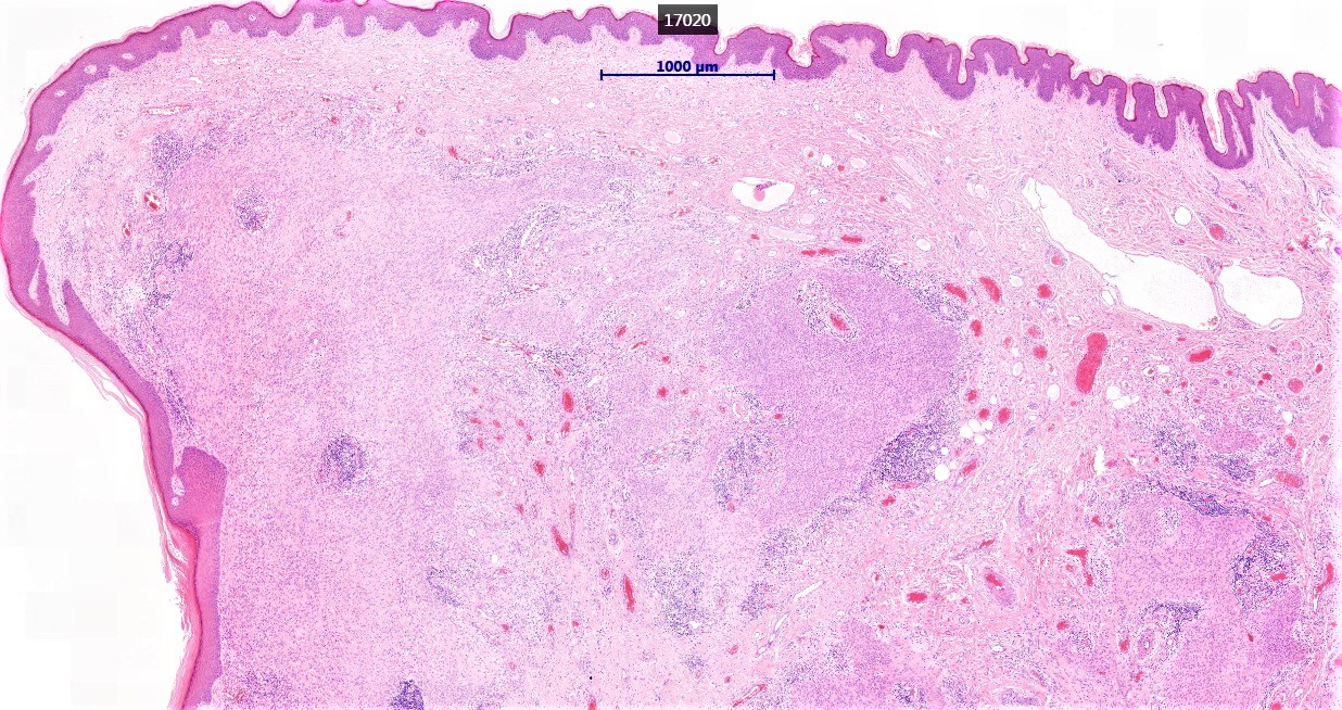
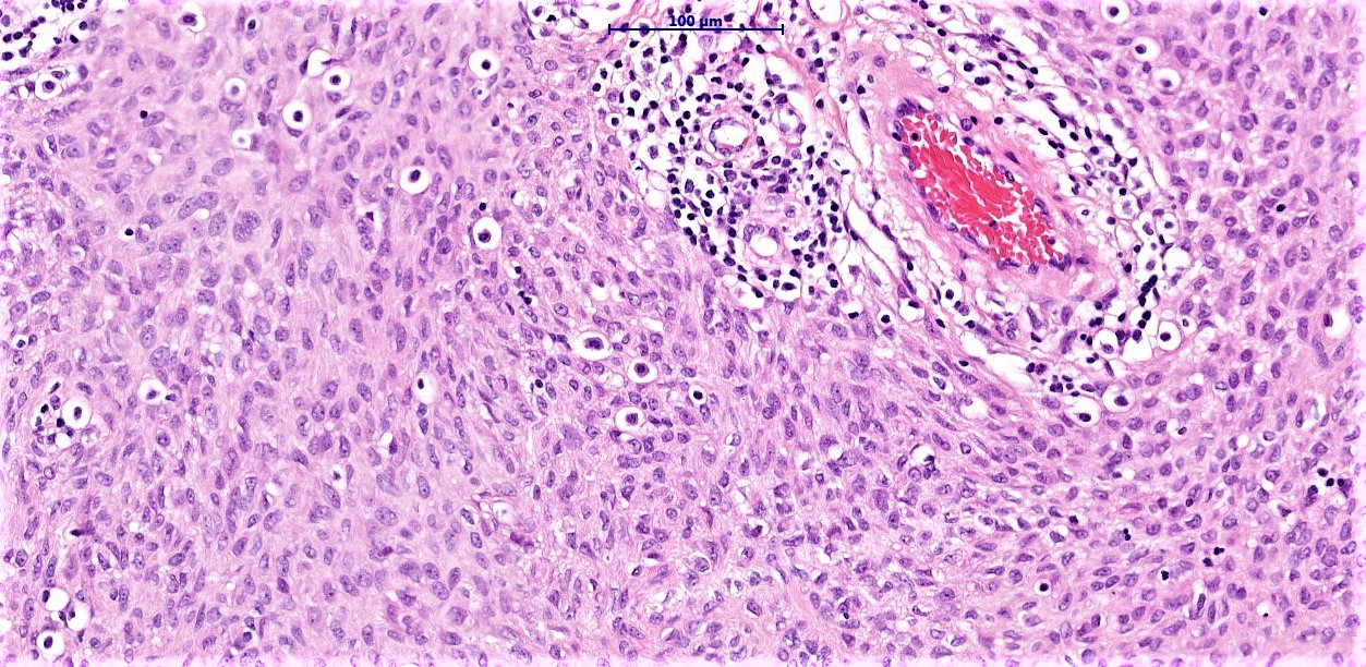
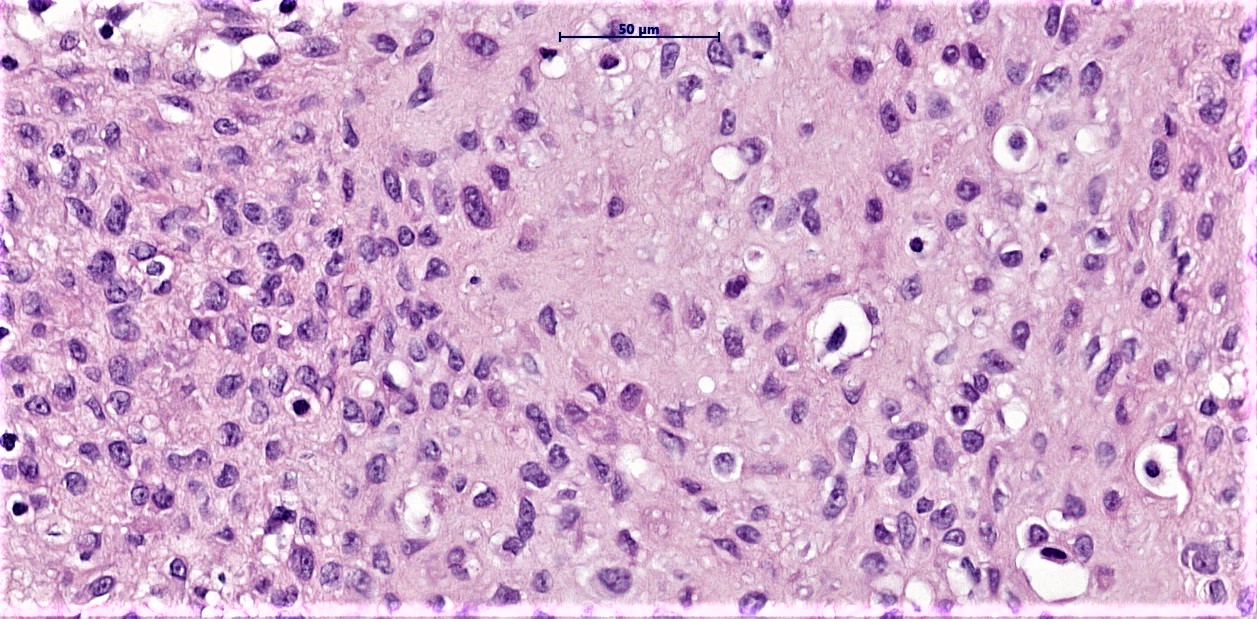
36/M, ?squamous papilloma, right shoulder

Edited by Admin_Dermpath


  Report Record

User Feedback


vincenzo polizzi

Posted

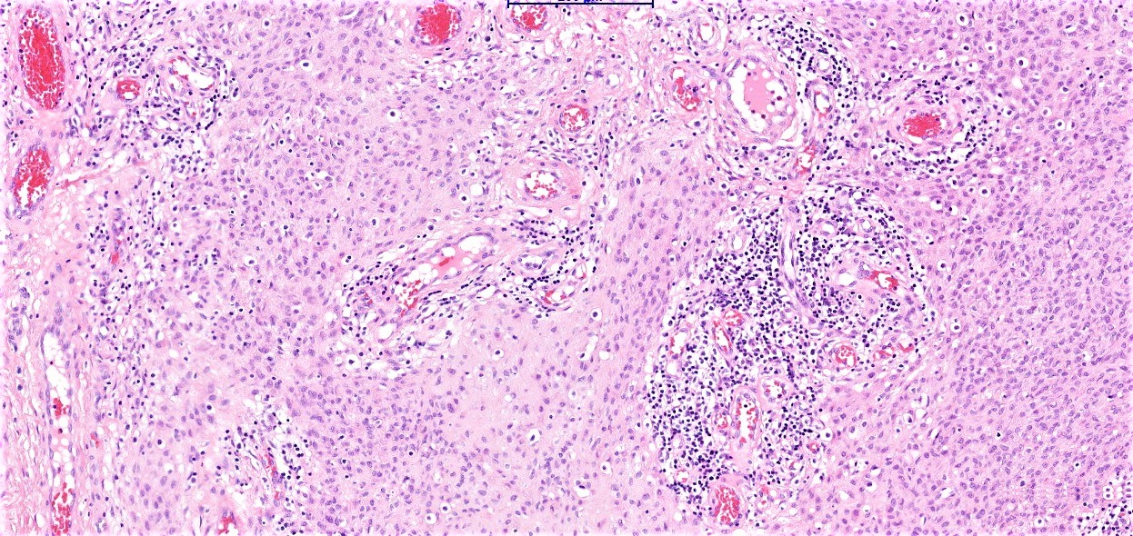
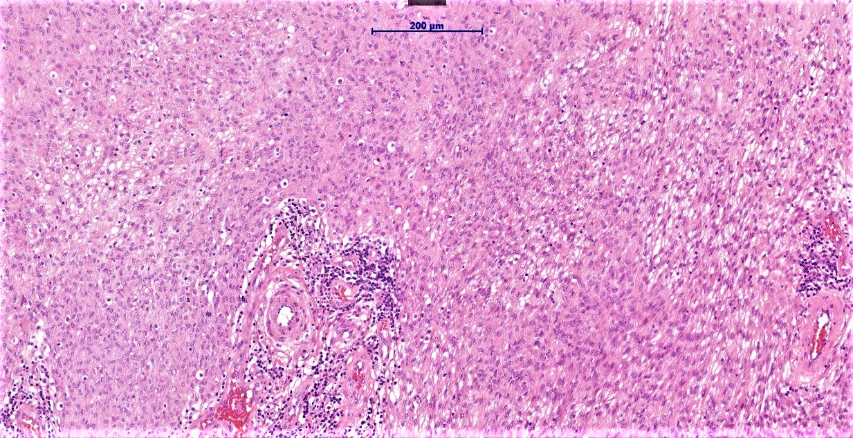
This is a well circumscribed, dermal based, syncytial growth of histiocytoid cells, with pale eosinophilic cytoplasm, reminiscent of something myoid. It's an intriguing case, and I'm thinking of a recently described lesion on the myoepithelial spectrum: Cutaneous Syncytial Myoepithelioma.

S100 and EMA could be useful in DD pathway.


Share this comment


Link to comment
share_externally

Raul Perret

Posted

I agree completely with Vincenzo, there seems to be some initial chondroid metaplasia in the last image. A similar case was published at the ASDP website recently, I find the perivascular lymphocytic infiltrates to be quite helpful for suspecting the entity.

Share this comment


Link to comment
share_externally

Dr. Mona Abdel-Halim

Posted

Agree with Vincenzo! 

Marvelous case... I did not know about this entity before but I read the case refered to by Raul and it is typical... Thanks Arti... educating case :-)

Share this comment


Link to comment
share_externally

Anil Patki

Posted

The presence of vessels and vacuoles in the cells is suggestive of cutaneous epithelioid angiomatous nodule

Share this comment


Link to comment
share_externally

Robledo F. Rocha

Posted

I couldn’t find those irregular vascular spaces lined by epithelioid endothelial cells, and extravasated erythrocytes, for epithelioid angiomatous nodule, maybe a variant of epithelioid hemangioma according to Omar Sangüeza. So, I agree with Vincenzo, this is a syncytial myoepithelioma.
Talking about Dr. Sangüeza, he is now presenting his lecture on novel immunohistochemical markers in Dermatopathology during SLAP Congress.

Share this comment


Link to comment
share_externally

Arti Bakshi

Posted

Yes, this is a syncytial myopeithelioma. The tumour was EMA and S100 positive. The sheet like growth with syncytial eosinophilic cytoplasm is characteristic of this variant of myoepithelioma. In the largest series of this variant (see link https://www.ncbi.nlm.nih.gov/pubmed/23588365 ), all cases behaved in a benign fashion (as opposed to greater risk of recurrence/mets in other variants of myopeitheioma).

A spirited defence of cutaneous angiomatous nodule by Anil! But it is not this entity due to the reasons outlined by Robledo and the immunos help to rule it out. A good differential nevertheless!

Thanks to Raul for the link to the case on ASDP. Enjoy your conference, Robledo, and keep sharing what u learn with us too!

Share this comment


Link to comment
share_externally


Join the conversation

You can post now and register later. If you have an account, sign in now to post with your account.

Guest
Add a comment...

×   Pasted as rich text.   Paste as plain text instead

  Only 75 emoji are allowed.

×   Your link has been automatically embedded.   Display as a link instead

×   Your previous content has been restored.   Clear editor

×   You cannot paste images directly. Upload or insert images from URL.

Loading...

×
×
  • Create New...