Jump to content
In this section we have spot diagnoses posted on a daily basis since June 2010, now over 4000! You can review the archived cases and read the suggested diagnoses by users and the final comment by the contributors.
Case are uploaded each week day by 10 am UK time with the correct diagnosis will generally be posted at 8 pm UK time. Why not view the most recent spot diagnosis and proffer a diagnosis?

Case Number : Case 1973 - 21 Dec 2017 Posted By: Guest

Please read the clinical history and view the images by clicking on them before you proffer your diagnosis.
Submitted Date :
   (0 reviews)

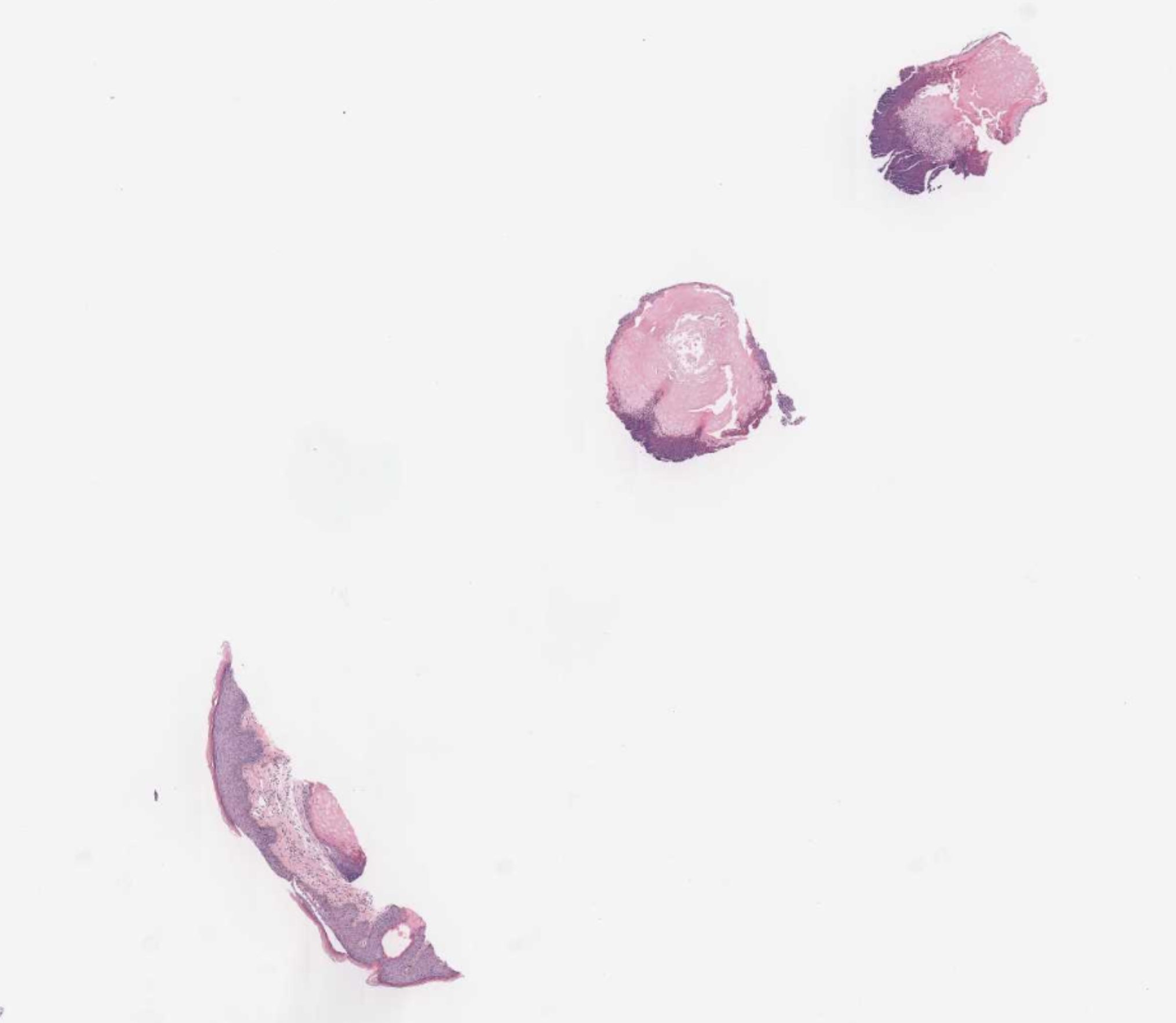
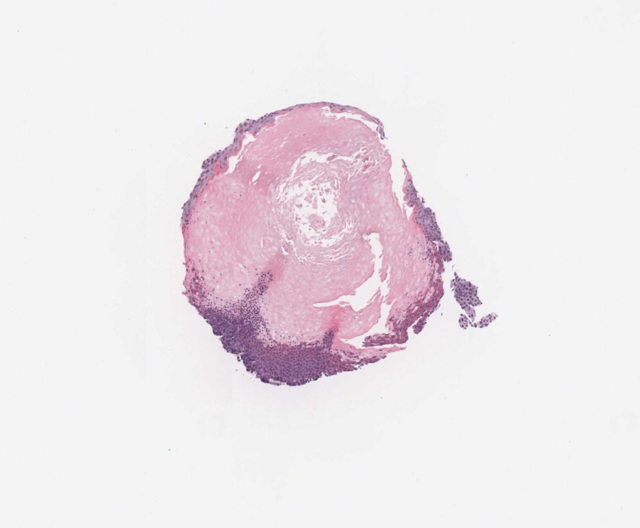
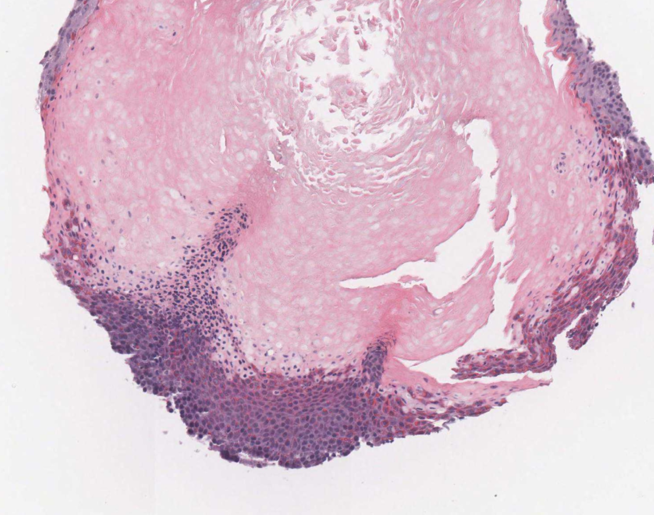
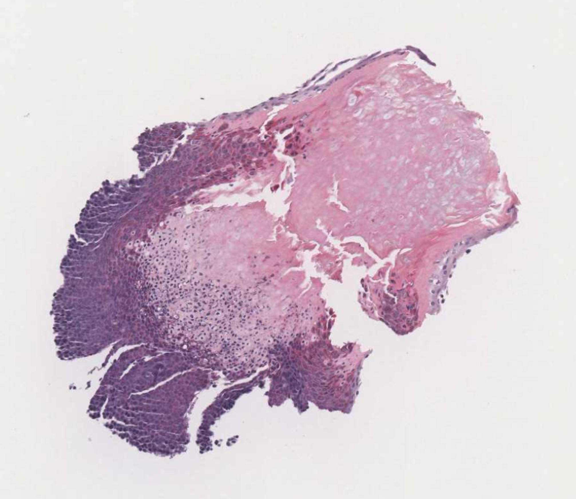
52 year old HIV + female with renal failure. Several year history of lesions on nose.

Edited by Iskander H. Chaudhry


  Report Record

User Feedback


Admin_Dermpath

Posted

Dear All 

Dr Chaudhry posted immuno on yesterday's case:

Best wishes

DPro Admin

Share this comment


Link to comment
share_externally

Admin_Dermpath

Posted

Dr Chaudhry posted his diagnosis to:


Share this comment


Link to comment
share_externally

Oops, dark cells and shadow cells- it's a pilomatricoma!

Share this comment


Link to comment
share_externally

Uma Sundram

Posted

I'm not sure because the images are so low power. I would have said pilomatricoma as well.

Share this comment


Link to comment
share_externally

Dr. Richard Carr

Posted

My thoughts are hair cortex comedo (Spot Diagnosis 837) or possibly as multiple & patient is immune suppressed  trichodysplasia spinulosa - perhaps both!

Share this comment


Link to comment
share_externally

small biopsy with areas showing features of inner root sheath differentiation and matrix differentiation. no other hair follicle parts differentiation seen (such as infundibulum and outer root sheath) in the superfical biopsy. still could be a superfical panfolliculoma.

Share this comment


Link to comment
share_externally

John Zhang

Posted

Must be a shrewd observation, Henry. Although I can't clearly appreciate trichohyalin granules to indicate inner root sheath differentiation, McKee and Michal believe that pilomatricoma "although predominantly a tumor with matrical differentiation, careful inspection of early or well-established lesions often will reveal foci of clear cells with ltrichohyalin granules (differentiation toward the lower portion of inner root sheath) and blue-gray cornified cells (upper root sheath differentiation)."

Share this comment


Link to comment
share_externally

Dr. Richard Carr

Posted

The orange hue in the epithelium cells more superficial will equate to IRS differentiation - you'd need a slightly higher magnification to resolve the individual granules than here but the appearance is typical.

Share this comment


Link to comment
share_externally

Admin_Dermpath

Posted

Hi Everyone,


Dr Phelp's final diagnosis is Trichodysplasia spinulosa. 

Share this comment


Link to comment
share_externally


Join the conversation

You can post now and register later. If you have an account, sign in now to post with your account.

Guest
Add a comment...

×   Pasted as rich text.   Paste as plain text instead

  Only 75 emoji are allowed.

×   Your link has been automatically embedded.   Display as a link instead

×   Your previous content has been restored.   Clear editor

×   You cannot paste images directly. Upload or insert images from URL.

Loading...

×
×
  • Create New...