Jump to content
In this section we have spot diagnoses posted on a daily basis since June 2010, now over 4000! You can review the archived cases and read the suggested diagnoses by users and the final comment by the contributors.
Case are uploaded each week day by 10 am UK time with the correct diagnosis will generally be posted at 8 pm UK time. Why not view the most recent spot diagnosis and proffer a diagnosis?

Case Number : Case 1974 - 22 Dec 2017 Posted By: Dr. Richard Carr

Please read the clinical history and view the images by clicking on them before you proffer your diagnosis.
Submitted Date :
   (0 reviews)

No Clinical History given

Edited by Admin_Dermpath


  Report Record

User Feedback


Dr. Mona Abdel-Halim

Posted

Subepithelial conjunctival nevus?


Share this comment


Link to comment
share_externally

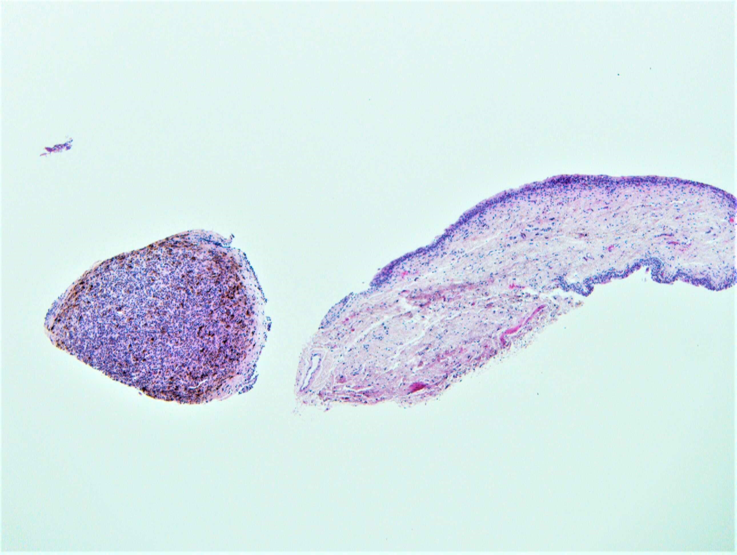
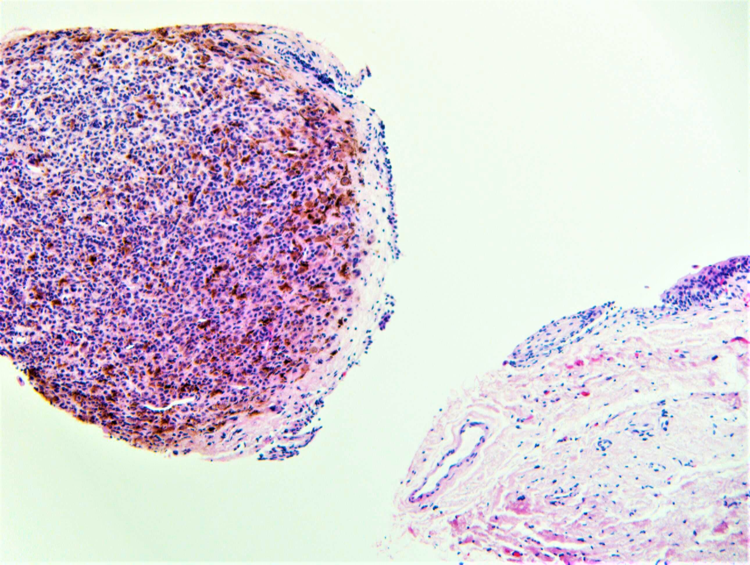
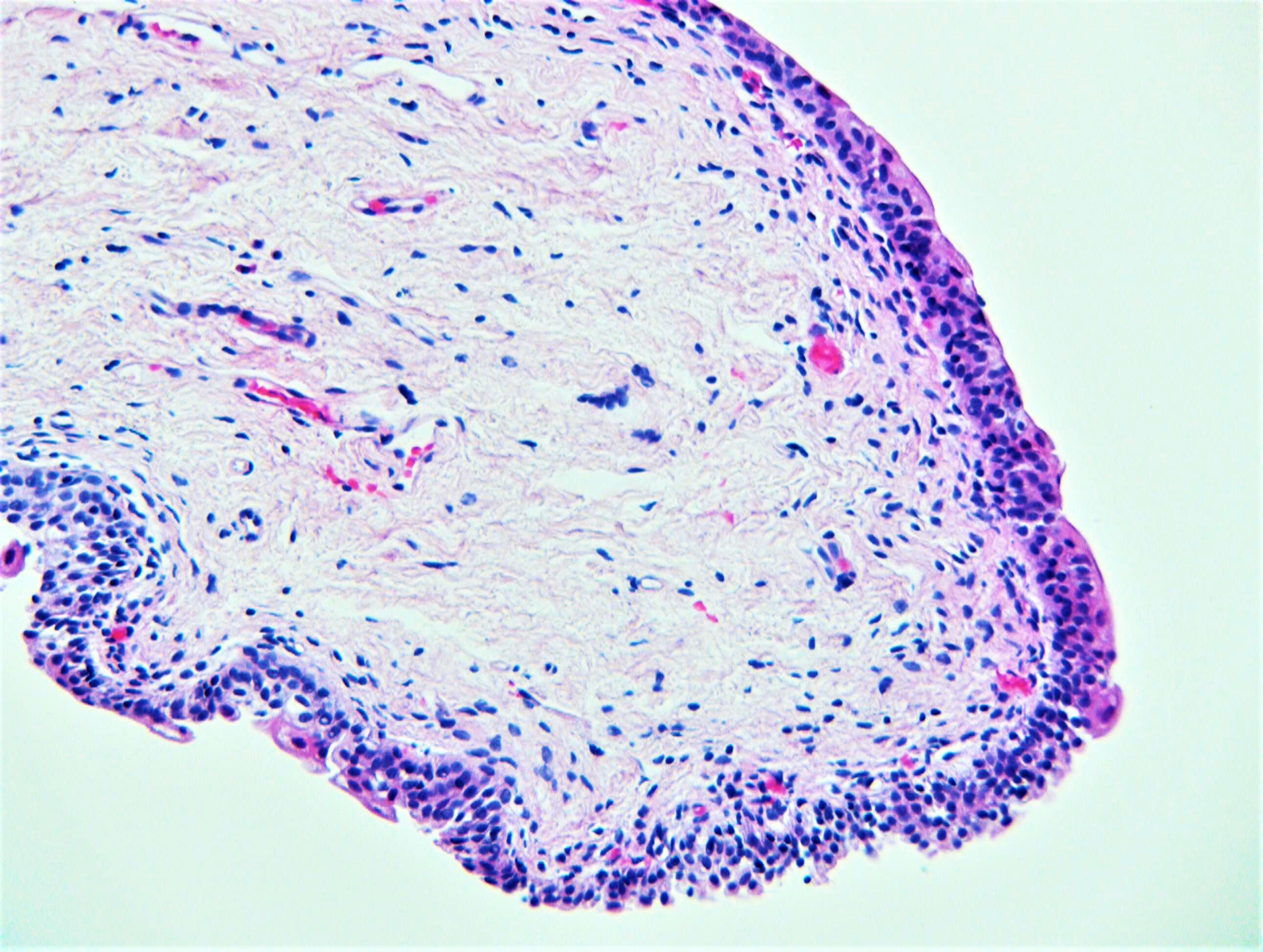
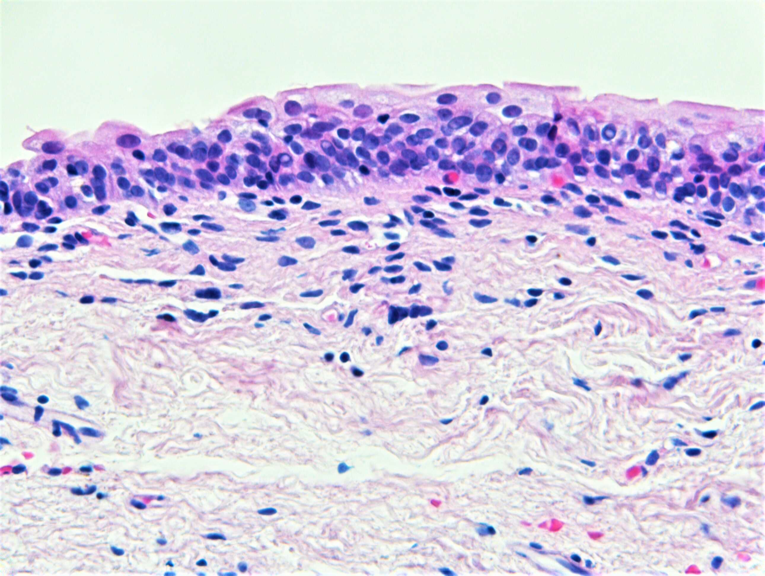
That was my first impression too: some kind of melanocytic lesion of the conjunctiva. But upon close inspection of the epithelial lining, I thought I saw umbrella cells on top and I don't really appreciate goblet cells in the lining as commonly seen in the conjunctiva. So I would suggest a melanocytic lesion of the urethra or ureter as an alternative.

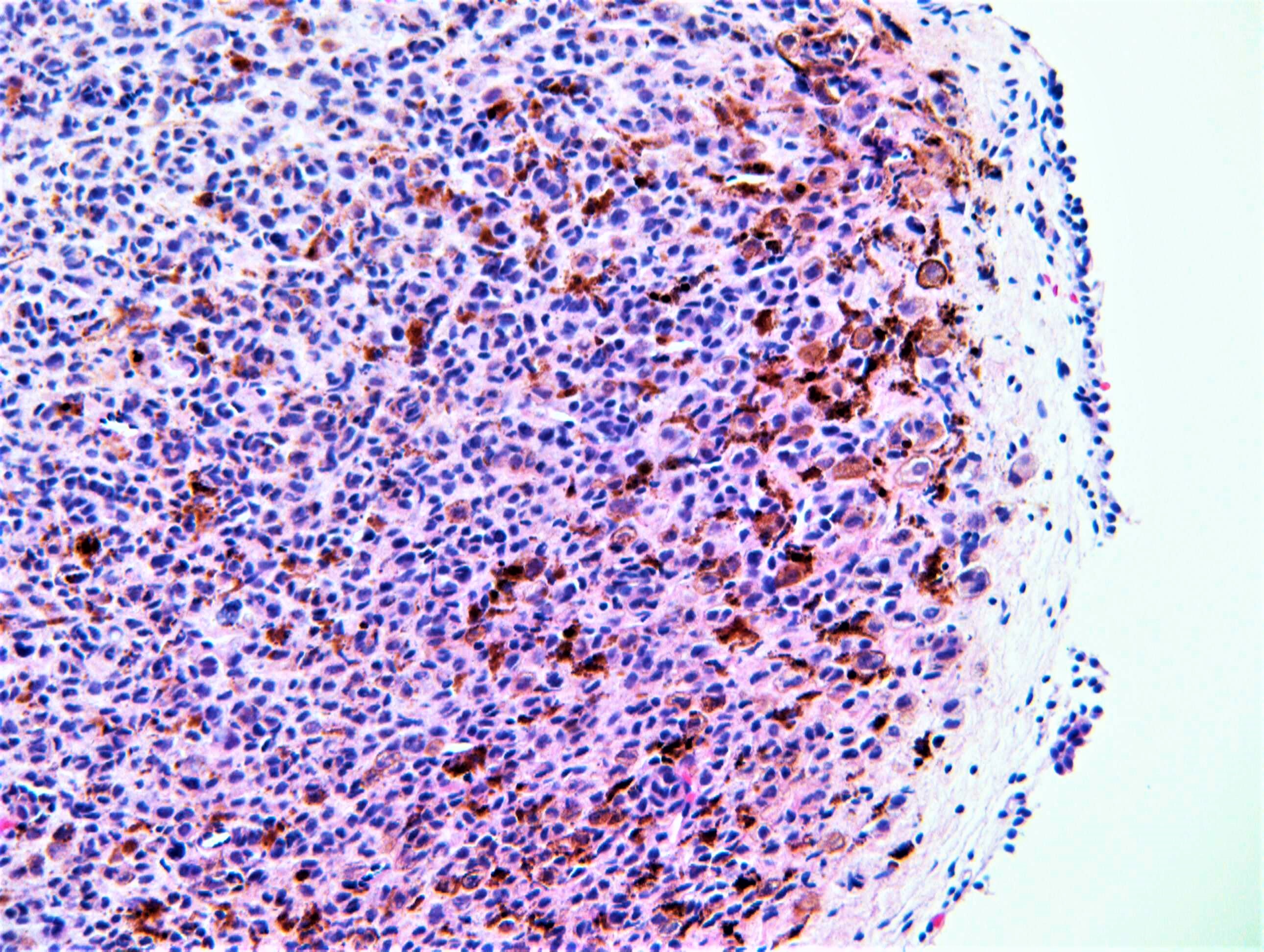
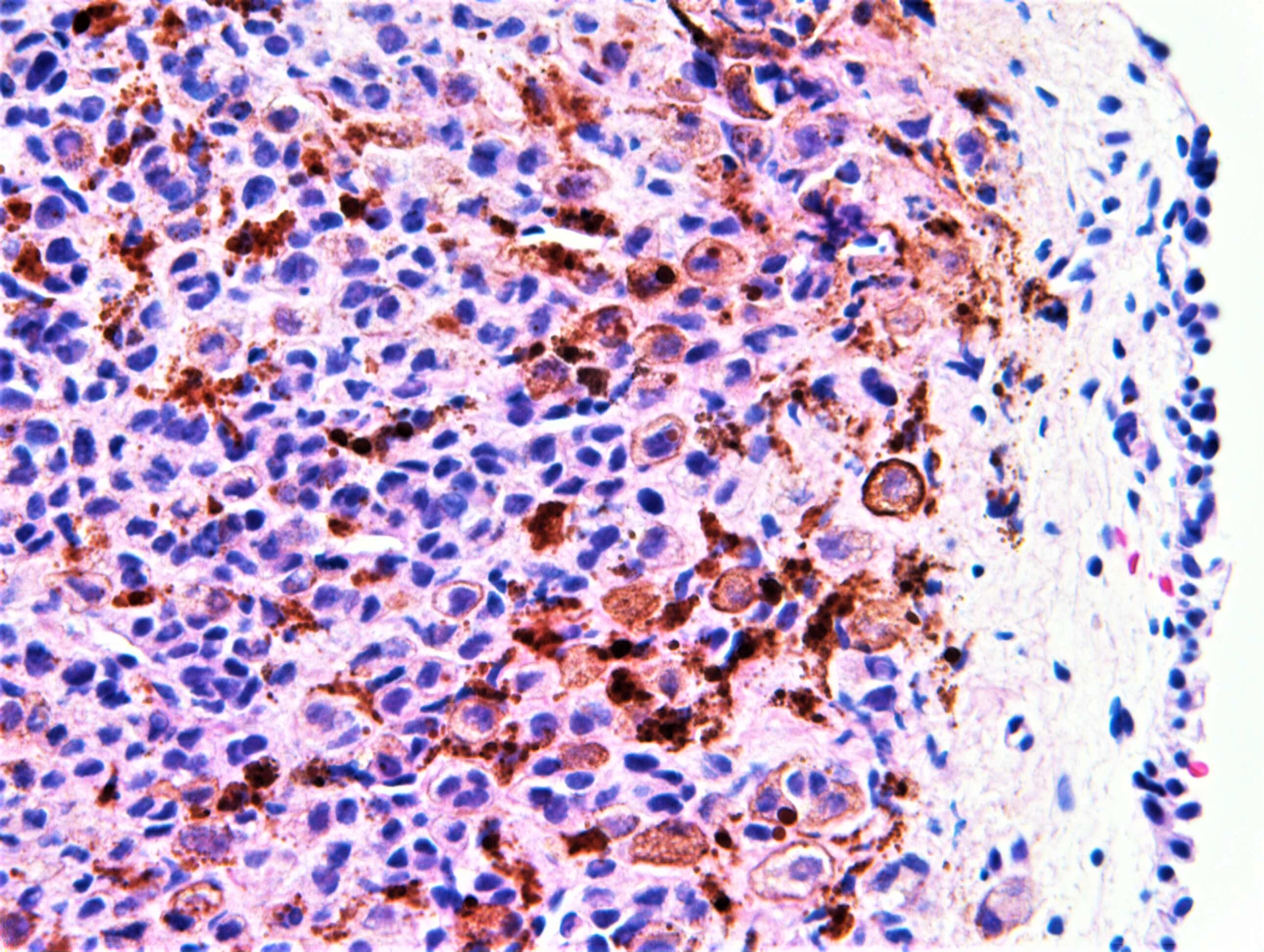
As what the melanocytic lesion represents, it is very difficult just tonmake the diagnosis on these figures. I do not see any melanoma in situ component in the epithelium. The nodule of melanocytes is quite cellular and pigmented. I can't tell whether there are mitoses or not. The melanocytes are quite monotonous without clear cut maturation. If this is a conjunctival lesion, I do not see any epithelial cysts that we usually see in benign nevi. So I'm kind of worried that this could be a melanoma of the urethra? A cellular blue nevus also crossed my mind but I need to rule out melanoma first, particularly in this location. Clinical impression might help.


Share this comment


Link to comment
share_externally

vincenzo polizzi

Posted

Agree with above. I was thinking of some association between urethral female caruncle and melanocytic pigmented nevus ( but maybe he is a men, who knows...)

Share this comment


Link to comment
share_externally

Dr. Richard Carr

Posted

It's bladder and there is a history of ocular/retinal melanoma 3 years previously. Patient underwent cystoscopy for haematuria. We reported as most likely metastatic melanoma. Well done Anh!!

Share this comment


Link to comment
share_externally

Dr. Richard Carr

Posted

I've posted the answer to 1969 (last Friday's). Seasons greetings.

Share this comment


Link to comment
share_externally

3 hours ago, Dr. Richard Carr said:

It's bladder and there is a history of ocular/retinal melanoma 3 years previously. Patient underwent cystoscopy for haematuria. We reported as most likely metastatic melanoma. Well done Anh!!

Thanks

Share this comment


Link to comment
share_externally


Join the conversation

You can post now and register later. If you have an account, sign in now to post with your account.

Guest
Add a comment...

×   Pasted as rich text.   Paste as plain text instead

  Only 75 emoji are allowed.

×   Your link has been automatically embedded.   Display as a link instead

×   Your previous content has been restored.   Clear editor

×   You cannot paste images directly. Upload or insert images from URL.

Loading...

×
×
  • Create New...