Jump to content
In this section we have spot diagnoses posted on a daily basis since June 2010, now over 4000! You can review the archived cases and read the suggested diagnoses by users and the final comment by the contributors.
Case are uploaded each week day by 10 am UK time with the correct diagnosis will generally be posted at 8 pm UK time. Why not view the most recent spot diagnosis and proffer a diagnosis?

Case Number : Case 1976 - 28 Dec 2017 Posted By: Dr. Richard Carr

Please read the clinical history and view the images by clicking on them before you proffer your diagnosis.
Submitted Date :
   (0 reviews)

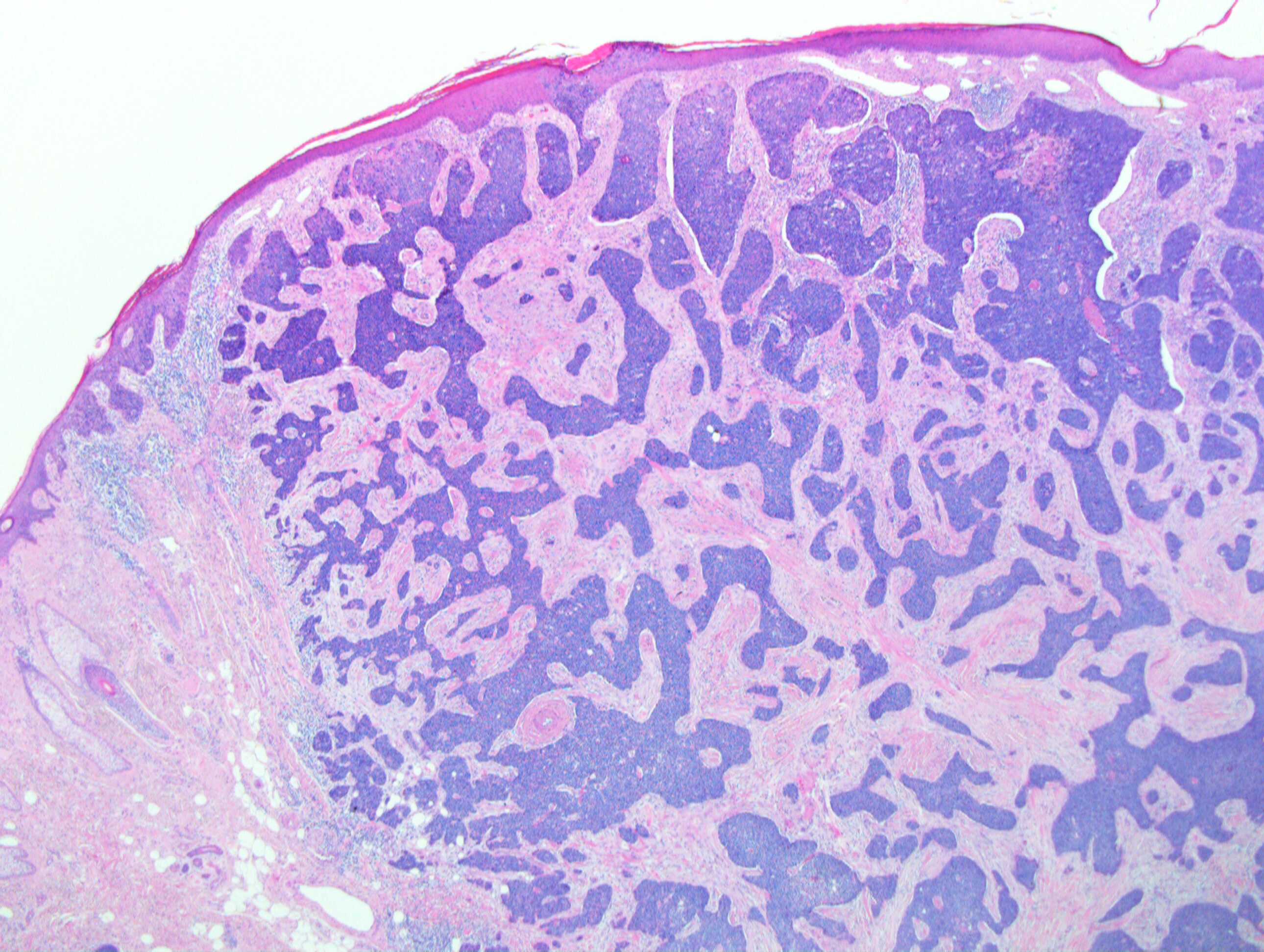
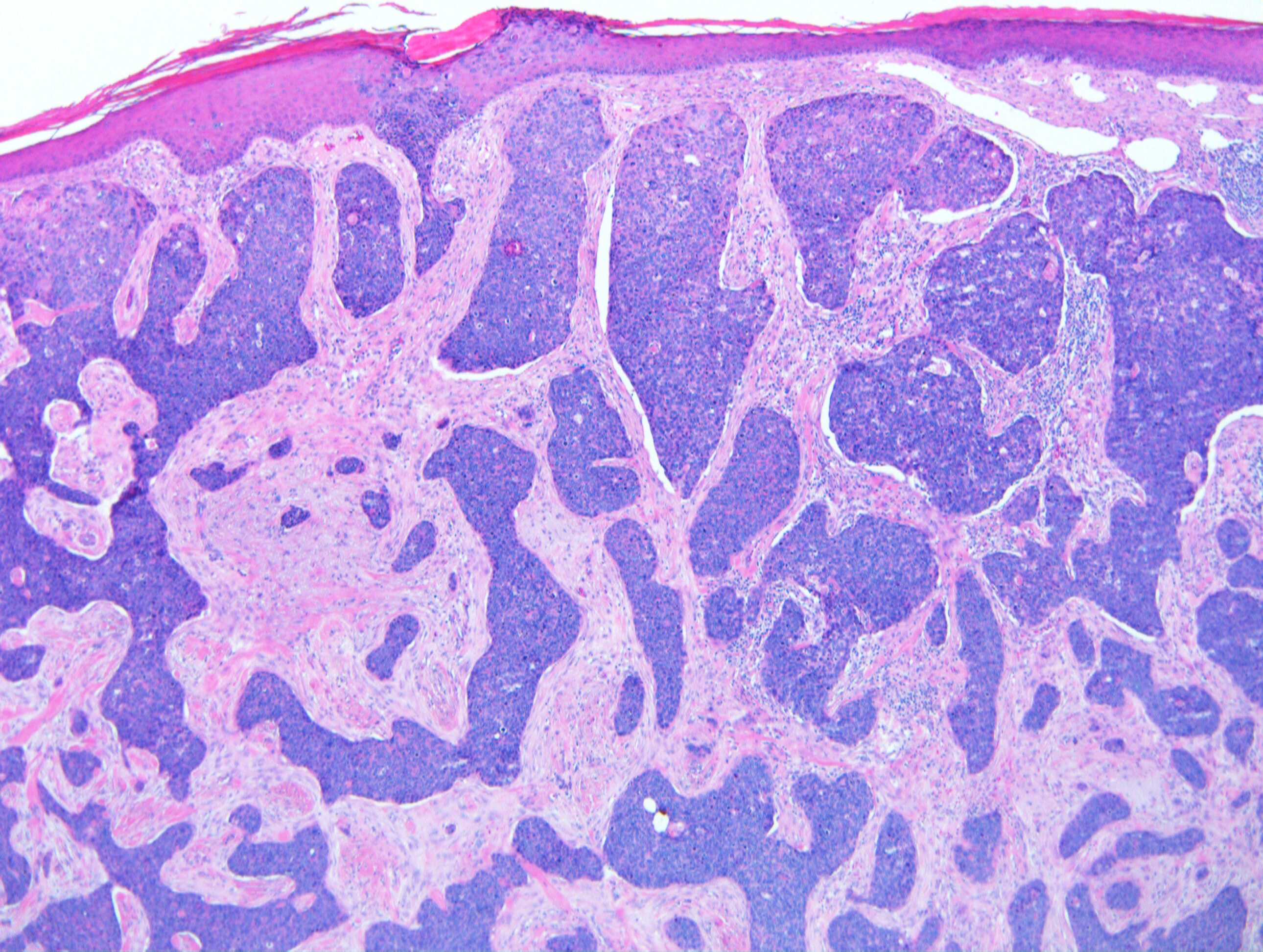
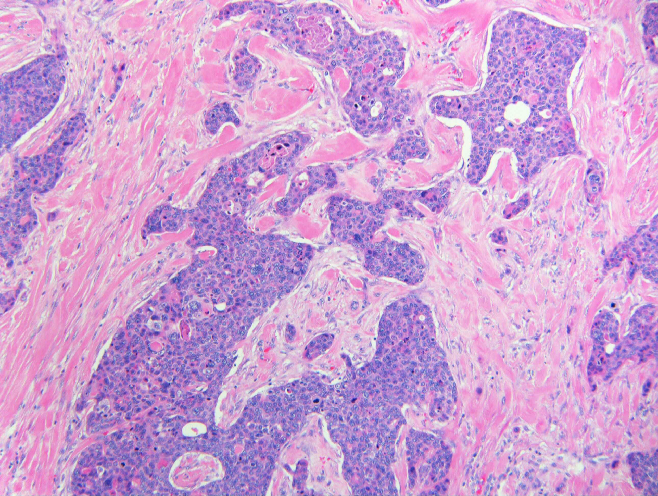
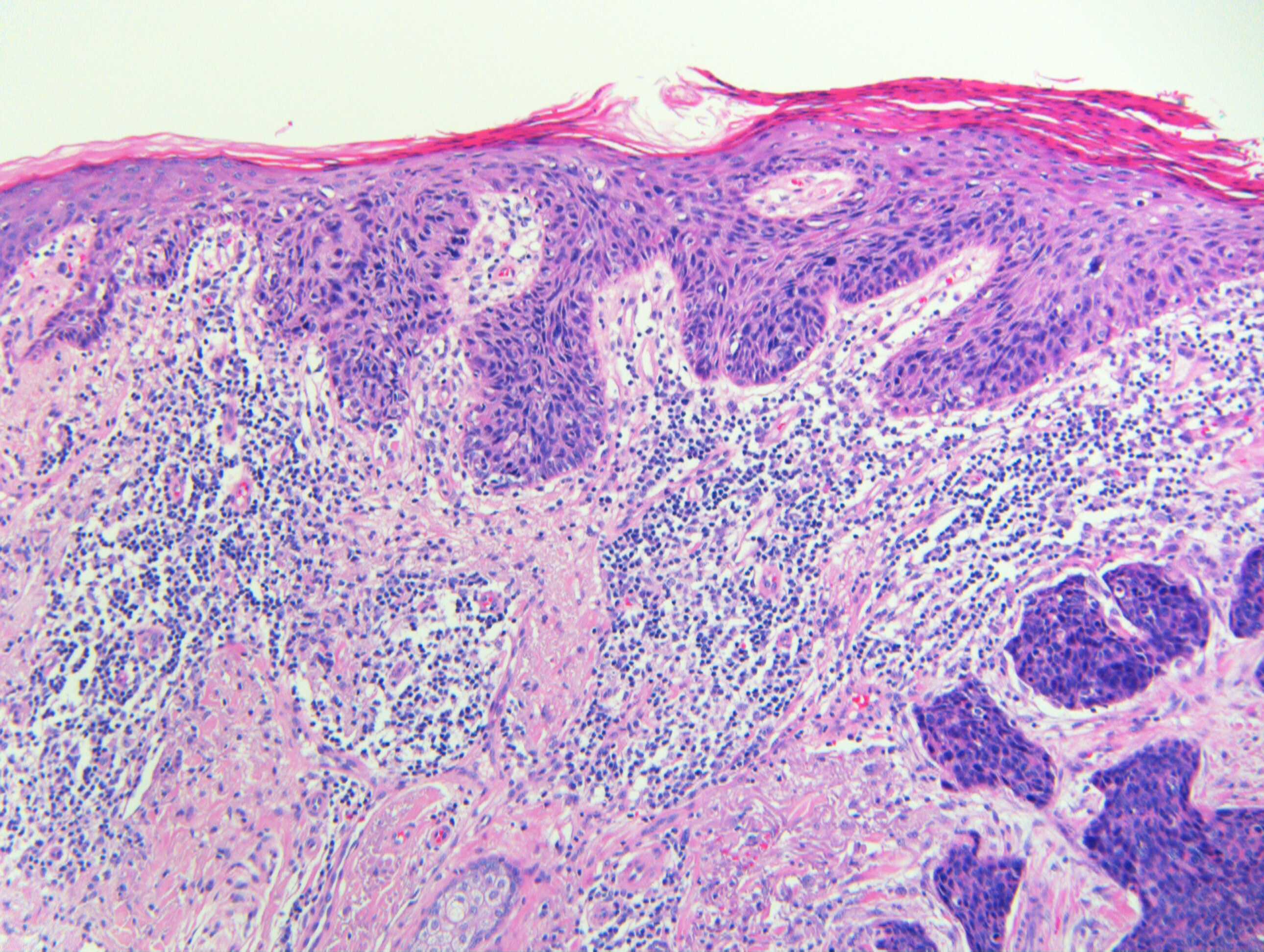
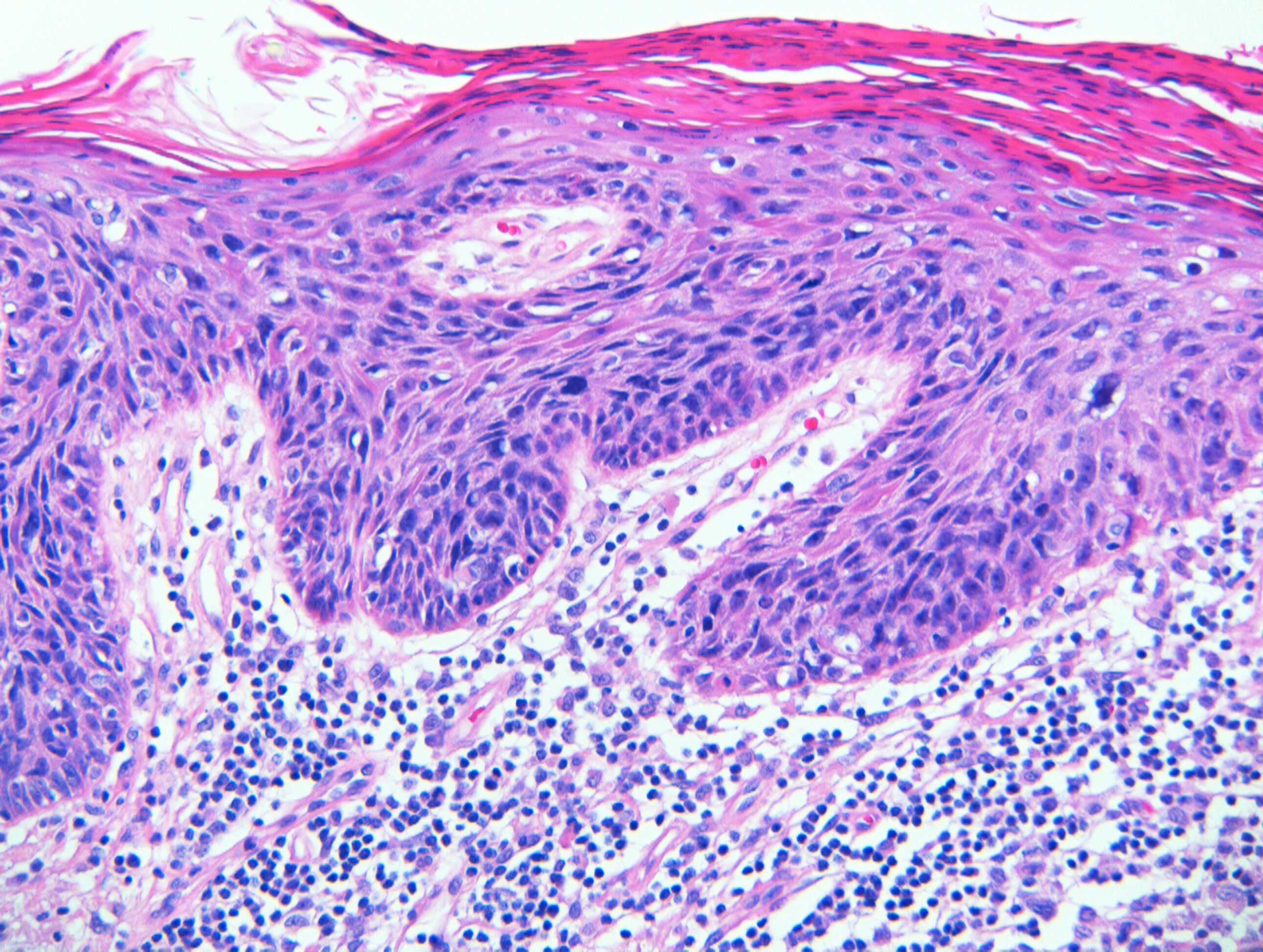
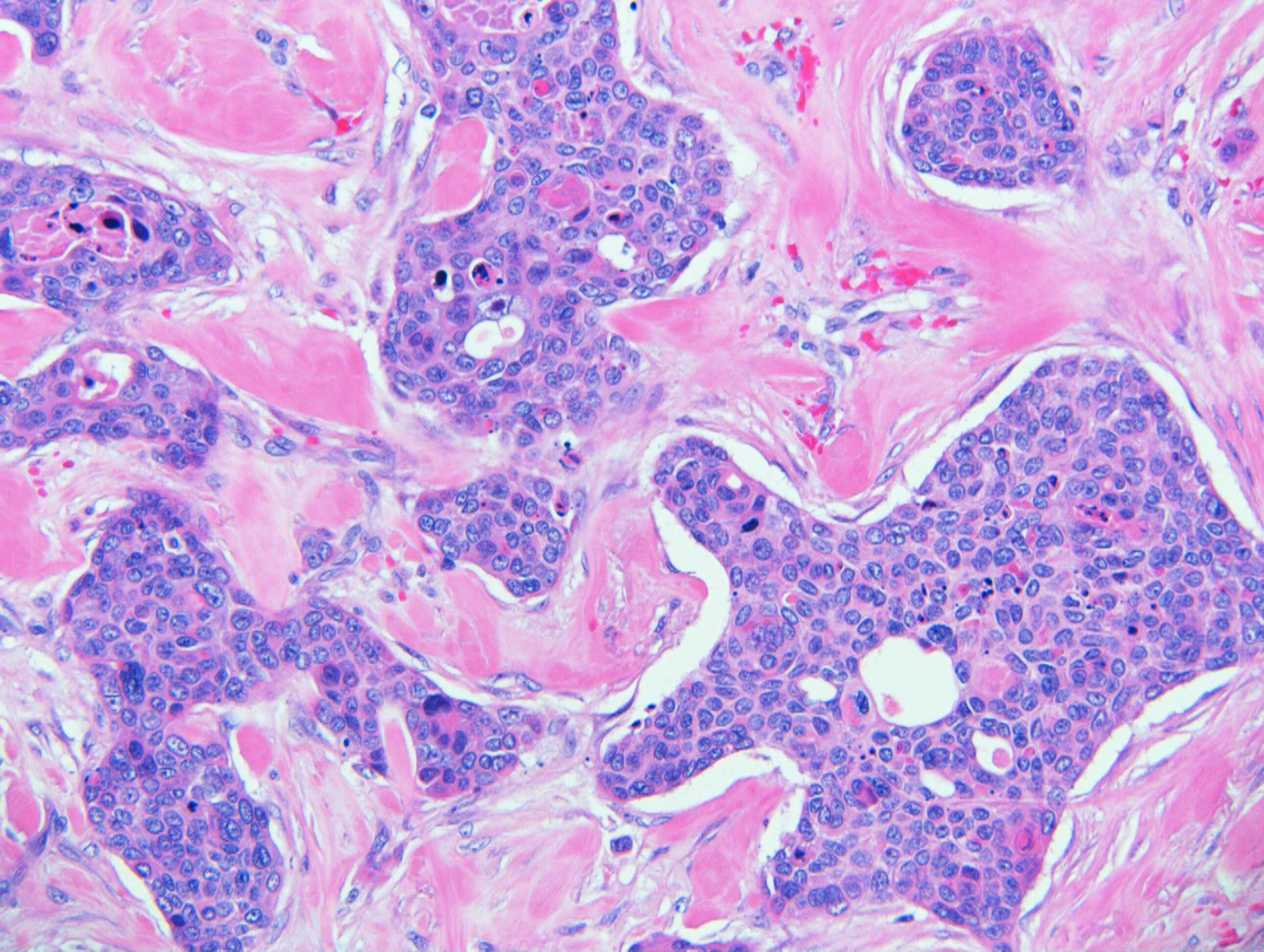
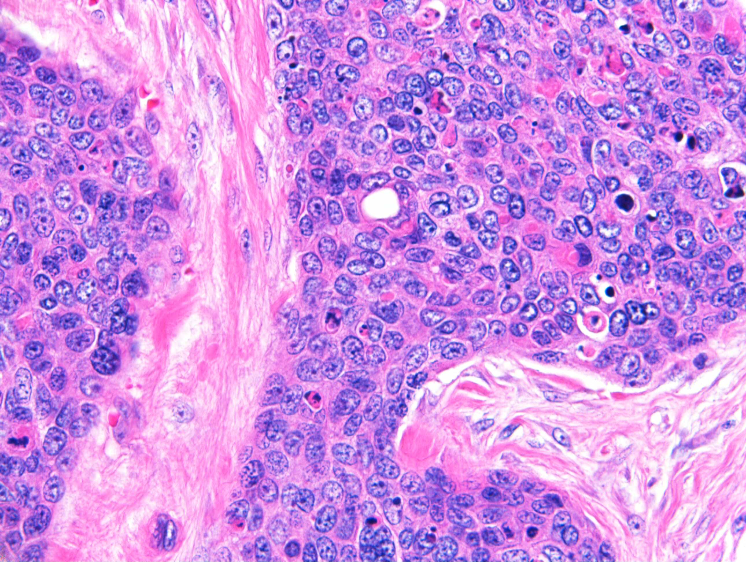
Clinical Details: M70. Ear.

Edited by Admin_Dermpath


  Report Record

User Feedback


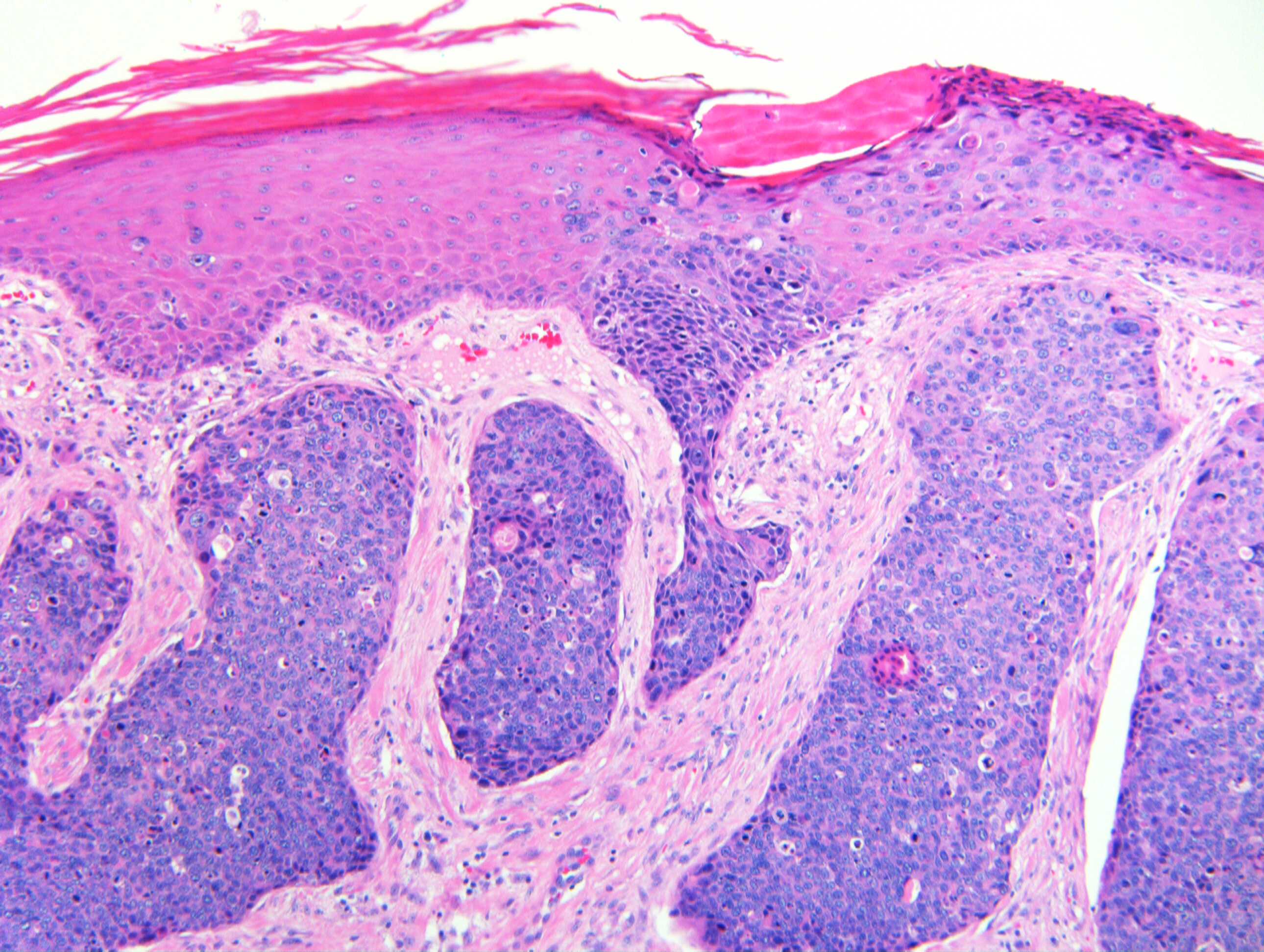
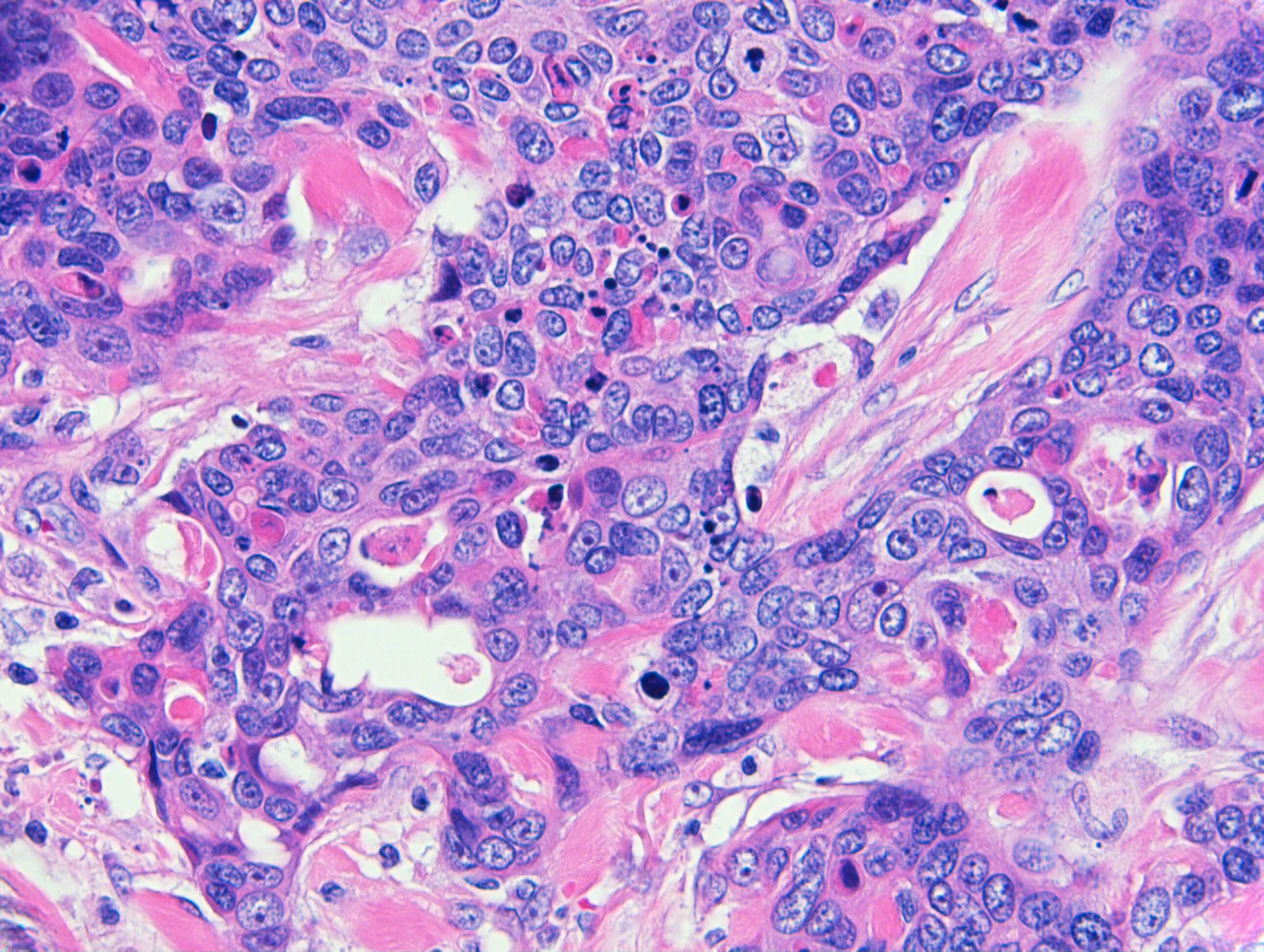
Looks like there is squamous cell carcinoma in situ. Infiltrative nests of malignant basaloid or squamous cell are seen in the dermis. The last two figures appear to show duct differentiation in the nests of tumor cells. 

Squamoid eccrine ductal carcinoma vs. basal cell carcinoma with ductal differentation. Favor the former.

Share this comment


Link to comment
share_externally

Agree with porocarcinoma. The clue that it is arising from the acrosyringium is in fig 4.

Share this comment


Link to comment
share_externally

Dr. Richard Carr

Posted

I'm awaiting some IHC to be posted.

Share this comment


Link to comment
share_externally

Dr. Richard Carr

Posted

I called it a high grade cutaneous carcinoma, basaloid (looks a little "bowenoid" i.e. undifferentiated to me) with focal ductal differentiation. I don't argue with calling it a porocarcinoma (high grade) or adenosquamous carcinoma (high grade).  Not sure if the focus of Bowen's at the edge is separate incidental as it was p16 rather diffuse c/w less staining in the rest of the tumour. I will ask again for the IHC to be posted (I did try!).

Share this comment


Link to comment
share_externally

Dr. Richard Carr

Posted

Looks like the IHC is now posted. On p63 we seen the central ductal differentiation losses the staining and is focally positive for Cam5.2 (low molecular keratins that are NOT normally seen in cutaneous SCC. I managed to loose the BerEP4 slide which also showed focal ductal luminal staining). Bowen's or bowenoid actinic keratosis is almost invariably diffusely nuclear p16 positive not the case in the invasive tumour here although both lesions are "positive" for p53.

Share this comment


Link to comment
share_externally


Join the conversation

You can post now and register later. If you have an account, sign in now to post with your account.

Guest
Add a comment...

×   Pasted as rich text.   Paste as plain text instead

  Only 75 emoji are allowed.

×   Your link has been automatically embedded.   Display as a link instead

×   Your previous content has been restored.   Clear editor

×   You cannot paste images directly. Upload or insert images from URL.

Loading...

×
×
  • Create New...