Jump to content
In this section we have spot diagnoses posted on a daily basis since June 2010, now over 4000! You can review the archived cases and read the suggested diagnoses by users and the final comment by the contributors.
Case are uploaded each week day by 10 am UK time with the correct diagnosis will generally be posted at 8 pm UK time. Why not view the most recent spot diagnosis and proffer a diagnosis?

Case Number : Case 1746 - 6 February - Dr Richard A Carr Posted By: Guest

Please read the clinical history and view the images by clicking on them before you proffer your diagnosis.
Submitted Date :
   (0 reviews)

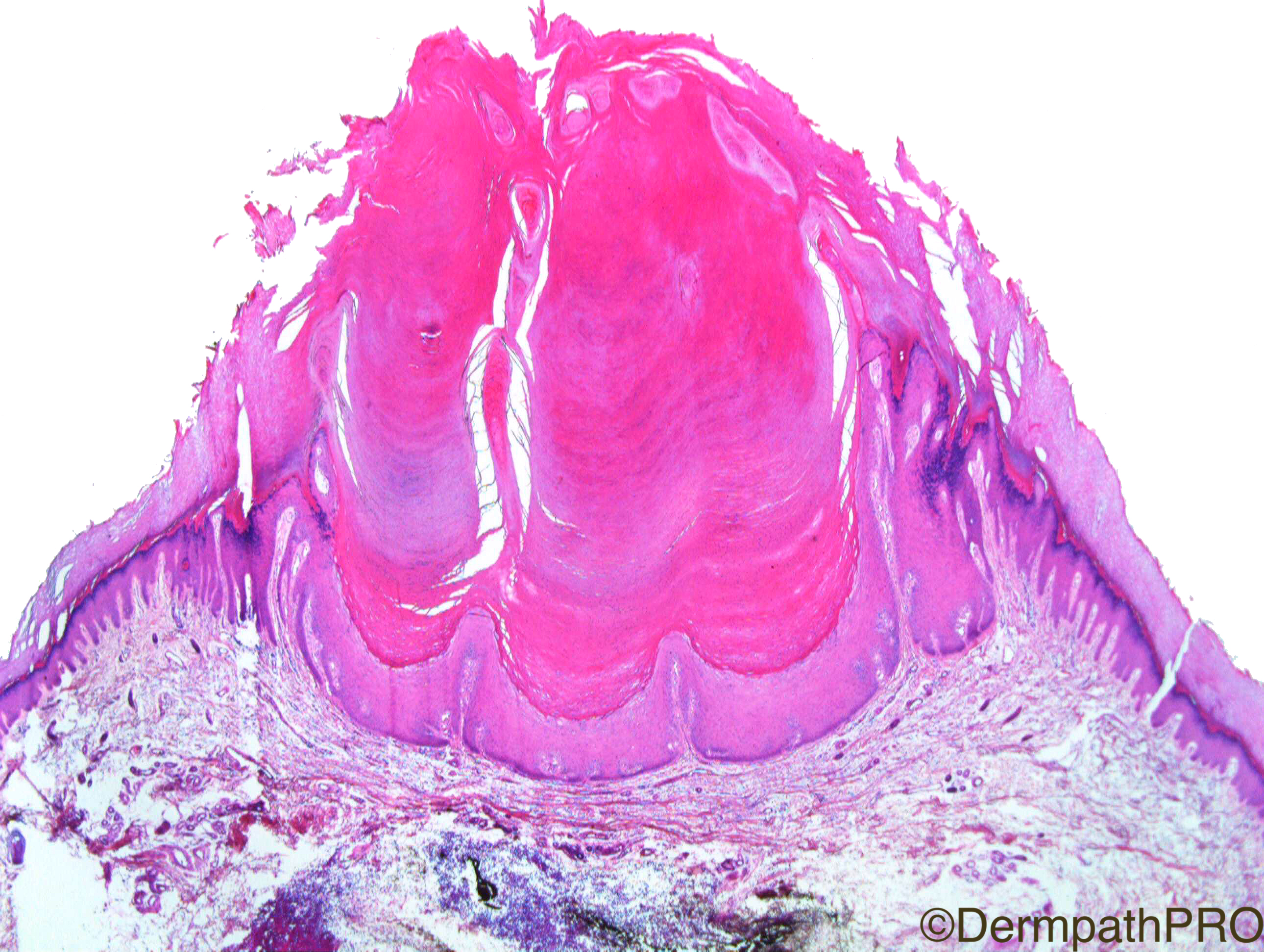
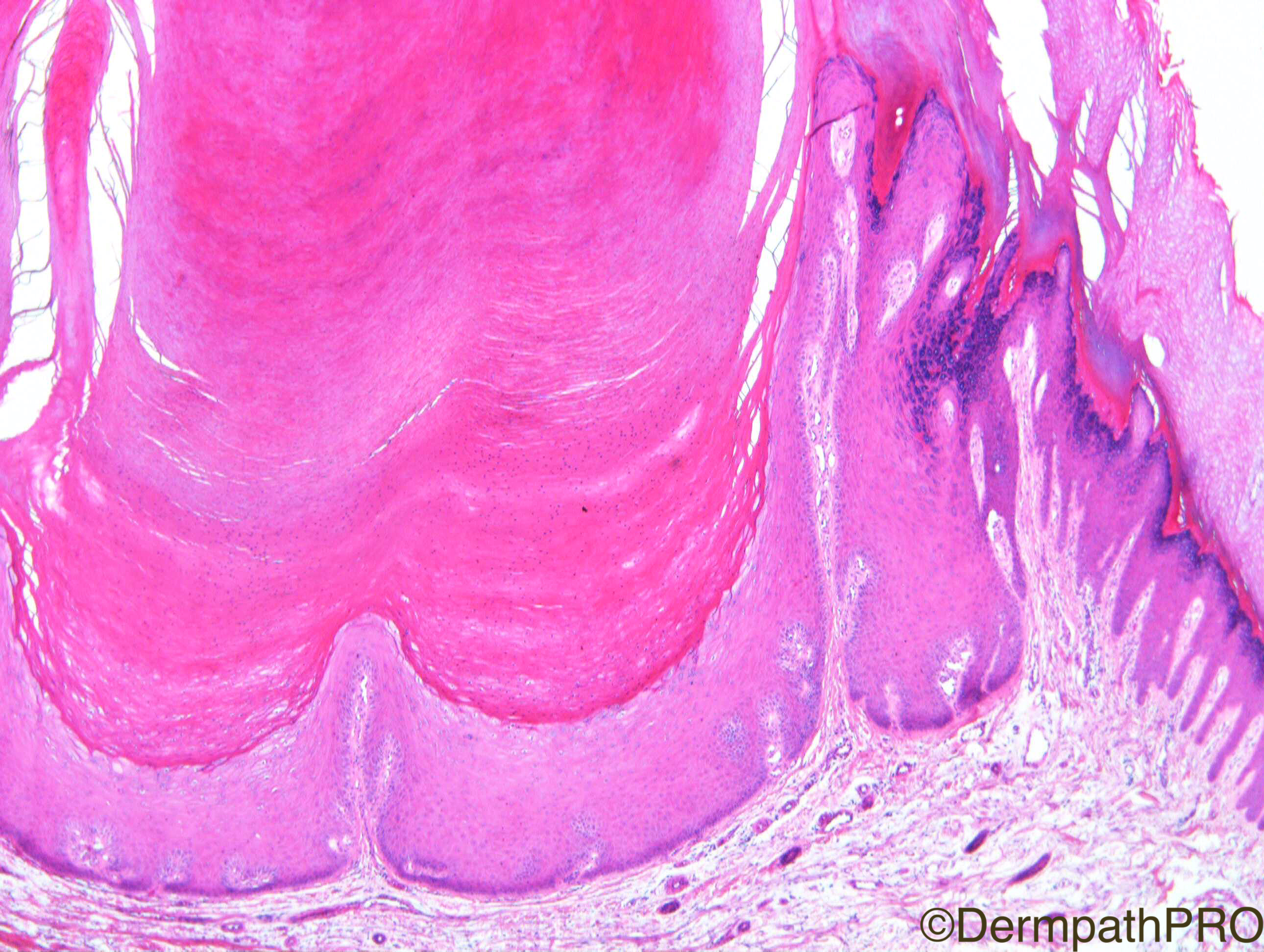
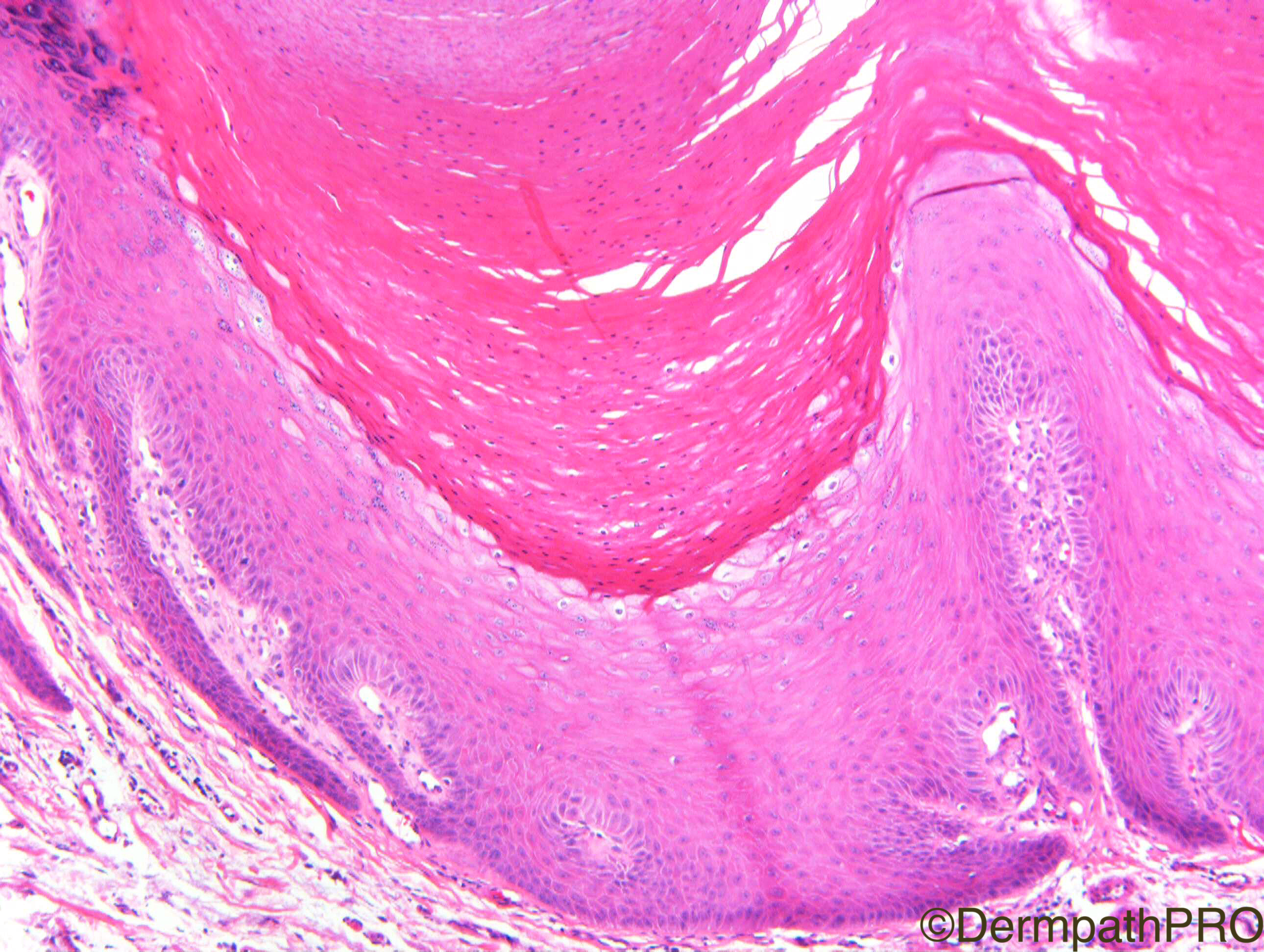
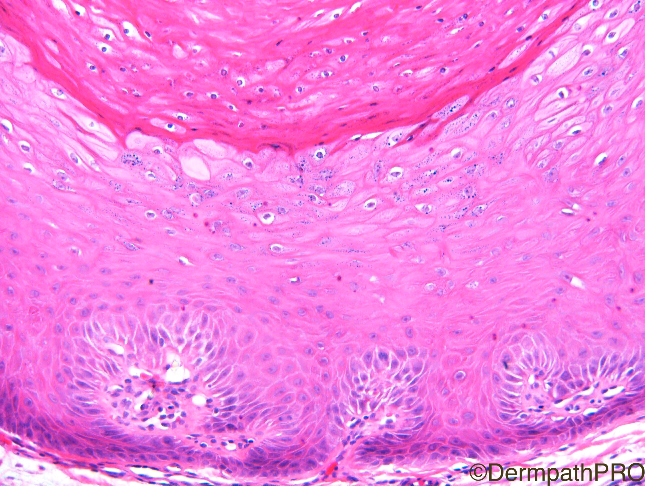
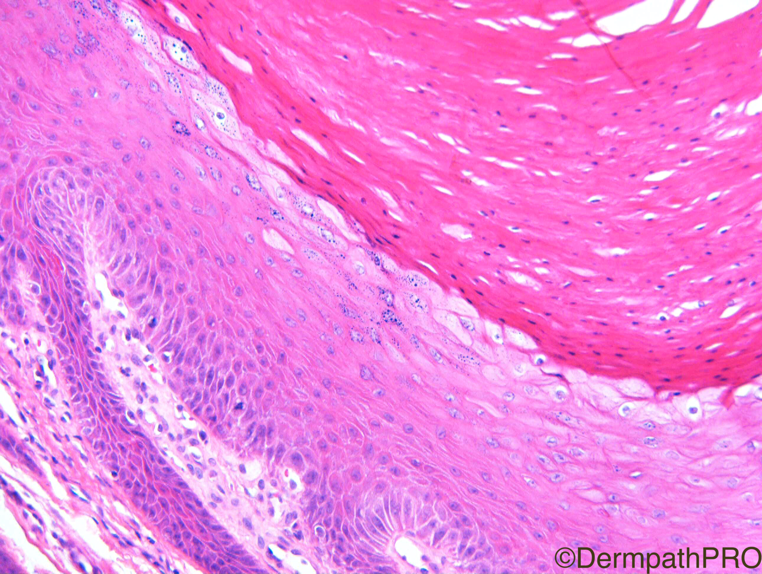
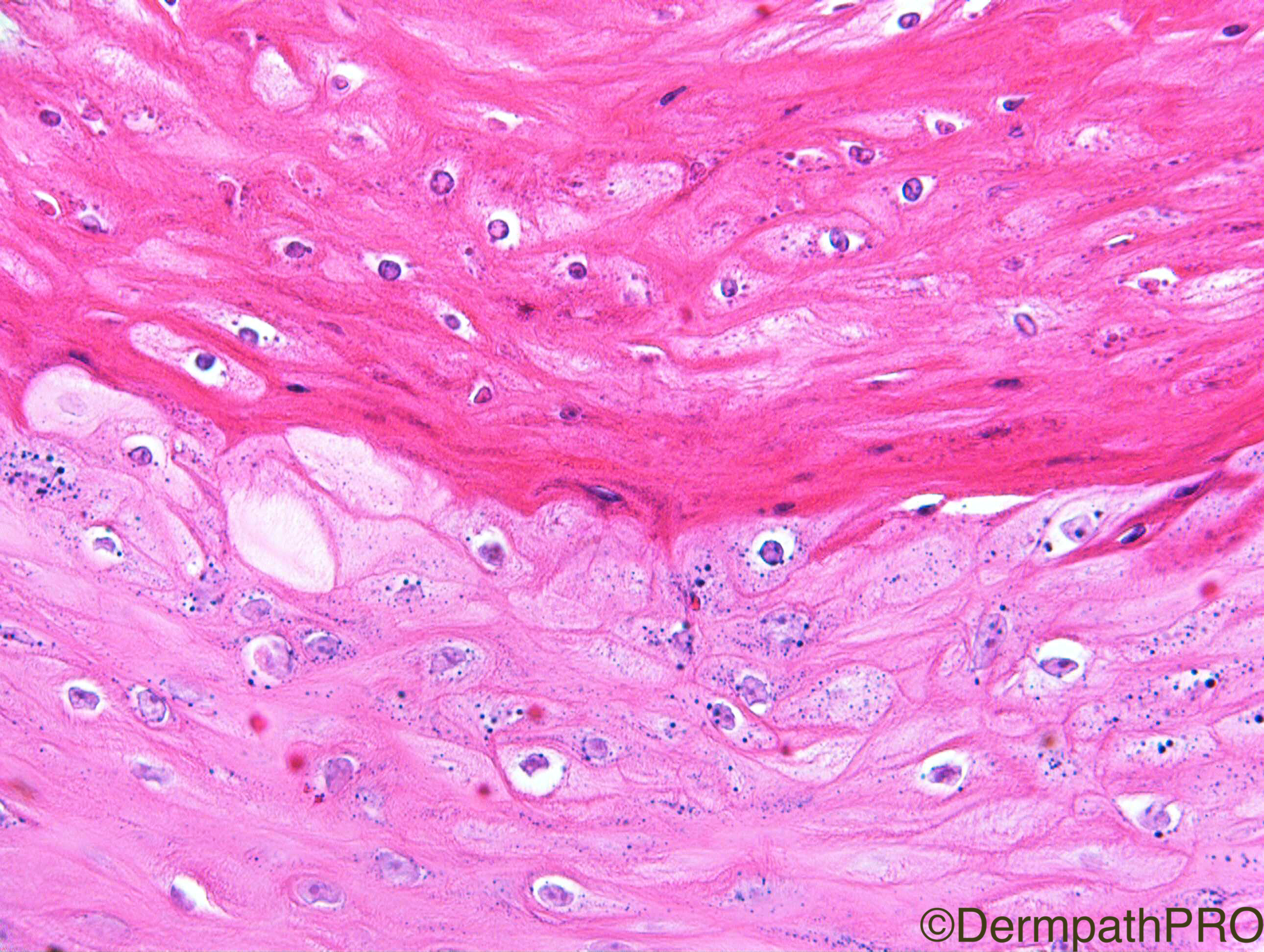
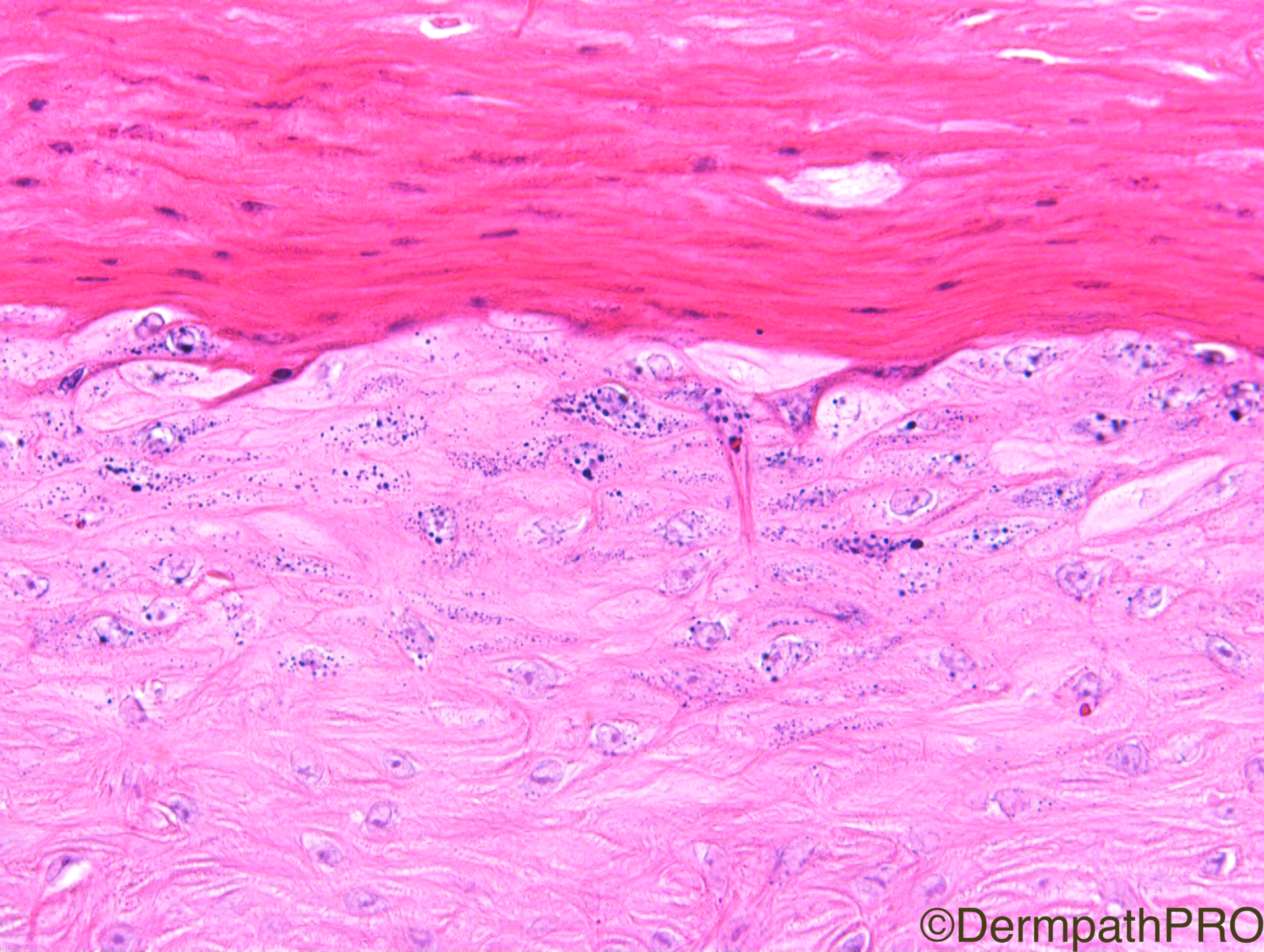
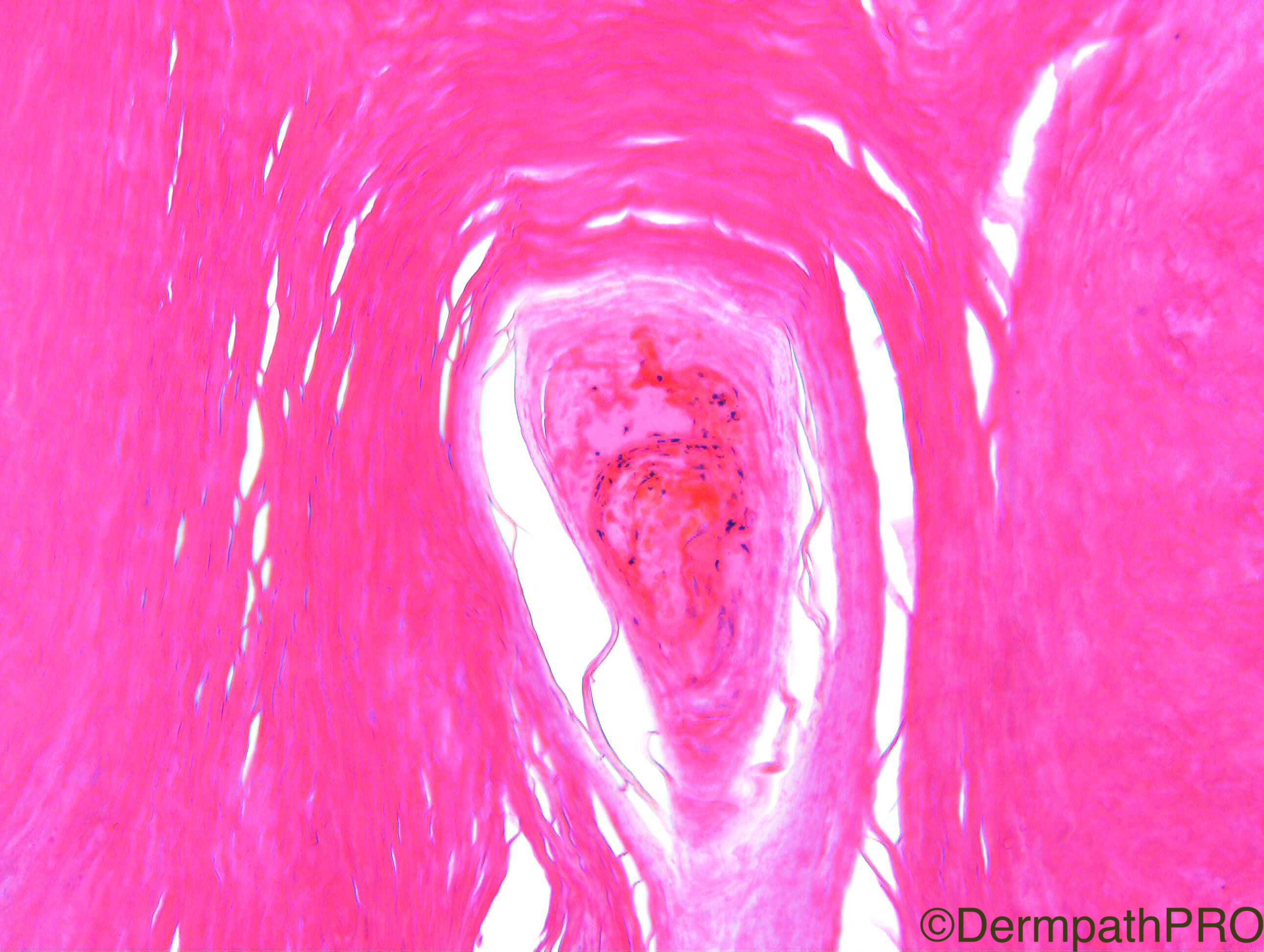
Clinical History: F80. 8 week history of keratotic nodule, dorsum left hand.  Clinically KA.

Case Posted by Dr Richard A Carr

  Report Record

User Feedback


Admin_Dermpath

Posted

Get your week off to a great start with this lovely case from Dr Richard A Carr


Geoff Cross - DermpathPRO Projects

Share this comment


Link to comment
share_externally

urmilapandey

Posted (edited)

crateriform lesion, thickened epidermis with upper layers showing HPV-like features, haemorrage in stratum corneum, no inflammation, no obvious elastic fibres in the epidermis and I couldn't see any convincing epidermodysplasia. considered regressed KA but to me this looks like an HPV induced lesion ?odd verruca.

Edited by urmilapandey

Share this comment


Link to comment
share_externally

Raul Perret

Posted

only differential i can think of is trichilemmal horn although for me this case is better classified as a verruca as you guys mentioned

Share this comment


Link to comment
share_externally

vincenzo polizzi

Posted

Eosinophilic nuclear inclusions of keratinocytes of stratum corneum and infolding of elongated rete ridges towards a virtual central point of the base are in keeping with an old Viral Wart, I think. Not atypia nor glassy cytoplasmic changes as clues to KA, nor dermal scarring

Share this comment


Link to comment
share_externally

Nitin Khirwadkar

Posted

Agree. Thought about an EV acanthoma, but the cytoplasm lacks the slate grey colour.

Share this comment


Link to comment
share_externally

Dr. Richard Carr

Posted

This lesion reminded me of a subset of the epidermal proliferative lesions we see in the patients on BRAF inhibitors for metastatic melanoma. I think as you all agreed it's just a viral wart and I also think it probably is HPV associated. However I do find the term "benign verrucous keratosis" quite an appealing appellation for such lesions especially when one cannot always force it easily into a viral wart. I think there is clearly a subset of sporadic verrucous keratoses (akin to the lesions seen in BRAFi receiving patients) that are probably HPV associated but that don't always have clearly diagnostic cytological features for HPV with certainty.  It seems the BRAFi associated lesions are often associated with the Beta type HPVs so perhaps Urmilla and Nitin are on to something.  Obviously this lesion grew rapidly so it is clinically mimicking the pattern seen in BRAFi induced lesions. I wonder if it might also have a mutation in a RAS or epidermal growth receptor gene etc.

https://www.ncbi.nlm.nih.gov/pmc/articles/PMC4452444/

Share this comment


Link to comment
share_externally


Join the conversation

You can post now and register later. If you have an account, sign in now to post with your account.

Guest
Add a comment...

×   Pasted as rich text.   Paste as plain text instead

  Only 75 emoji are allowed.

×   Your link has been automatically embedded.   Display as a link instead

×   Your previous content has been restored.   Clear editor

×   You cannot paste images directly. Upload or insert images from URL.

Loading...

×
×
  • Create New...