Jump to content
In this section we have spot diagnoses posted on a daily basis since June 2010, now over 4000! You can review the archived cases and read the suggested diagnoses by users and the final comment by the contributors.
Case are uploaded each week day by 10 am UK time with the correct diagnosis will generally be posted at 8 pm UK time. Why not view the most recent spot diagnosis and proffer a diagnosis?

Case Number : Case 1761 - 27 February - Dr Limin Yu Posted By: Guest

Please read the clinical history and view the images by clicking on them before you proffer your diagnosis.
Submitted Date :
   (0 reviews)

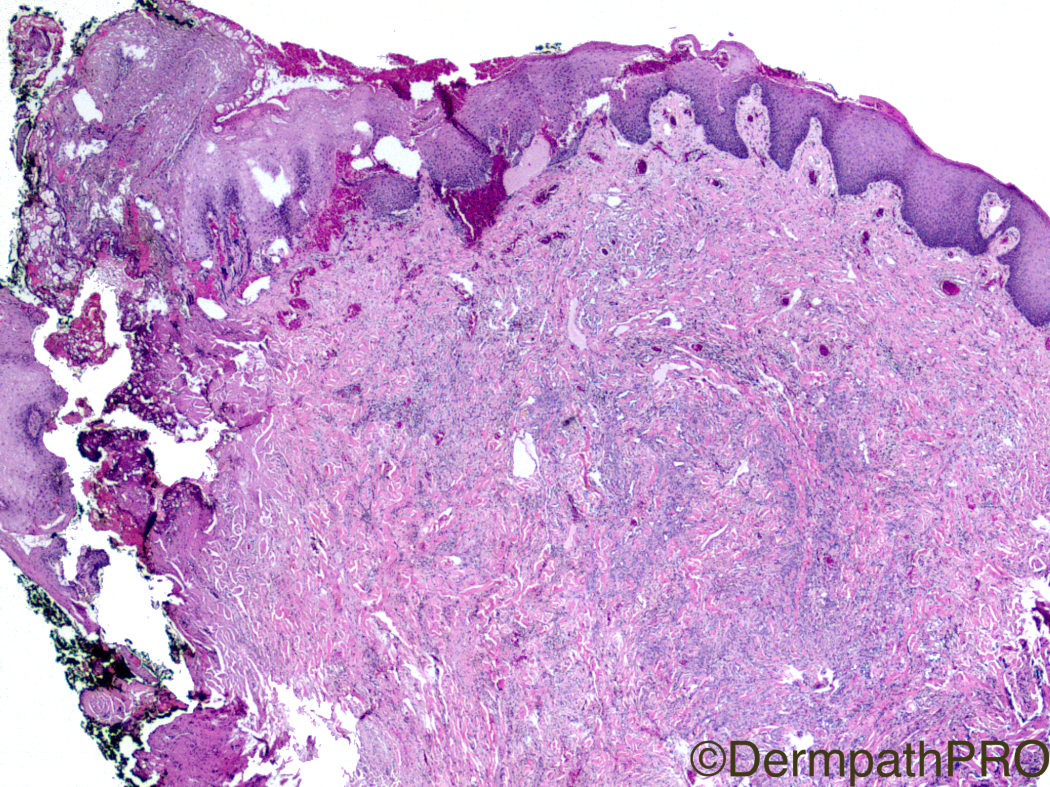
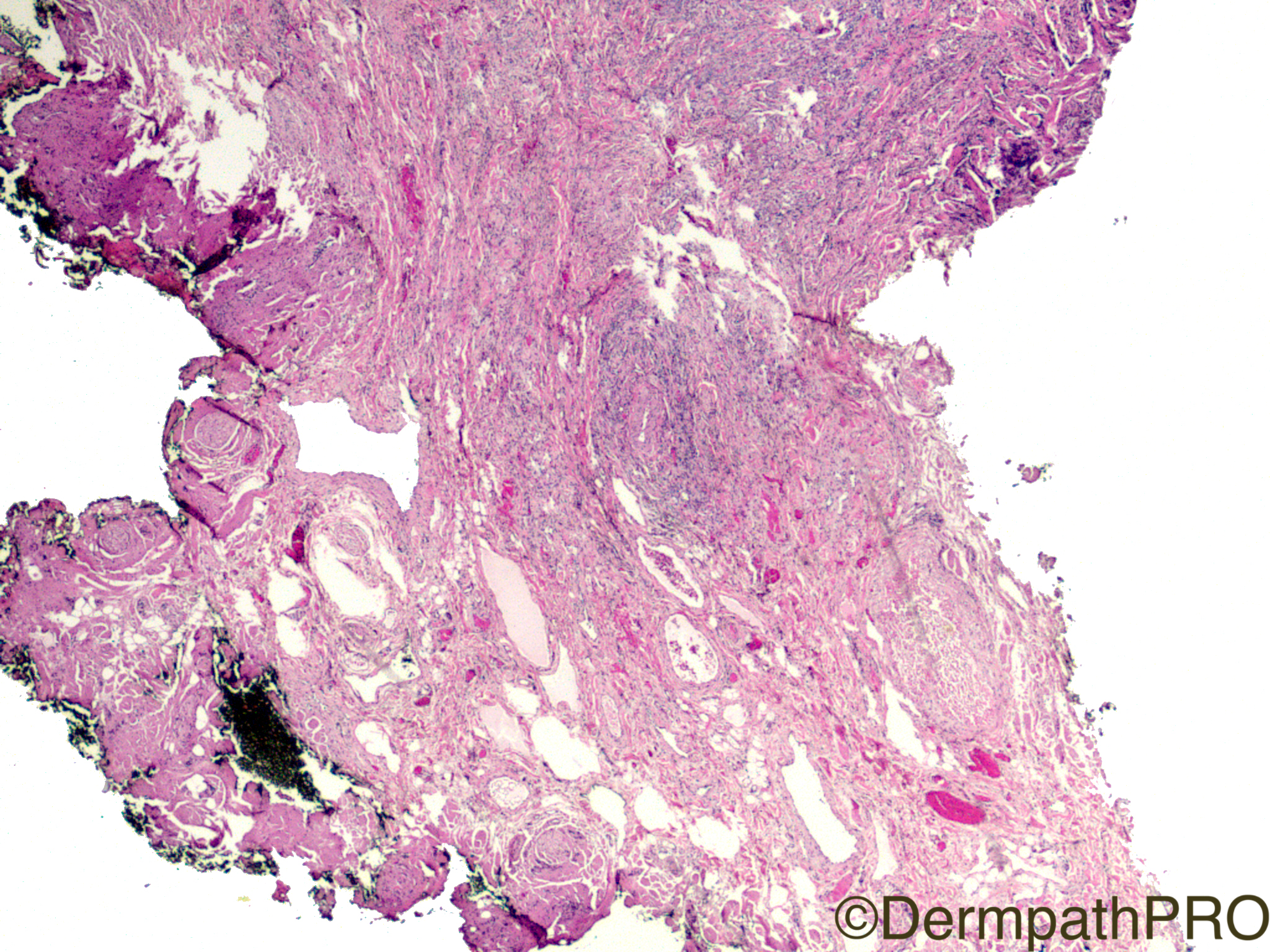
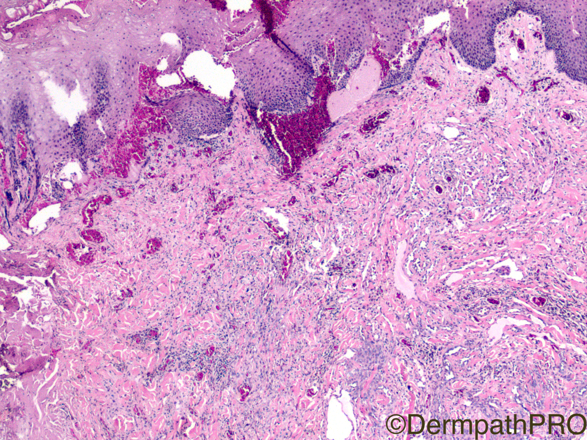
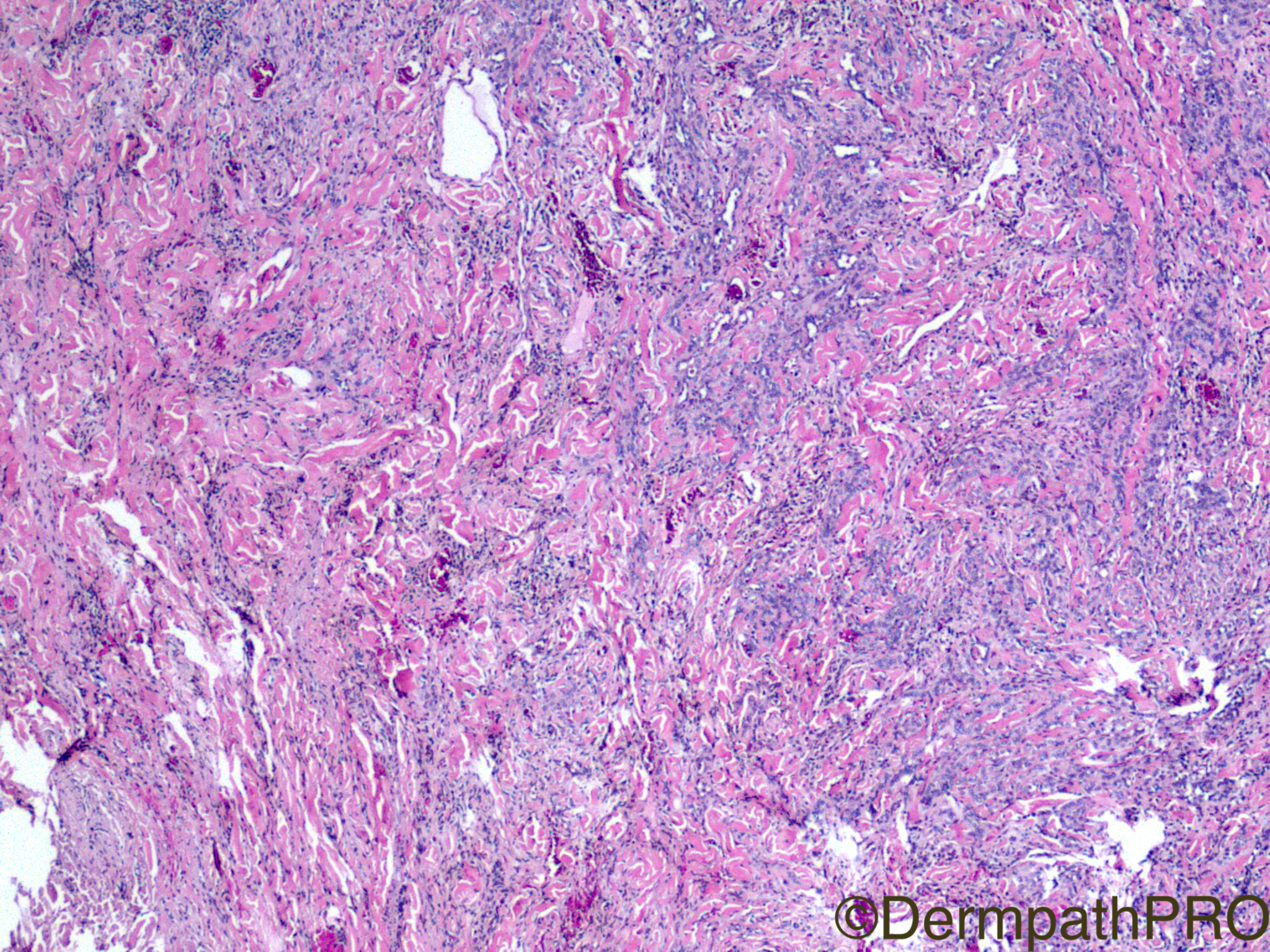
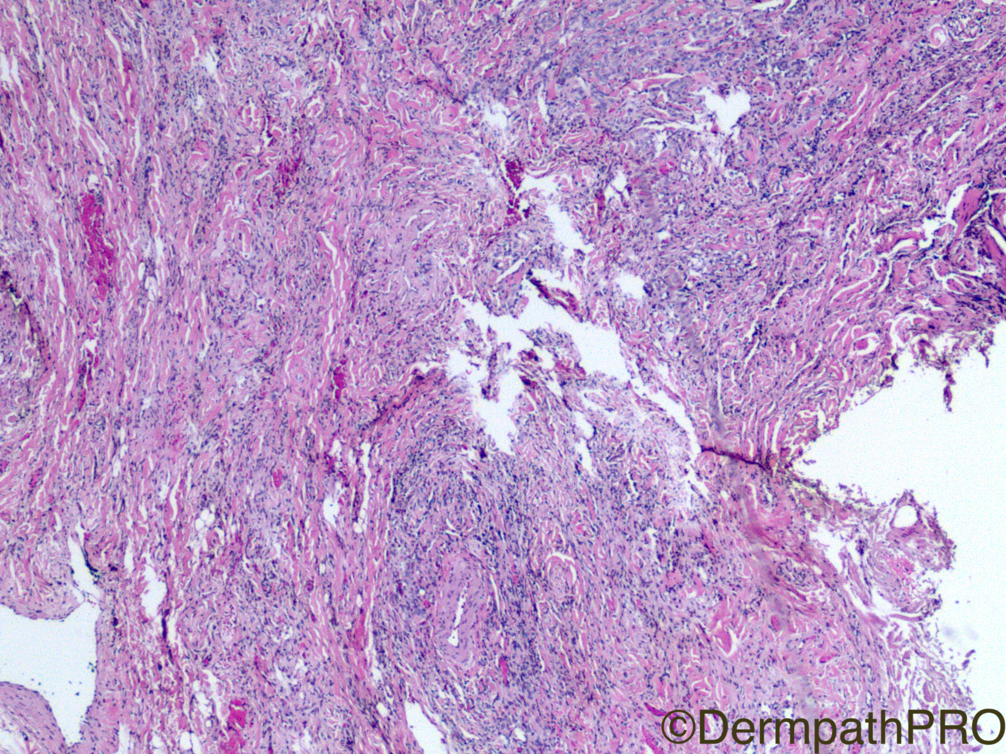
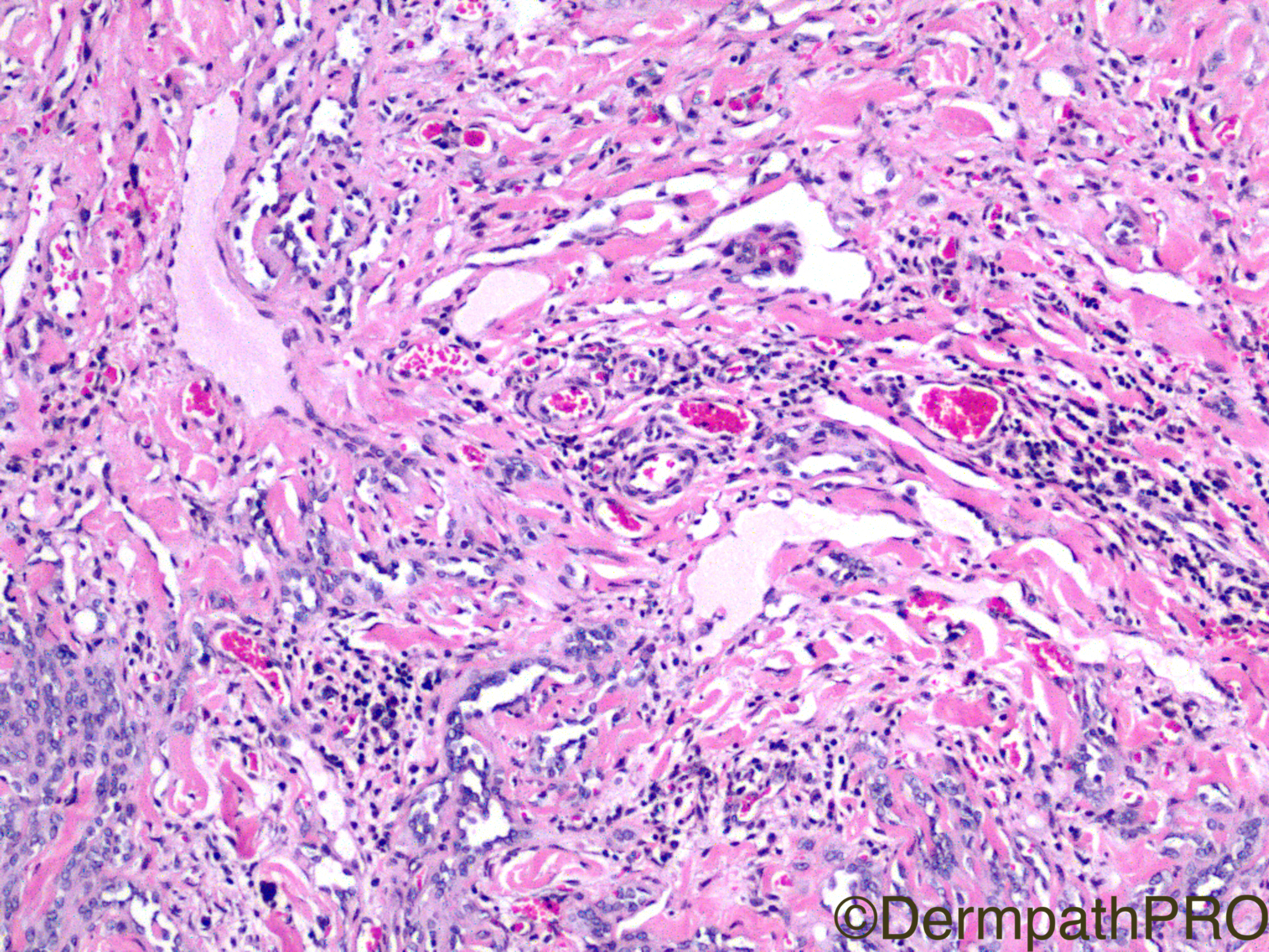
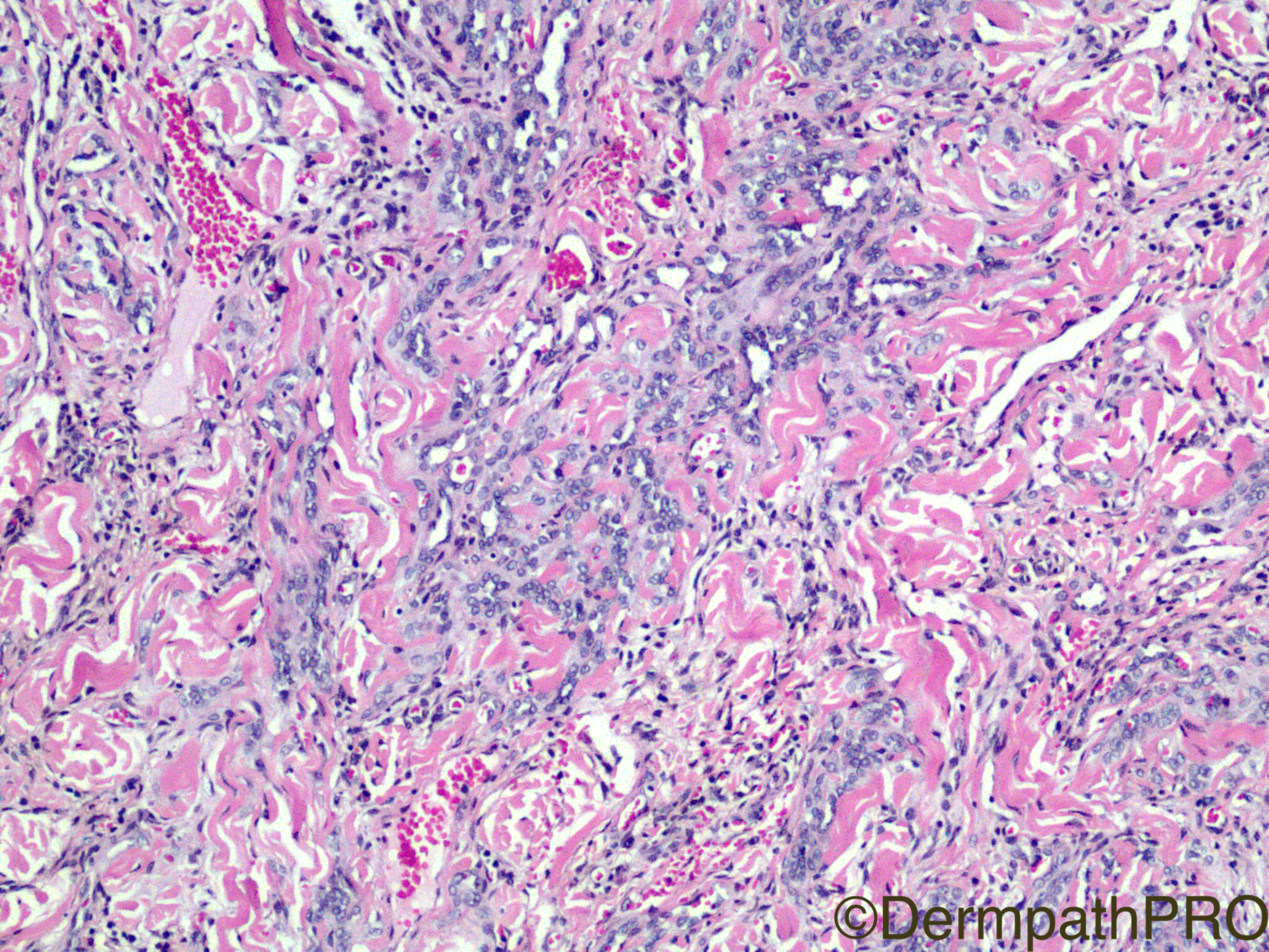
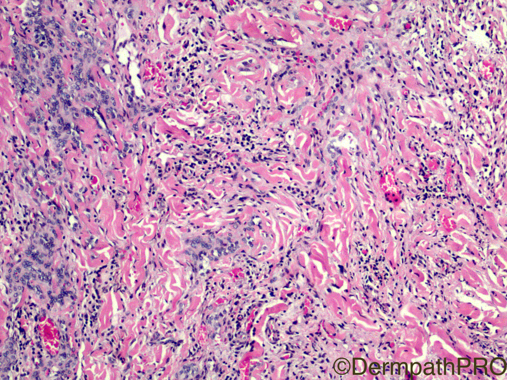
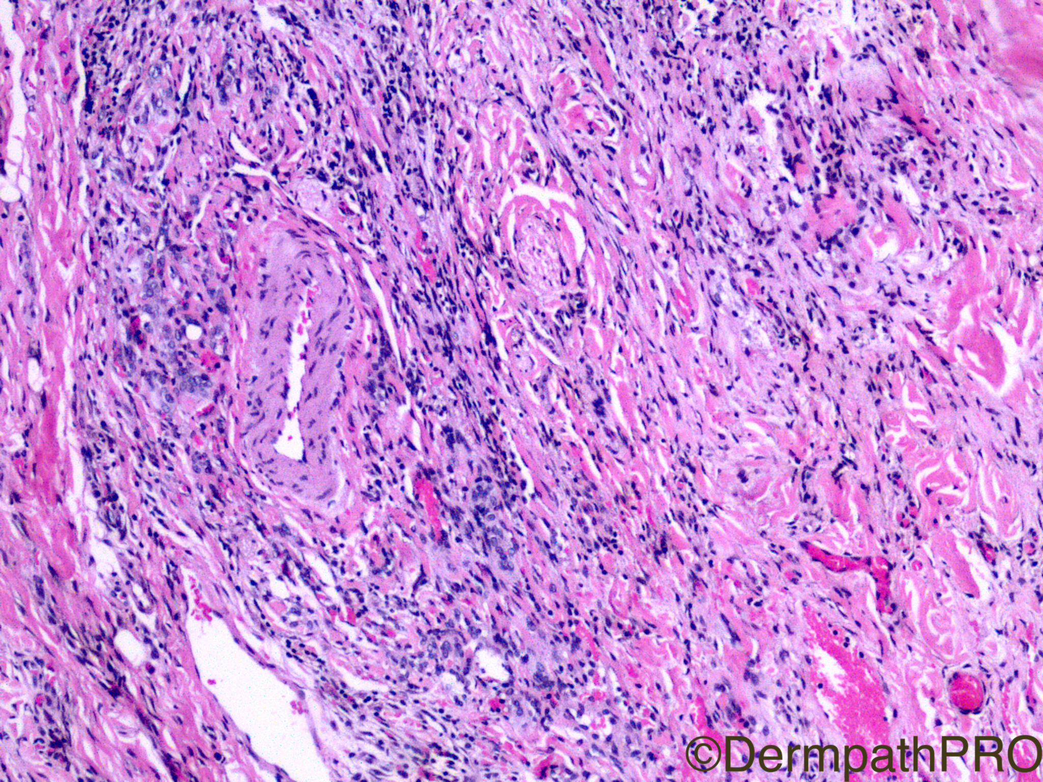
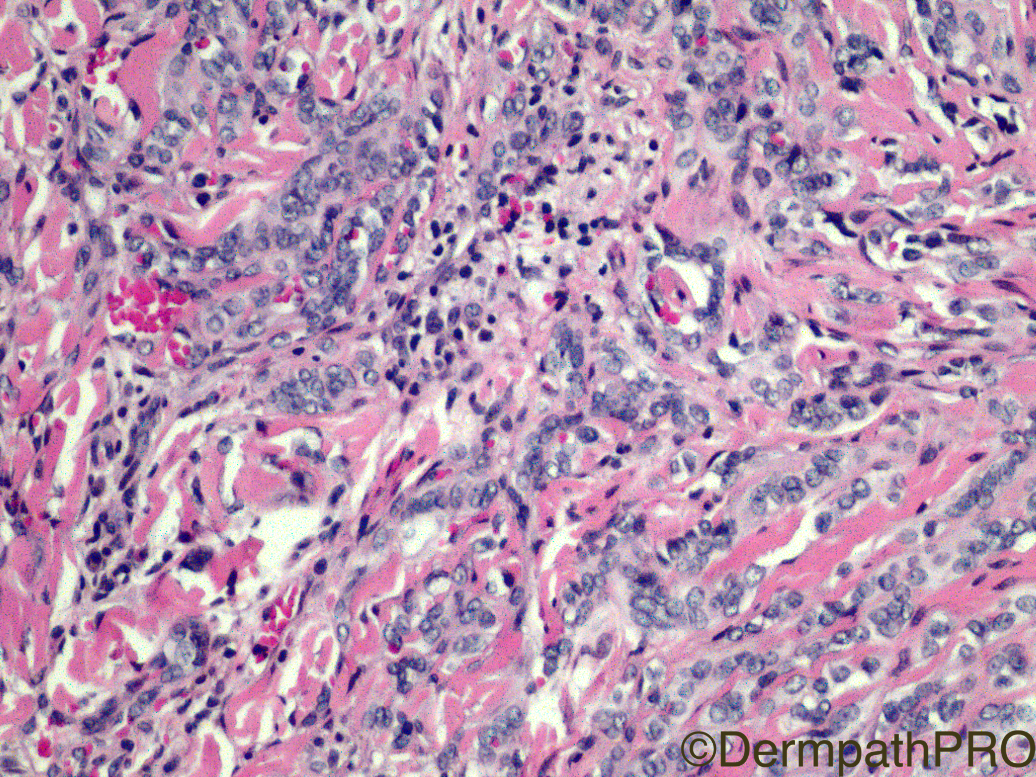
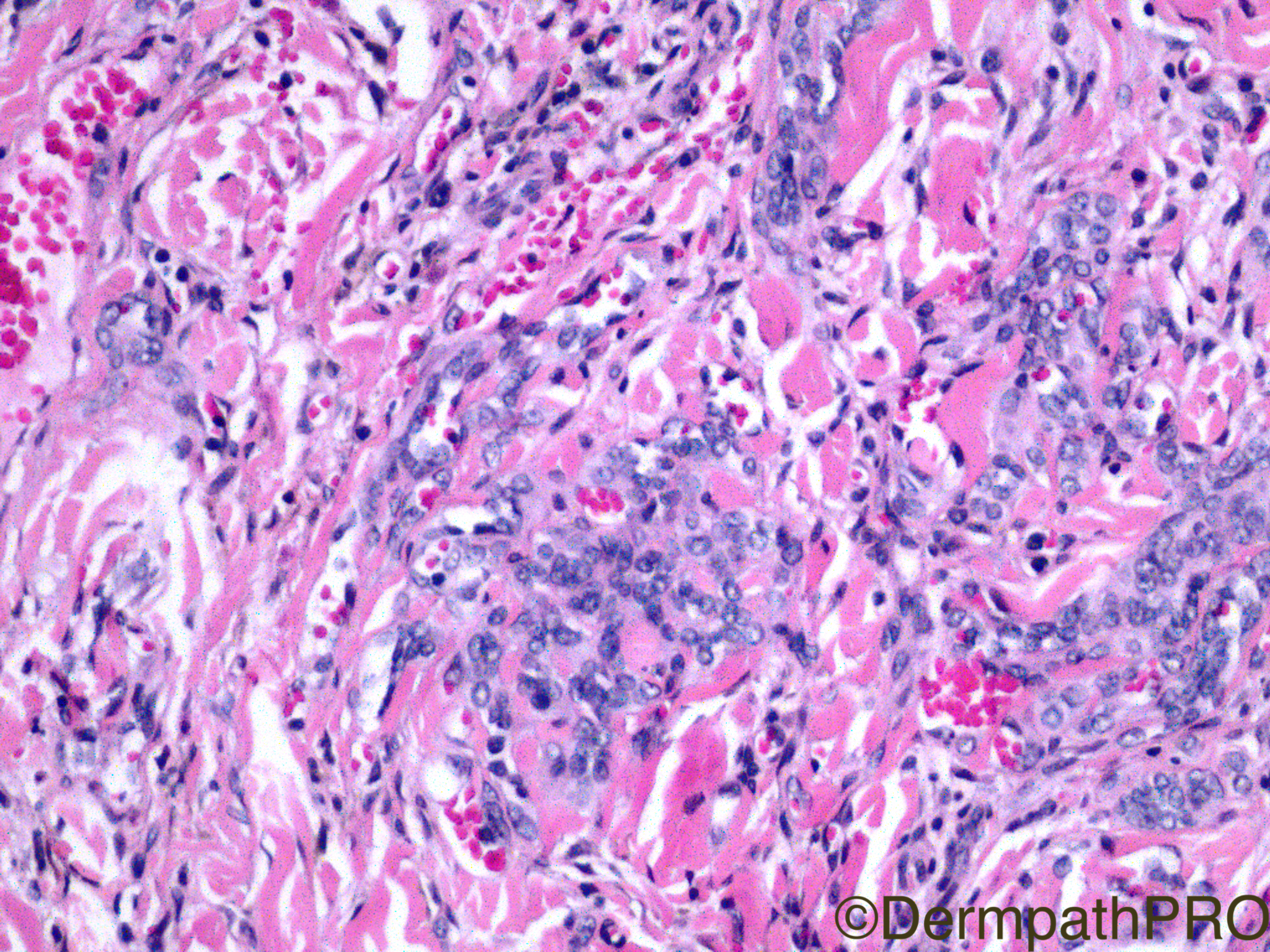
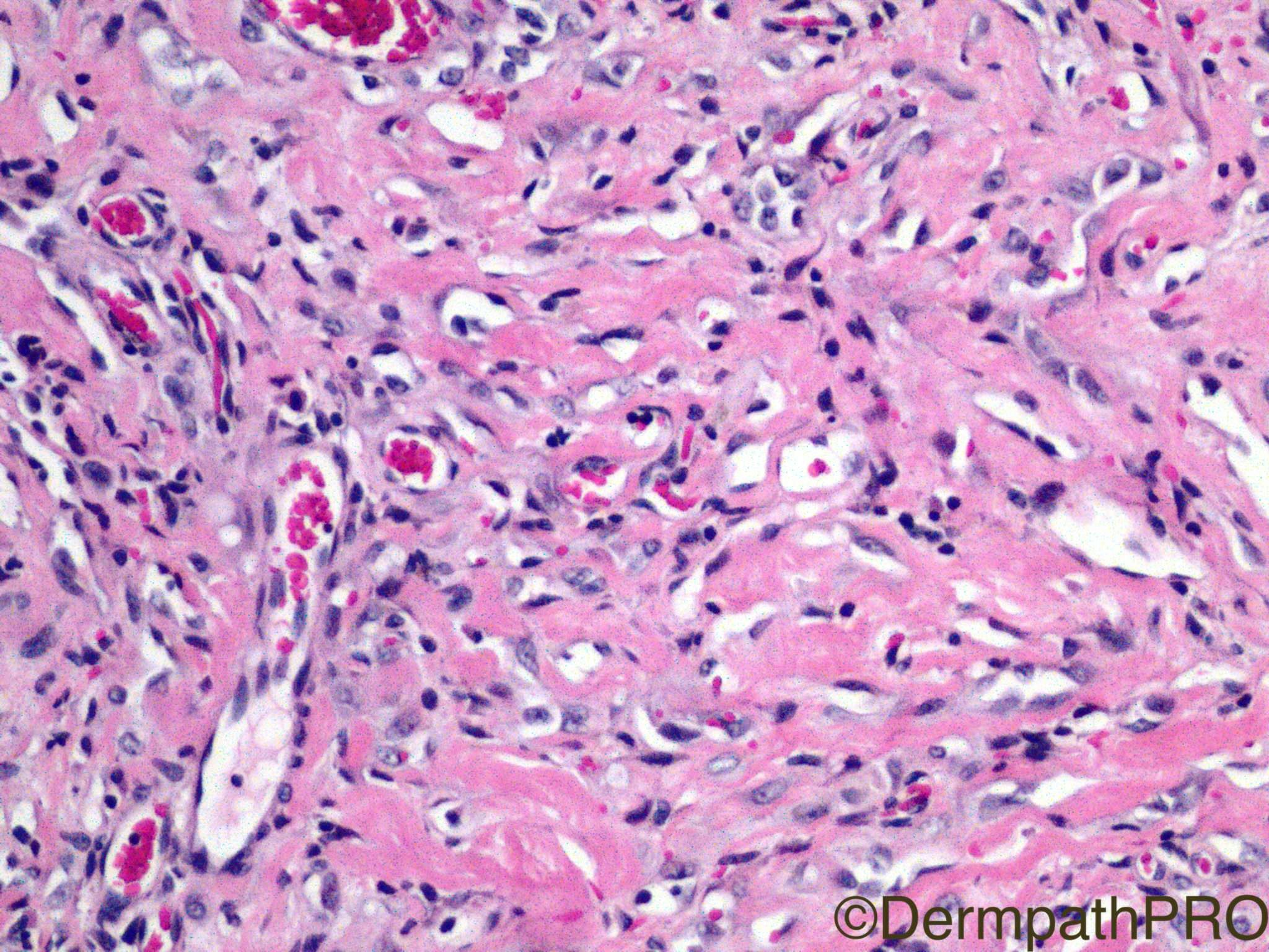
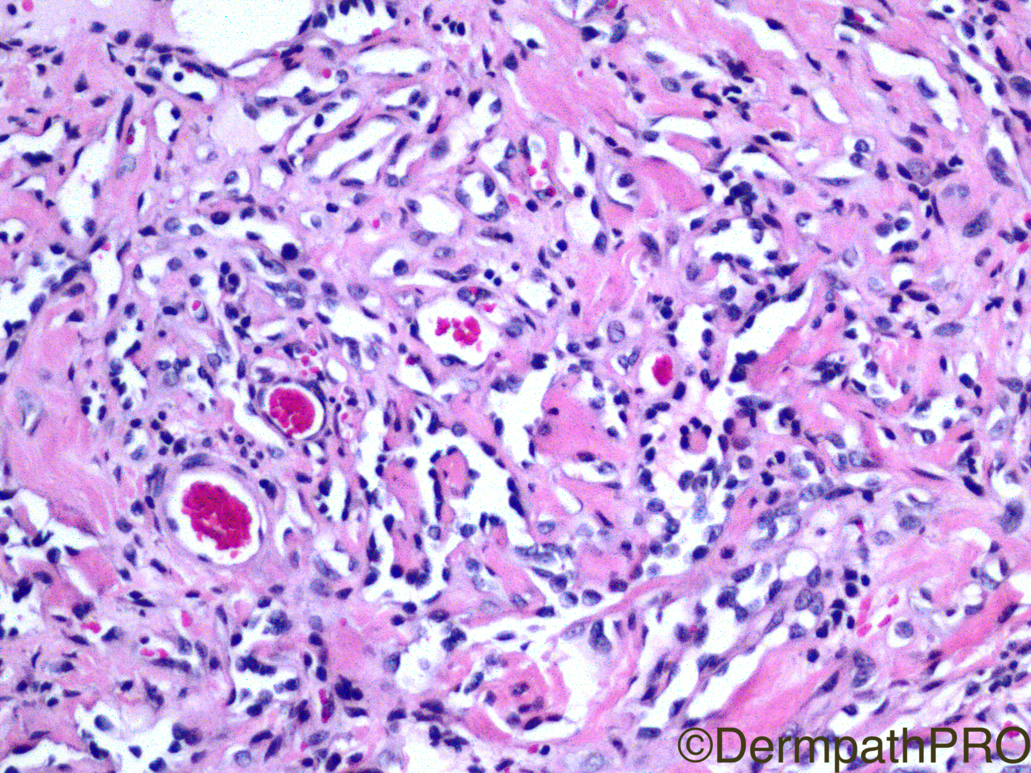
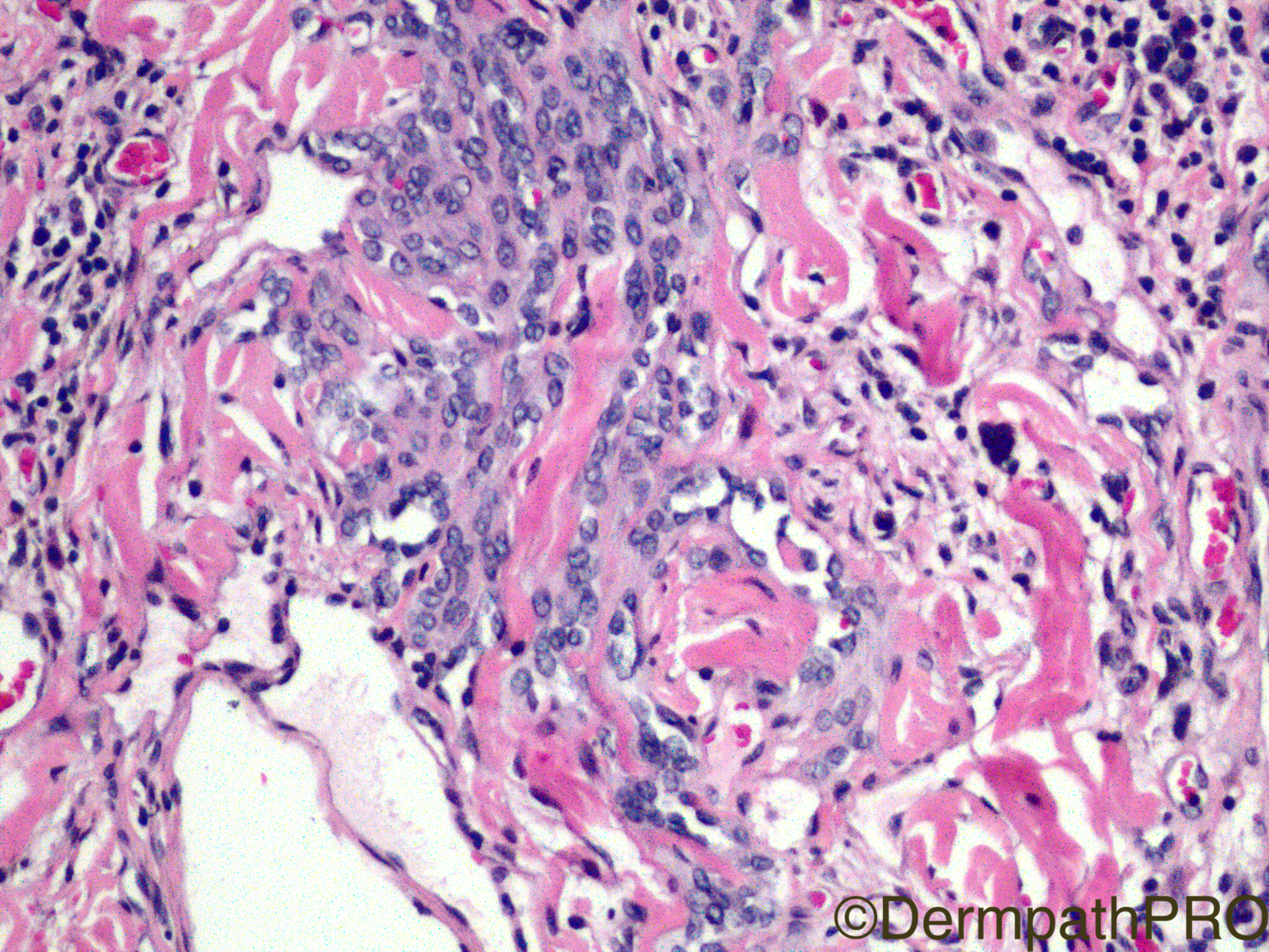
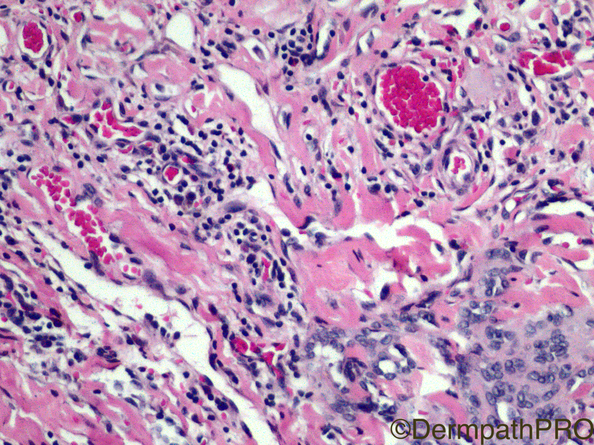
Clinical History: This is a biopsy from a 8yo girl's lower lip.

Case Posted by Dr Limin Yu
Edited by Admin_Dermpath


  Report Record

User Feedback


Admin_Dermpath

Posted

Dr Limin Yu has got a whole host of images for today's Spot Diagnosis Case.


Geoff Cross - DermpathPRO Projects

Share this comment


Link to comment
share_externally

Alice Roberts

Posted

Clinical info would be helpful, but angiosarcoma favored. Considered acroangiodermatitis but looks like dissecting vessels and endothelial atypical are too much.

Share this comment


Link to comment
share_externally

This is a biopsy from a 8yo girl's lower lip. 


Share this comment


Link to comment
share_externally

Dr. Mona Abdel-Halim

Posted

12 minutes ago, Limin Yu said:

This is a biopsy from a 8yo girl's lower lip. 


Oops !!! 

Then may be just a strawberry hemangioma or NICH ?? 

Share this comment


Link to comment
share_externally

Sasi Attili

Posted

 :) Tricky case. I wasn't convinced of angiosarcoma in the images. 8 year old girl makes it even more unlikely I guess. The last but one image looks a bit retiform + there are a few inflammatory cells within the tumour. I am not so great with vascular tumours but DABSKA/PILA?

Share this comment


Link to comment
share_externally

vincenzo polizzi

Posted

I know only another lesion able to simulate angiosarcoma so strictly: the meningothelial hamartoma of the scalp, but that should be "offside"...The Hobnail Hemangioma is a theoretical option, but I haven't any spot diagnosis now. I have a spot question instead: could be a paediatric angiosarcoma? 


Share this comment


Link to comment
share_externally

Raul Perret

Posted

For me this case is challenging too. I thought angiosarcoma could be a consideration but it did not convince me 100%. In my opinion the lesion looks retiform and infiltrative (as Sassi remarked), some of the cells have round nuclei with prominent nucleoli and they dissect collagen bundles. In other areas we see some sclerosis and a lymphocytic infiltrate is also evident. For me this tumor goes morphologically better with retiform haemangioendothelioma although as we know this tumor is more frequent in the adult population and can have significant overlap with Dabska's tumor

Share this comment


Link to comment
share_externally

Raul Perret

Posted

Wonder what Eduardo Calonje's opinion of this case would be...

Share this comment


Link to comment
share_externally

Robledo F. Rocha

Posted

A hard case, but I tend to interpret as something in the spectrum of hobnail (Dabska-retiform) hemangioendothelioma this vascular tumor with infiltrative growth pattern comprising vessels lined by cuboidal endothelium that are embedded in a hyaline stroma infiltrated by lymphocytes.

Share this comment


Link to comment
share_externally

Arti Bakshi

Posted

Agree, on the spectrum of hobnail haemangioendothelioma....retiform architecture is more noticeable than intravascular papillae, so would favour retiform haemangioenothelioma (although completely agree that the 2 are related entities).

Share this comment


Link to comment
share_externally

Nitin Khirwadkar

Posted

Would go for a retiform haemangioendothelioma. However, this occurs more on limbs. 

Share this comment


Link to comment
share_externally

Guest Csbrprasad

Posted

I favour kaposi's sarcoma. One of the images shoe promontary sign.

Share this comment


Link to comment
share_externally

Sasi Attili

Posted

On 27/02/2017 at 21:36, Limin Yu said:

This is a biopsy from a 8yo girl's lower lip. 


Eagerly waiting for the answer :)

Share this comment


Link to comment
share_externally

Dr. Richard Carr

Posted

Great case. Definitely not for mere mortals in soft tissues like me!  My thoughts were some type of haemangioendothelioma (composite of Dabska/PILA, retiform and even some slightly epithelioid cells). Also have to exclude KS - easily done with HHV8. Given clinical scenario likely to be benign behaviour I guess.

Share this comment


Link to comment
share_externally

Limin Yu

Posted

Thank you for the intense and insightful discussion. This is an indeed challenging case. The case was sent to Dr. Fletcher for consultation and his opinion was composite hemangioendothelioma.  His letter is attached. 

Capture.JPG

Share this comment


Link to comment
share_externally

Raul Perret

Posted

What a great case thank you dr. Yu

Share this comment


Link to comment
share_externally


Join the conversation

You can post now and register later. If you have an account, sign in now to post with your account.

Guest
Add a comment...

×   Pasted as rich text.   Paste as plain text instead

  Only 75 emoji are allowed.

×   Your link has been automatically embedded.   Display as a link instead

×   Your previous content has been restored.   Clear editor

×   You cannot paste images directly. Upload or insert images from URL.

Loading...

×
×
  • Create New...