Jump to content
In this section we have spot diagnoses posted on a daily basis since June 2010, now over 4000! You can review the archived cases and read the suggested diagnoses by users and the final comment by the contributors.
Case are uploaded each week day by 10 am UK time with the correct diagnosis will generally be posted at 8 pm UK time. Why not view the most recent spot diagnosis and proffer a diagnosis?

Case Number : Case 1725 - 6 January - Dr Richard A Carr Posted By: Guest

Please read the clinical history and view the images by clicking on them before you proffer your diagnosis.
Submitted Date :
   (0 reviews)

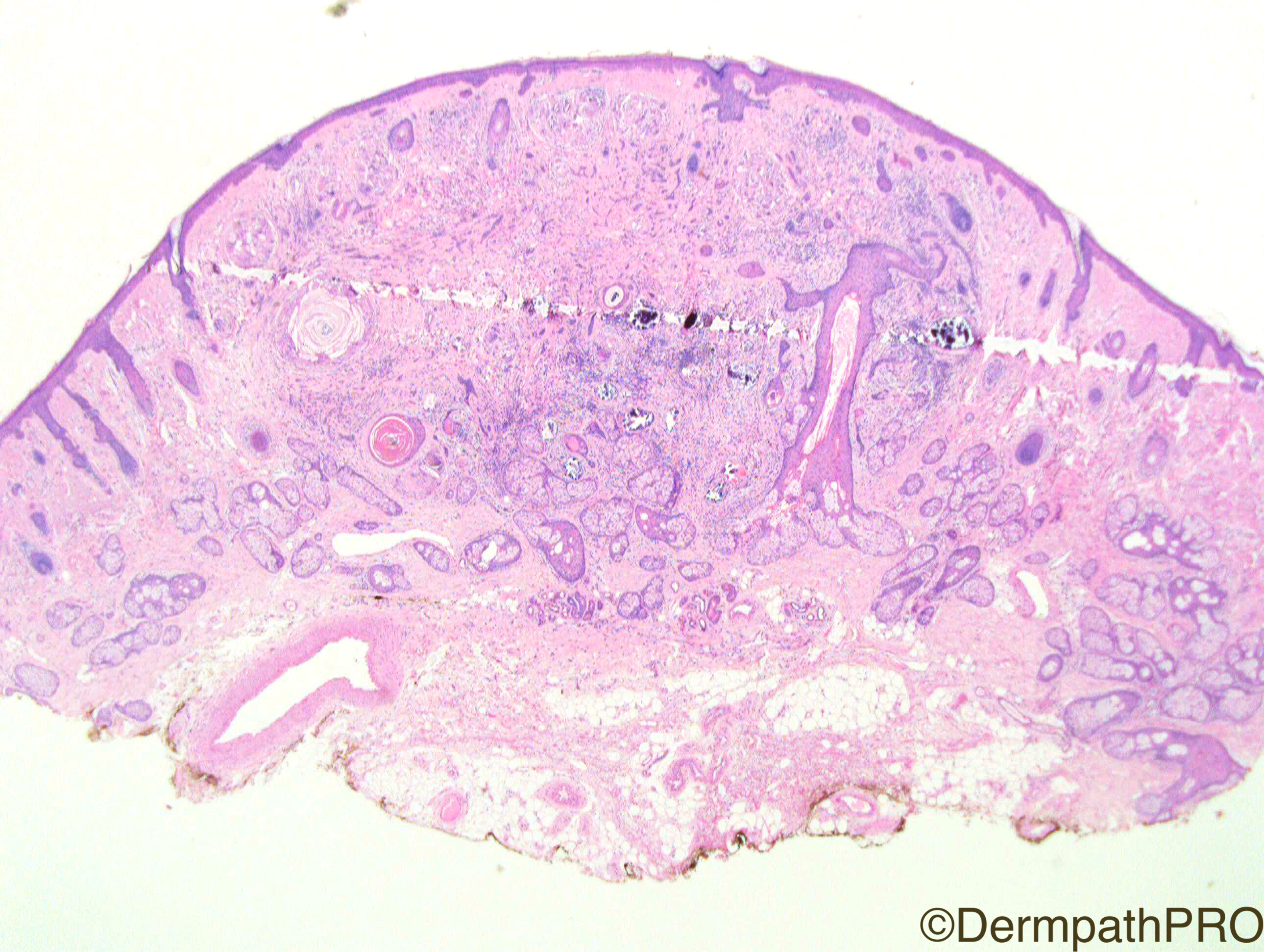
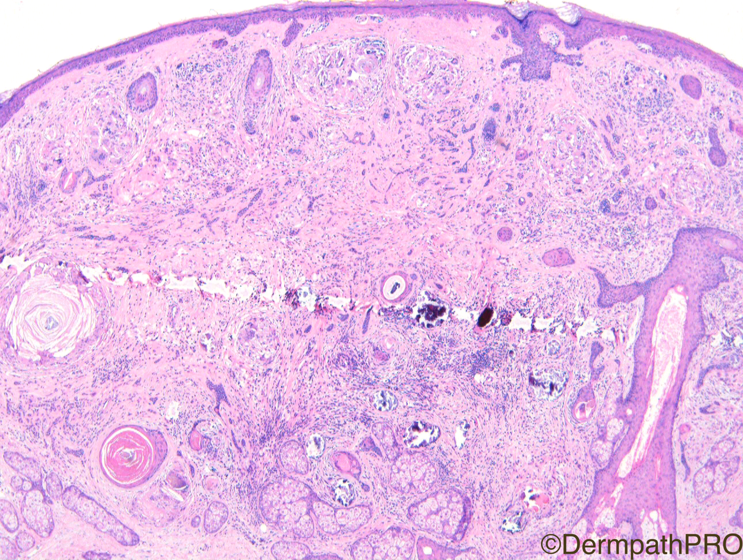
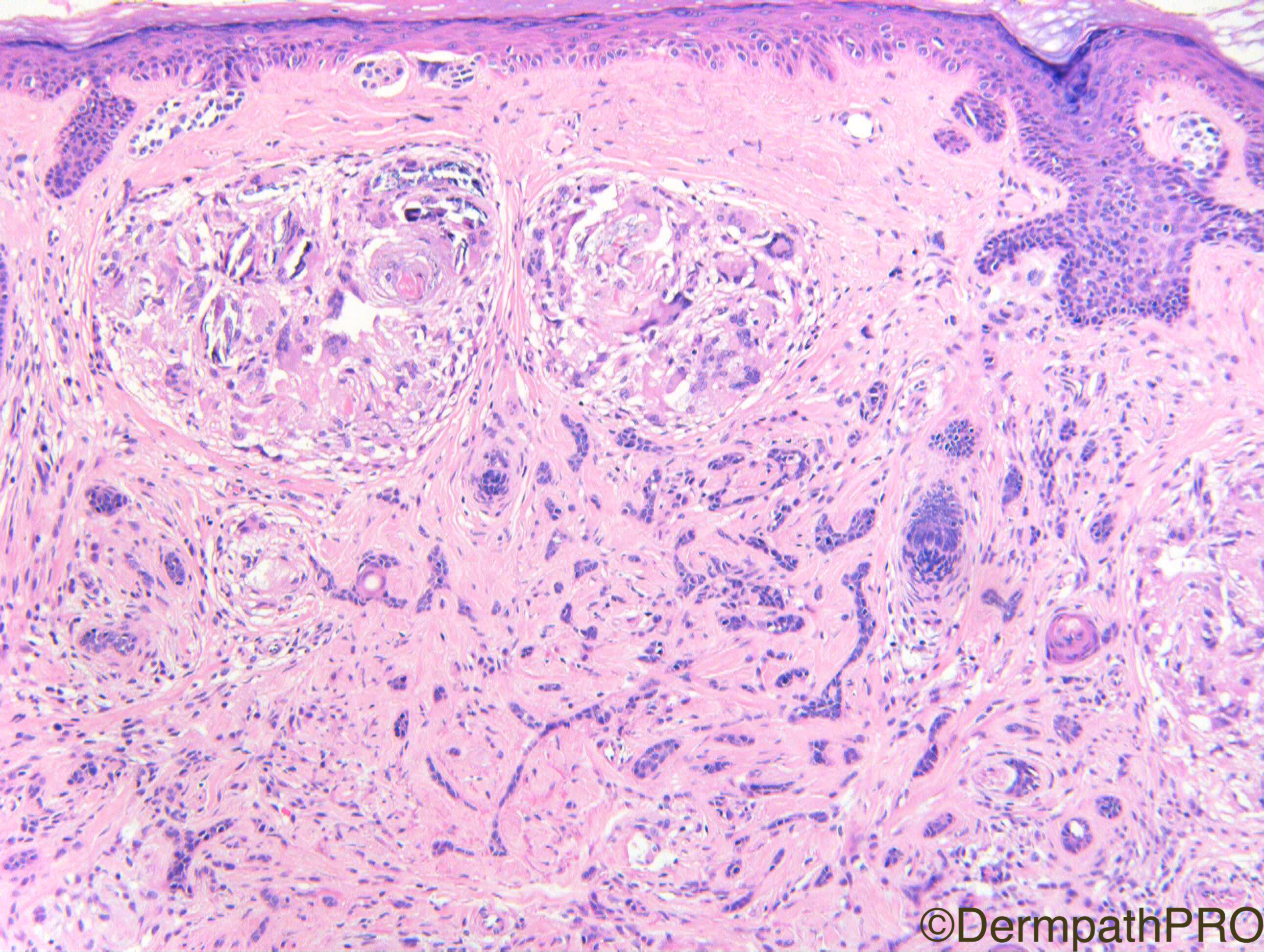
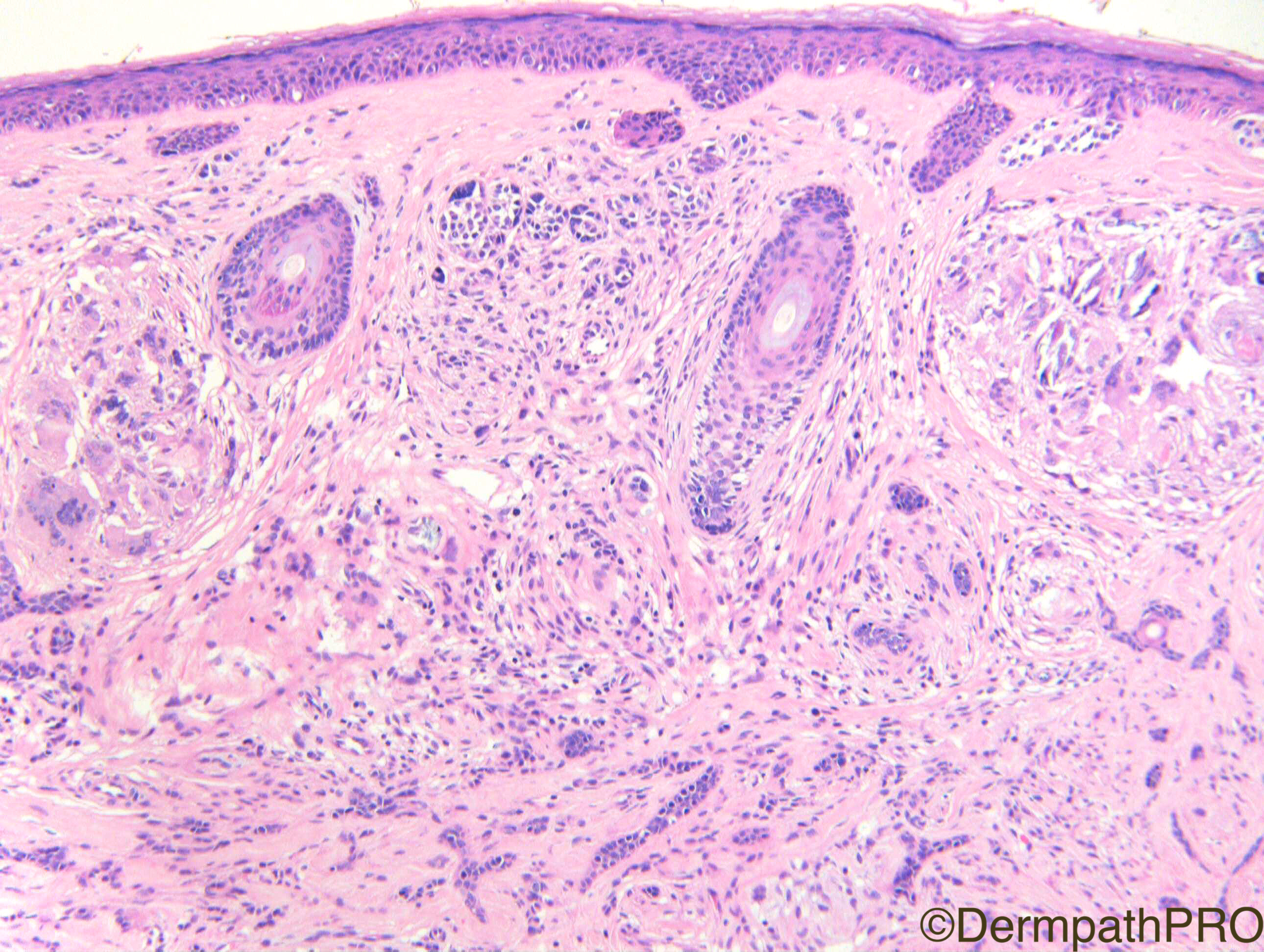
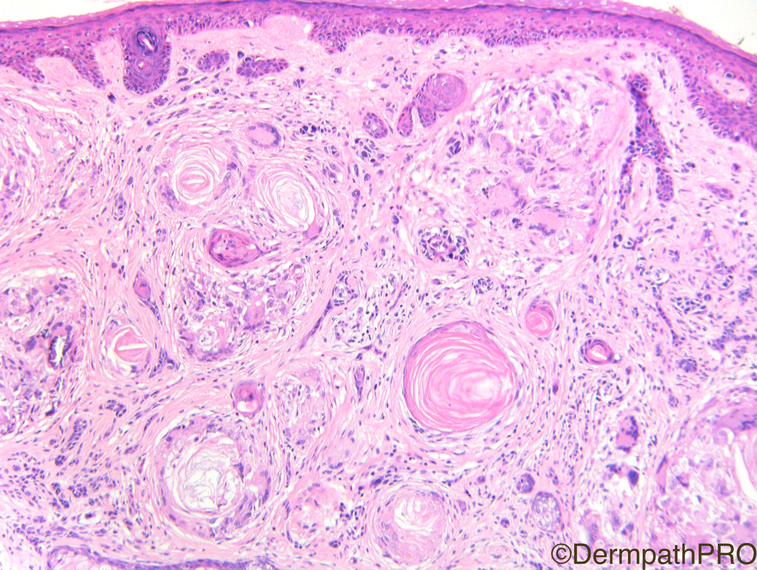
Clinical History: F60. Nasal tip. Lesion of long duration ?IDN. For newbies and trainees first please.

Case Posted by Dr Richard A Carr
Edited by Admin_Dermpath


  Report Record

User Feedback


Admin_Dermpath

Posted

Happy New Year everyone, here is a great case from Dr Richard A Carr, apparently it is a great example for Newbies and Trainees.


Ciao, Geoff Cross - DermpathPRO Projects

Share this comment


Link to comment
share_externally

Heba Rashad

Posted

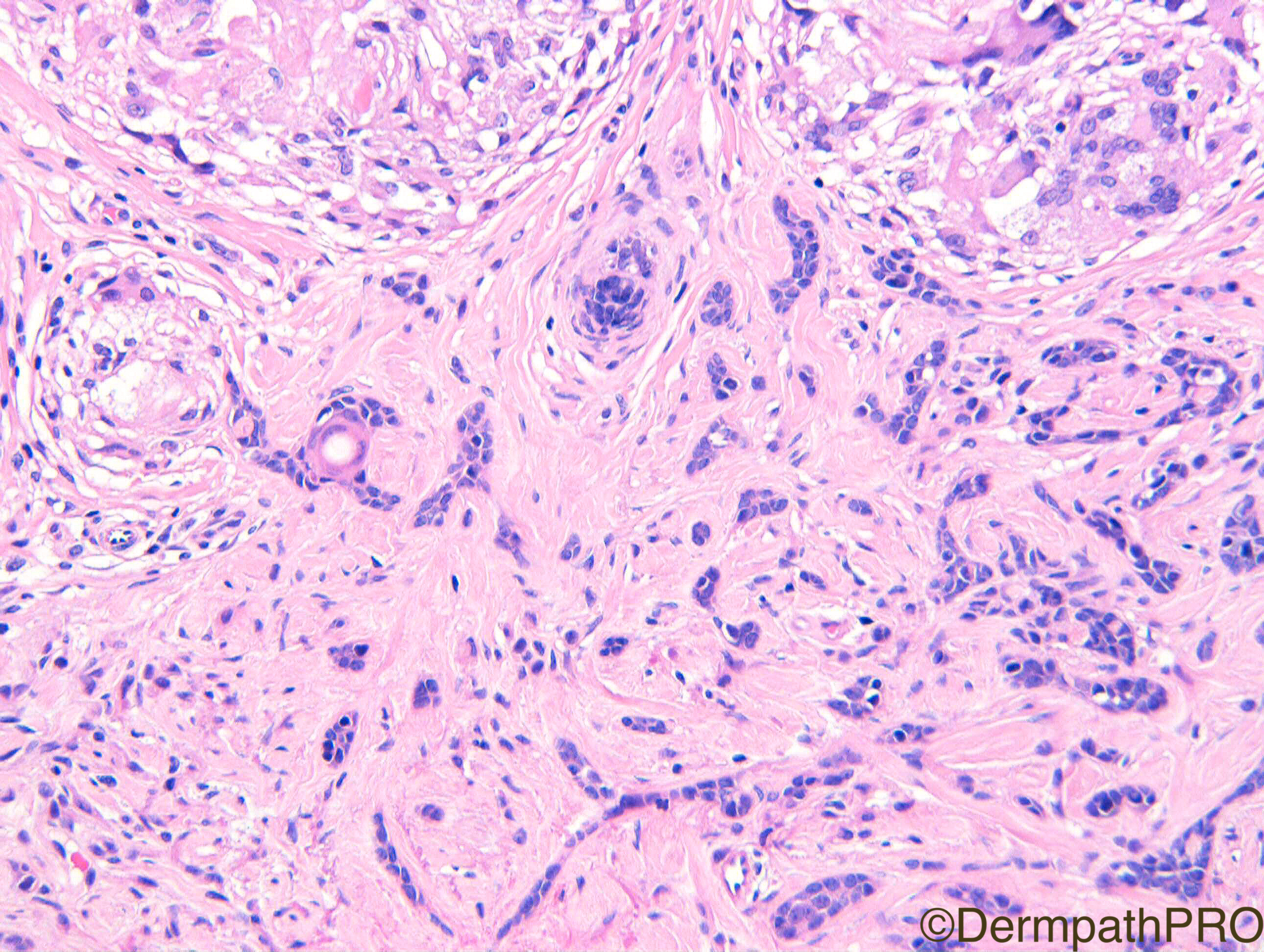
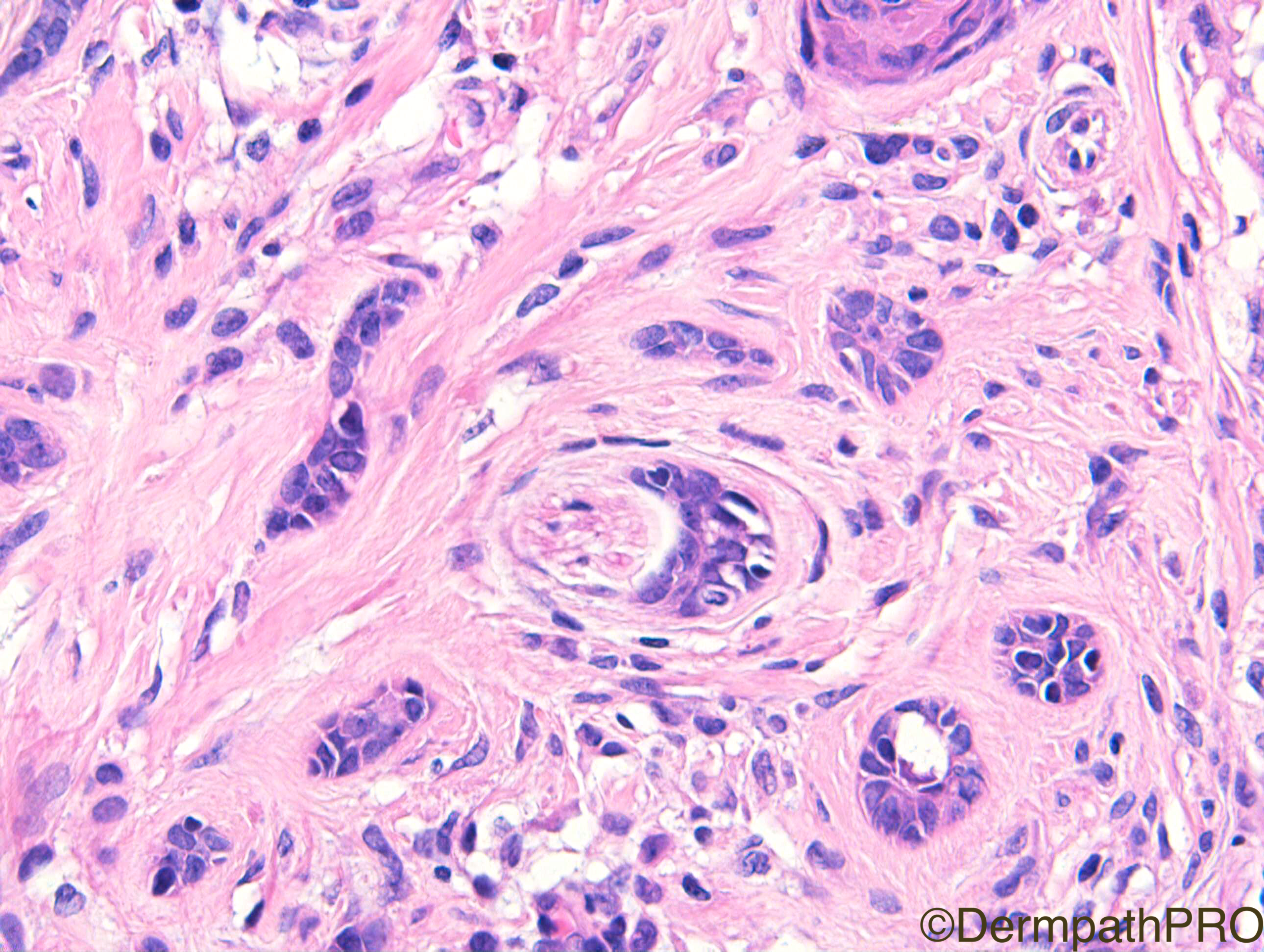
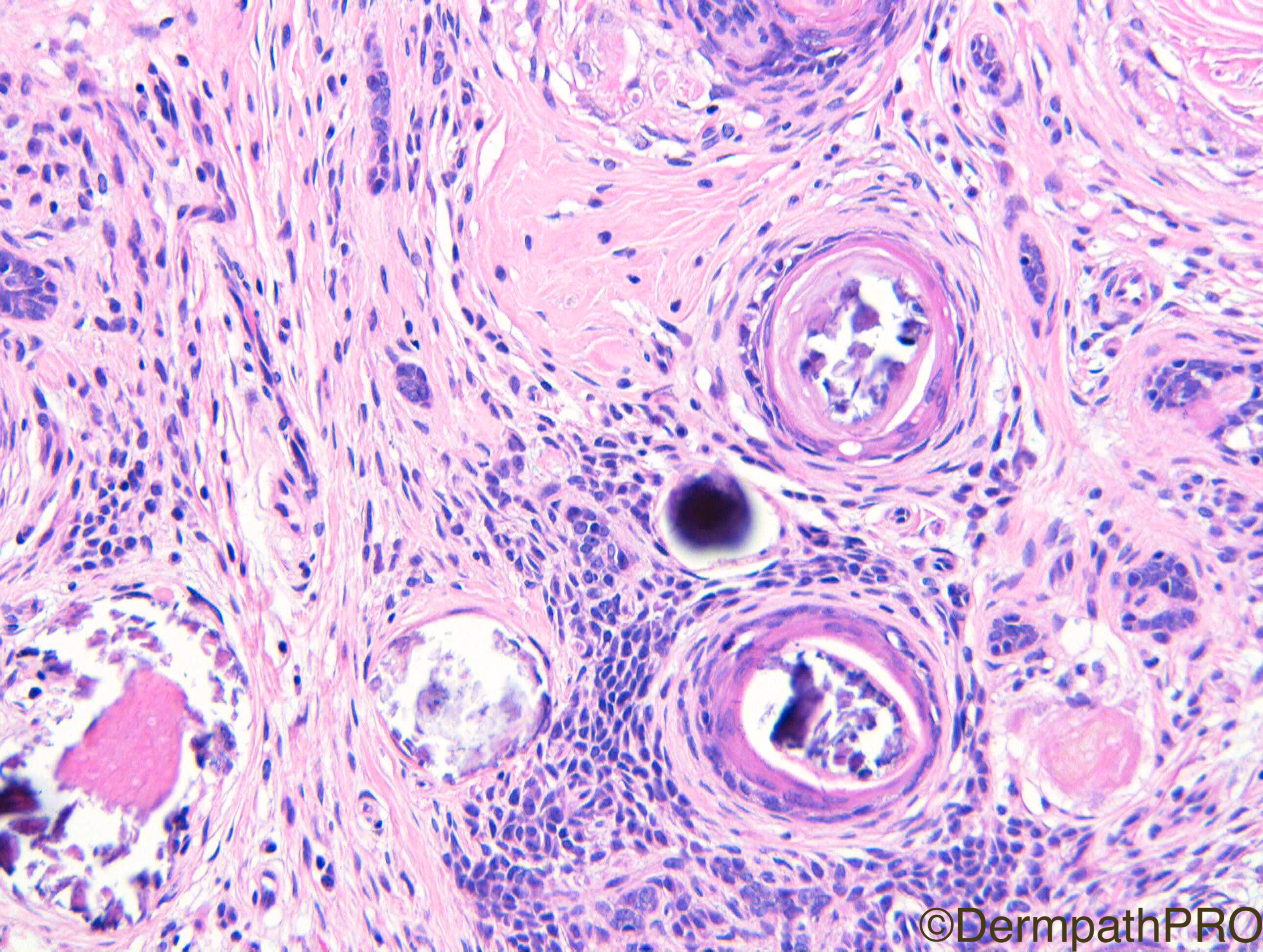
Desmoplastic trichoepithelioma with granulomatous reaction,calcification

Share this comment


Link to comment
share_externally

Dr. Mona Abdel-Halim

Posted

Beautiful DTE... waited till the end :-))

Share this comment


Link to comment
share_externally

vincenzo polizzi

Posted

...and a congenital nevus. 

Some pathological association are more frequent than others and make favor the epithelial induction way by melanocytes rather than the cancerization field theory or collision theory.  

Share this comment


Link to comment
share_externally

Dr. Richard Carr

Posted

Great to have some newbies contributing comments (welcome and please do keep contributing) and thanks for the stalwarts adding comments too.

Well you are pretty much all correct with the desmoplastic trichoepithelioma (DTE). Thanks Robledo for pointing out the intra-lesional perineural involvement which is well reported in DTE. Thanks to Vincenzo for mentioning the naevus and the fact that there may be a slight increase in the association between DTE and melanocytic naevus than we'd expect from pure chance.

Regarding perineural invasion in general I don't report it unless it is significantly extra-tumoural on an excision biopsy of a cancer case (like BCC, SCC or KA). If I do see intra-tumoural (i.e. within the main confines of the tumour) on a partial biopsy I comment of "uncertain significance". I have now seen intra-tumoural perineural tumour i.e. within the confines of DTE on several occasions but not ever extending beyond the borders / peripheral profile of the lesion.

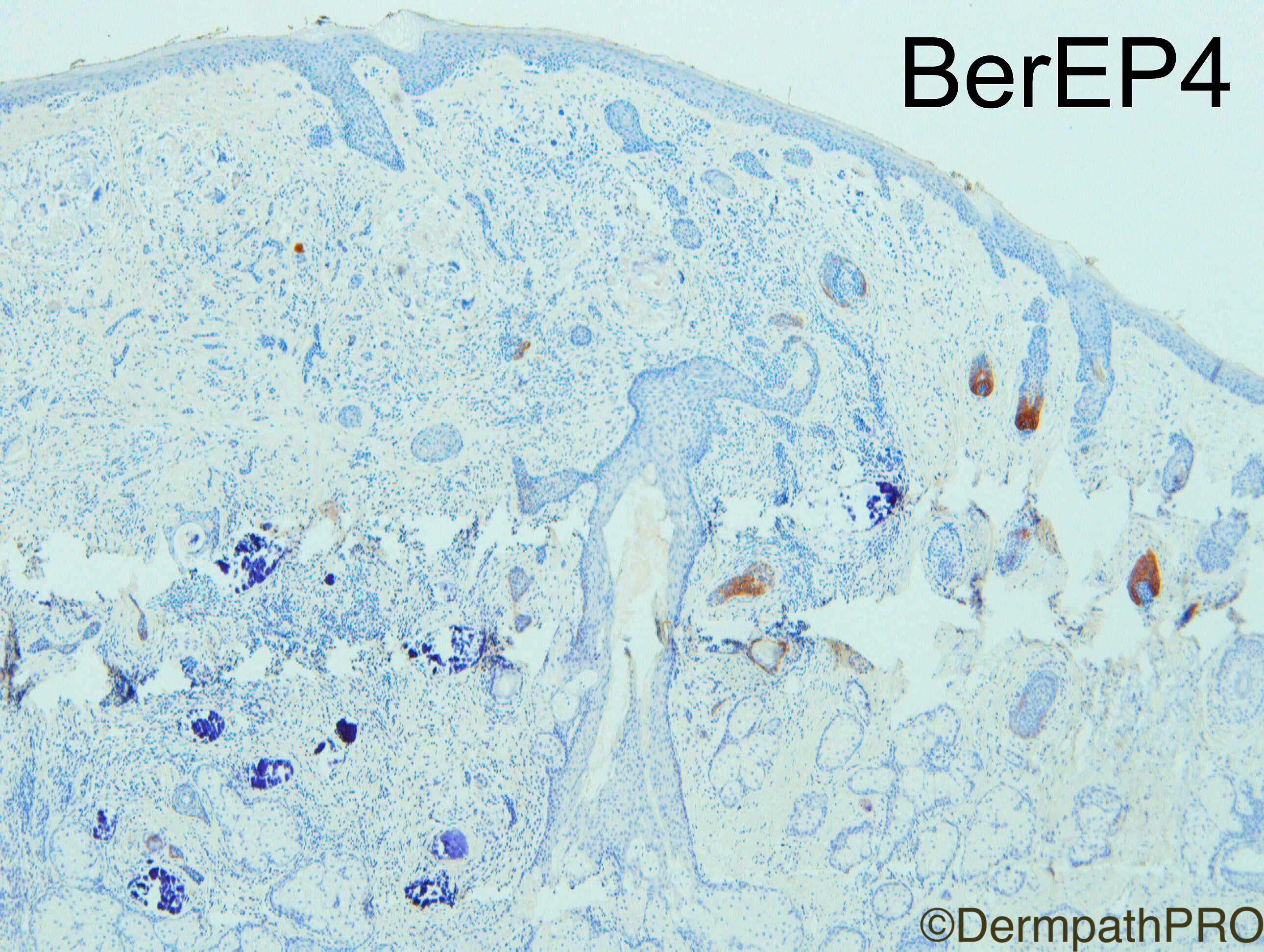
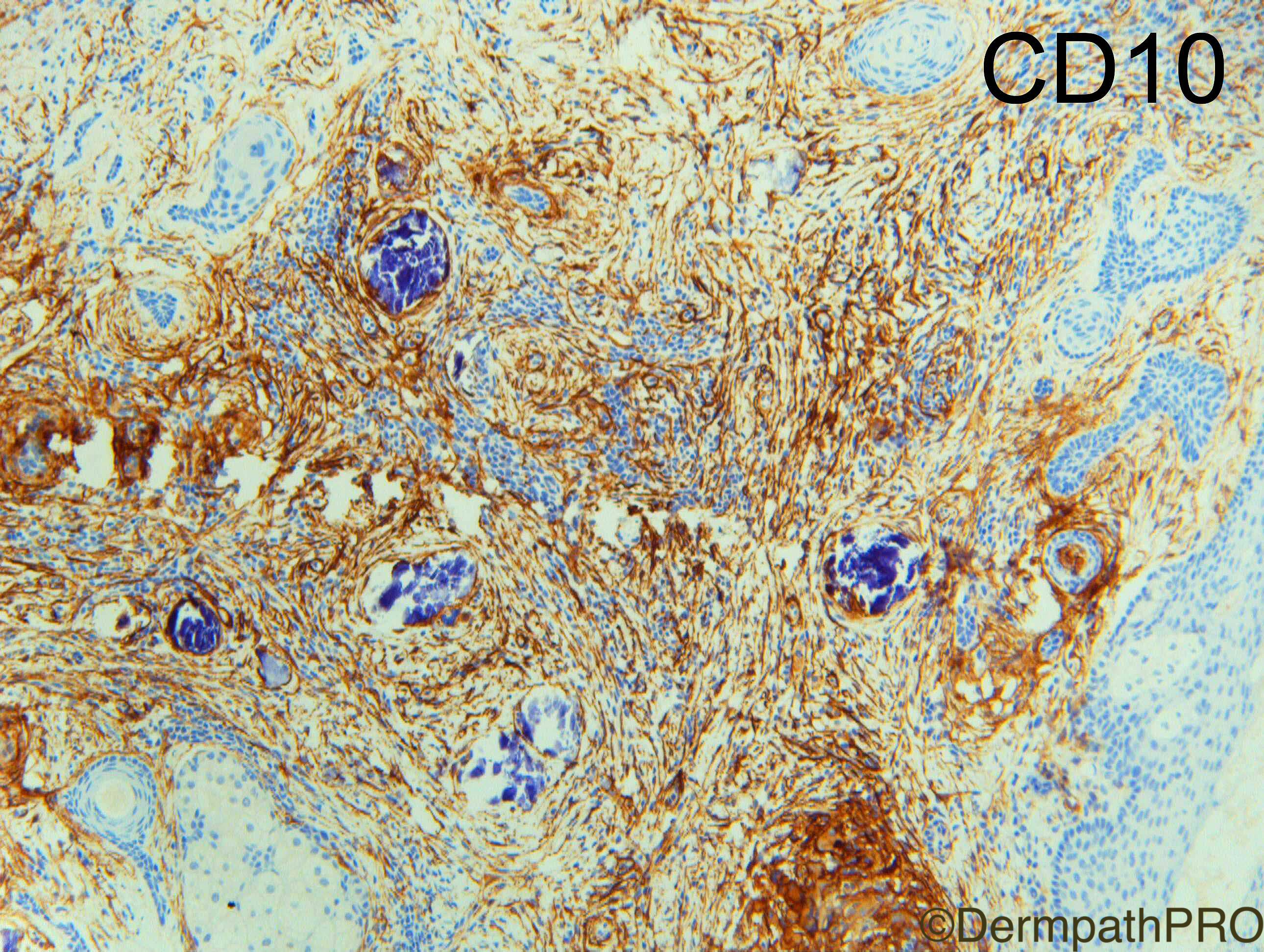
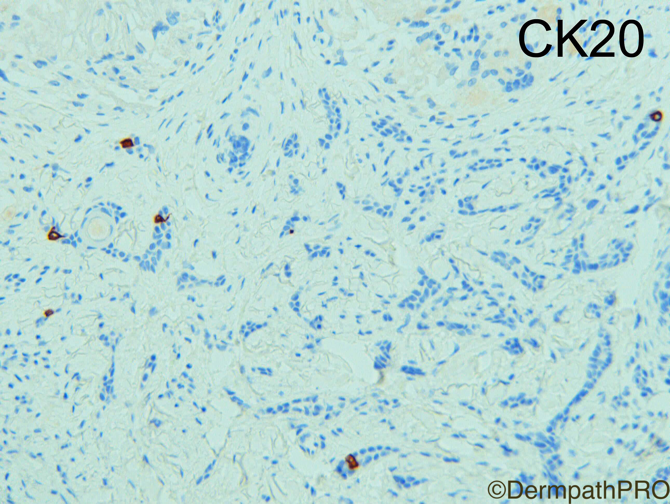
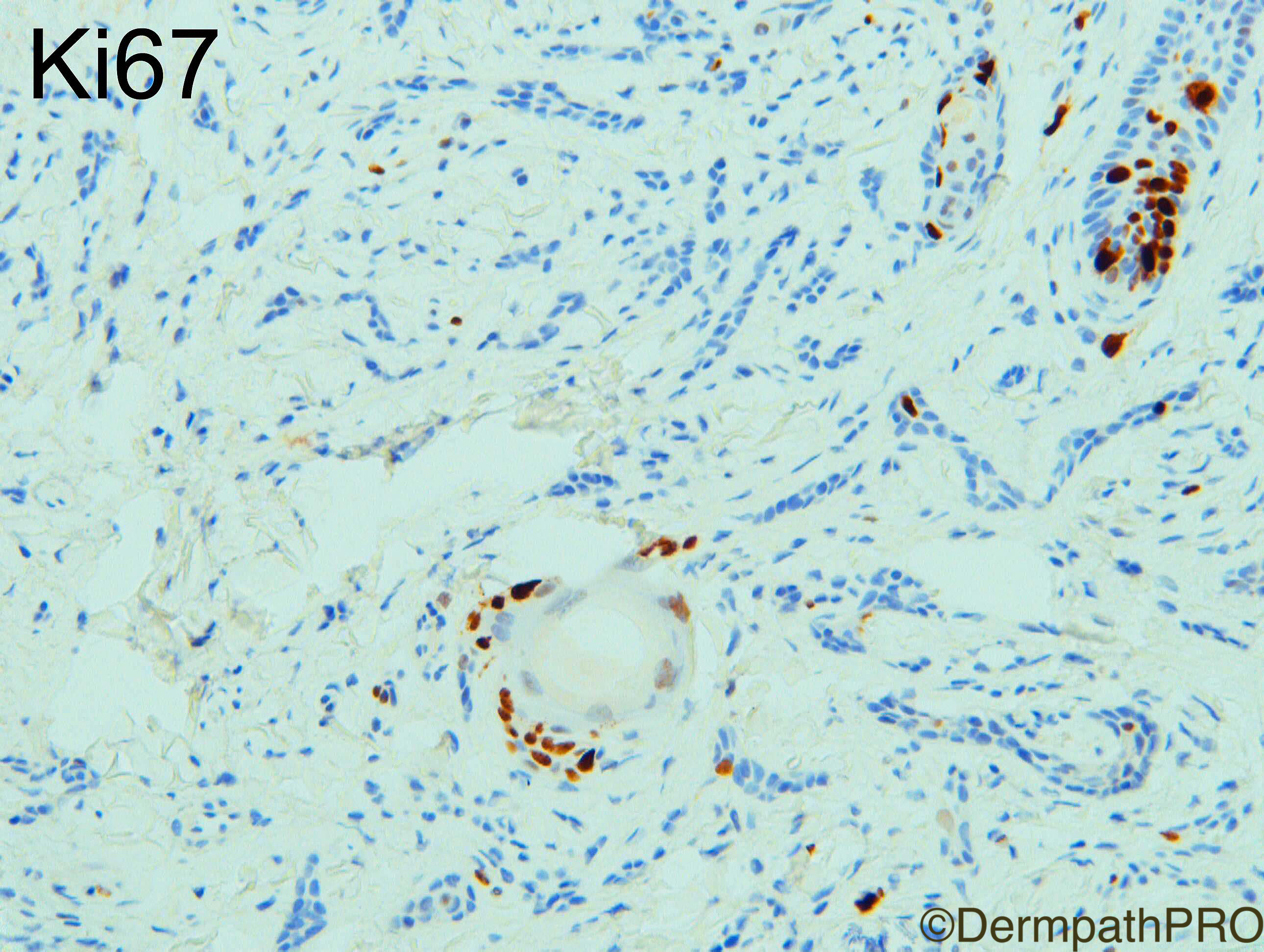
Regarding IHC in DTE I find BerEP4 is often weak to moderate and peripheral compared with invariable diffuse strong in infiltrative BCC and limited to duct lumina in MAC. CK20 usually scattered dendritic Merkel cells compared with negative infiltrative BCC and MAC. CD10 is not overly helpful in my experience as DTE rarely show nice papillary mesenchymal bodies although epithelial staining is often present in BCC I have not seen it in DTE to my memory. Ki67 should be very low in DTE up to 5% at most and is usually higher in BCC but quite subjective.  In summary CK20 can be very helpful in most cases (small biopsies may be an occasional exeption if you seen no dendritic Merkel cells) and BerEP4 is moderately helpful.

Enjoy your weekends.

Share this comment


Link to comment
share_externally

but Dr Carr, what is the difference between intratumoral/extratumoral perineural invasion

i think tumor cells once invaded around nerve fibers, it has a chance to travel a for distance; whether it is intra/extra tumoral

Share this comment


Link to comment
share_externally

Dr. Richard Carr

Posted

You might be right hence on partial biopsies I comment that I don't know the significance when I see it within the borders of the tumour. In a completely excised specimen I would not bother looking for or indeed reporting intra-tumoural perineural invasion as I don't think it carries any relevance. 

Obviously if you see it extending for some distance beyond the margins you might want to cut some levels to check clearance and warn clinicians of the possibility of local recurrence etc. Interestingly in SCC some work has been done relating to the size of the nerve (< or > or = 0.1mm diameter) with the latter having a worse prognosis. If margins are involved then this would certainly be an indication for margin control surgery e.g. Moh's and or adjuvant radiotherapy.

For melanoma the British guidelines refer only to PNI being "beyond the main bulk of the tumour" and is associated with an increased risk of local recurrence.

Share this comment


Link to comment
share_externally


Join the conversation

You can post now and register later. If you have an account, sign in now to post with your account.

Guest
Add a comment...

×   Pasted as rich text.   Paste as plain text instead

  Only 75 emoji are allowed.

×   Your link has been automatically embedded.   Display as a link instead

×   Your previous content has been restored.   Clear editor

×   You cannot paste images directly. Upload or insert images from URL.

Loading...

×
×
  • Create New...