Jump to content
In this section we have spot diagnoses posted on a daily basis since June 2010, now over 4000! You can review the archived cases and read the suggested diagnoses by users and the final comment by the contributors.
Case are uploaded each week day by 10 am UK time with the correct diagnosis will generally be posted at 8 pm UK time. Why not view the most recent spot diagnosis and proffer a diagnosis?

Case Number : Case 1732 - 17 January - Dr Uma Sundram Posted By: Guest

Please read the clinical history and view the images by clicking on them before you proffer your diagnosis.
Submitted Date :
   (0 reviews)

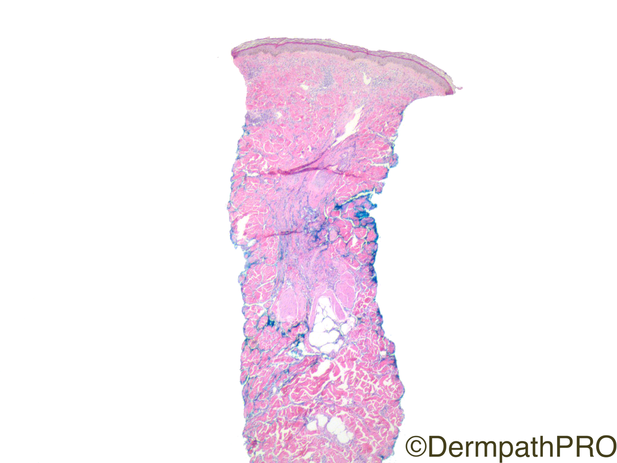
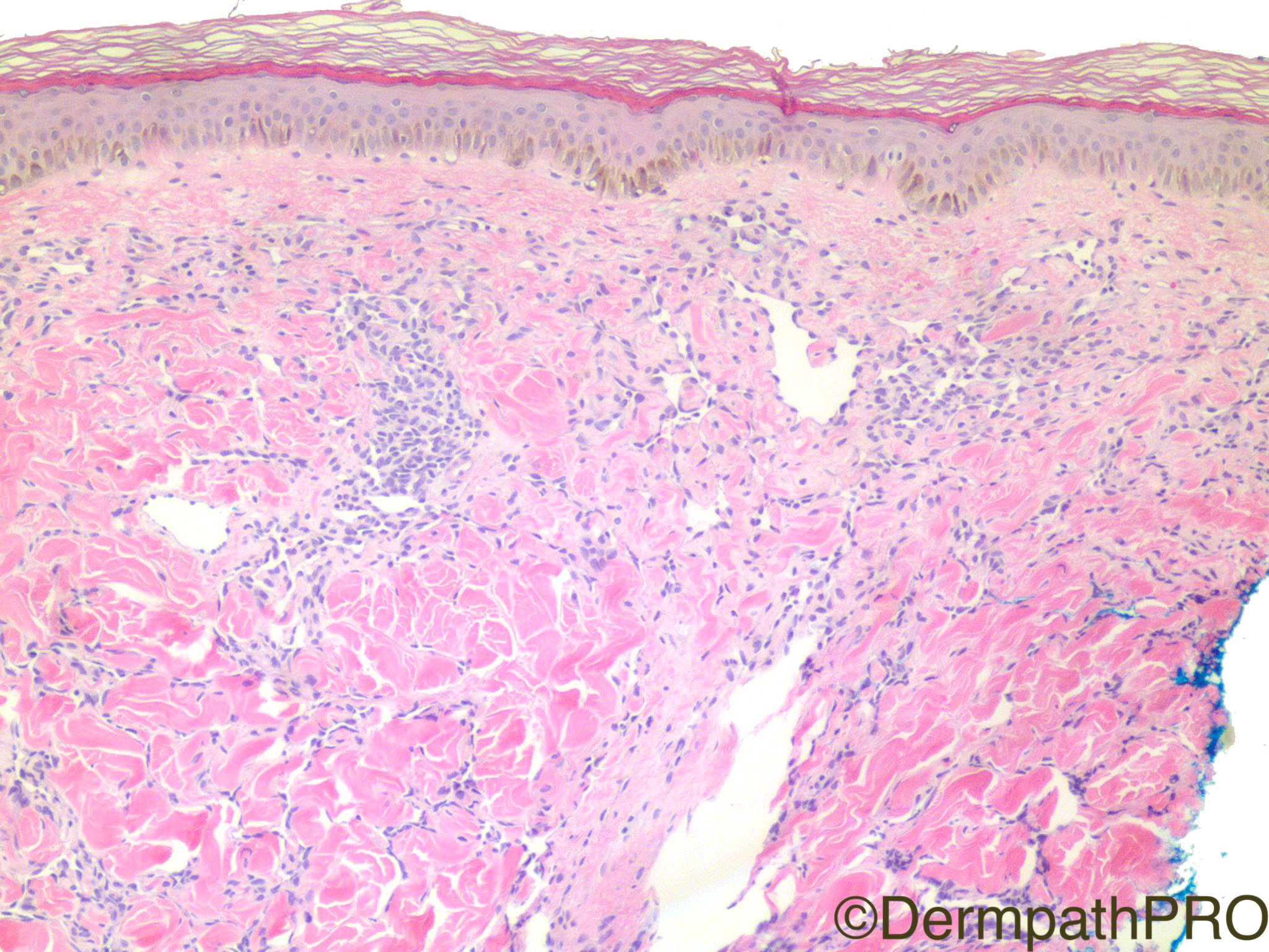
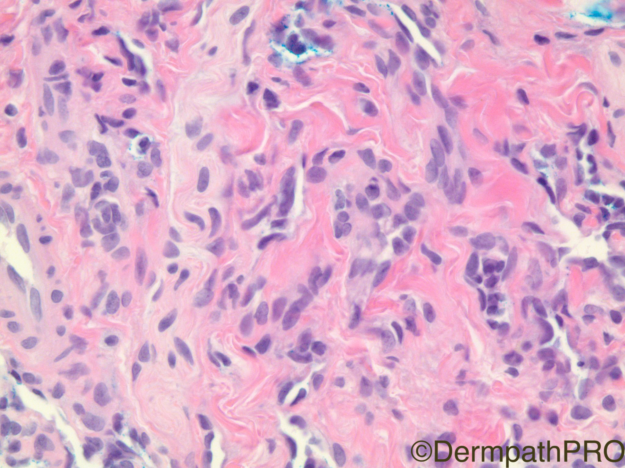
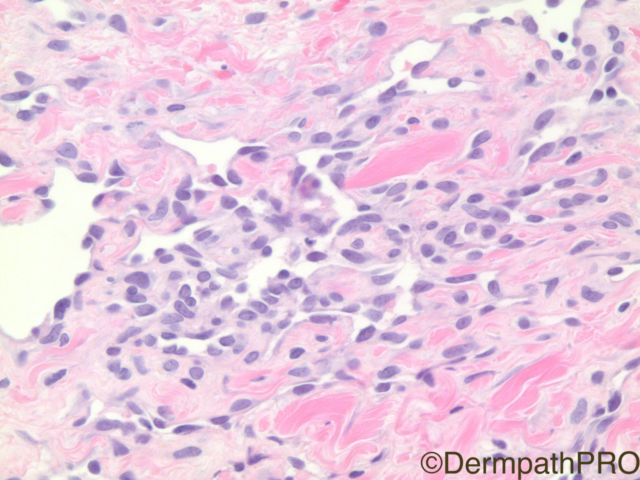
Clinical History: 65 year old woman with ‘large’ lesion on back.

Case Posted by Dr Uma Sundram


  Report Record

User Feedback


Admin_Dermpath

Posted

Today's Daily Spot Diagnosis Case is a biggy or should we now say bigly challenge for you all.


Cheers, Geoff Cross - DermpathPRO Projects

Share this comment


Link to comment
share_externally

vincenzo polizzi

Posted

Completely agree with Geoff diagnosis of biggy/bigly challenge.

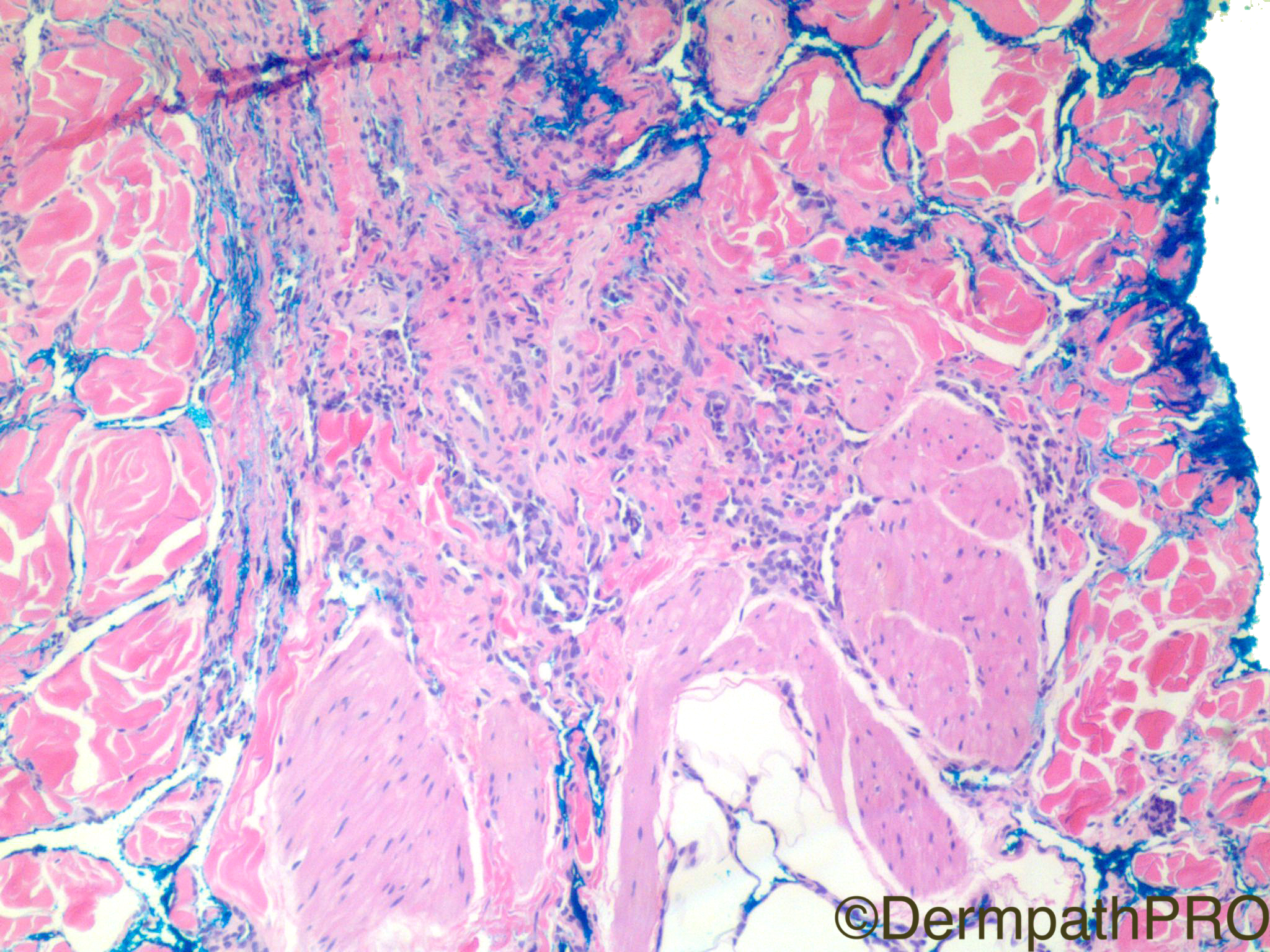
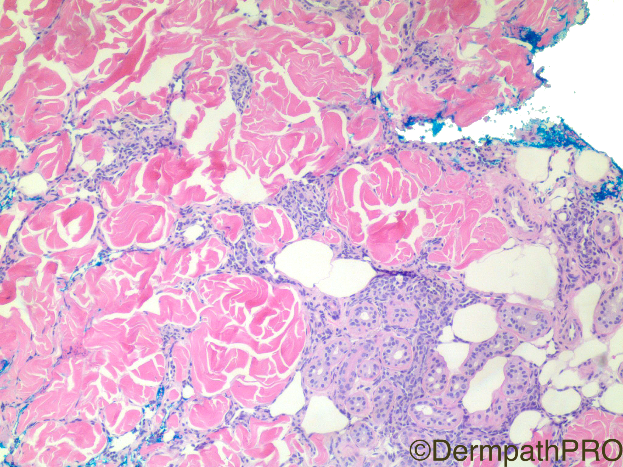
Dissecting pattern of endothelial cell growth, infiltrating superficial and deep dermis ( and subcutaneous tissue probably ), absent or very mild atypia with some sporadic piling up, no hemosiderin deposits...Last in  the second  picture there is a cellular crowding and there you can see two mitosis...What is it? A very early ( and very low grade ) angiosarcoma? A hobnail hemangioma without deep hemorragic component?

In real life I would claim an incisional biopsy, since the lesion is large!

Here i can just favor an angiosarcoma.


Share this comment


Link to comment
share_externally

Dr. Mona Abdel-Halim

Posted

Same concern of angiosarcoma and as Vincenzo said, I would love to see an incisional biopsy!

Share this comment


Link to comment
share_externally

Nitin Khirwadkar

Posted

Yes, infiltrative vascular tumour with crowding, endothelial atypia, angiosarcoma.

Share this comment


Link to comment
share_externally

Raul Perret

Posted

My impression was that this neoplasm looks relatively organized and lacks atypia, it bothered me the clinical description of (large) so its hard to rule out an angiosarcoma as everyone remarked. Nevertheless, I would favour a microvenular hemangioma only based on these images (this tumor can show some adnexal infiltration as in this case) would perform smooth muscle actin (to highlight pericytes), cd31, d2-40 and/or WT-1 as well as hhv-8 in this case. In real life also discuss with treating physician and asking for patients pictures

Share this comment


Link to comment
share_externally

Arti Bakshi

Posted

Hmm....difficult! Agree that a low grade angiosarcoma is first thought, but odd site!

The only other diffrential that come to mind is a microvenular haemangioma, which can show an infiltrative pattern of irregular branching small vessels. Involvement of arrector pili muscle is typical (?image 3)

Share this comment


Link to comment
share_externally

Arti Bakshi

Posted

Ha! Raul beat me to it!...but glad that we are on similar track!!

Share this comment


Link to comment
share_externally

Raul Perret

Posted

21 minutes ago, arti bakshi said:

Ha! Raul beat me to it!...but glad that we are on similar track!!

Me too, i thought I was the only one thinking of microv hemangioma!

Share this comment


Link to comment
share_externally

Dr. Richard Carr

Posted

I definitely was not thinking of a microvenular haemangioma (but easily ruled in/out with SMA). Looks highly irregular and infiltrative (agree minimal atypia).  I was thinking it might be a breast lesion (i.e. post-irradiation angiosarcoma / AVL) prior to seeing it was from the back. I favour angiosarcoma but agree with the cautious comments above - like to see the clinical images +/- larger biopsy before rendering a death sentance!

Share this comment


Link to comment
share_externally

My first thought would be Kaposi´s sarcoma, but well differentiated angiosarcoma also comes to mind. HHV-8 immunostain is necessary (as well as clinical history).

Share this comment


Link to comment
share_externally

Robledo F. Rocha

Posted

Seeing the images before read the clinical history, I interpreted those thin-walled vascular channels dissecting through collagen bundles and invading arrector pili muscles as microvenular hemangioma. However, after read the clinical history of a large lesion (if the adjective large between quotation marks really means large), I favor angiosarcoma.

Share this comment


Link to comment
share_externally

Uma Sundram

Posted

This is a truly difficult case and I am grateful for all of your instructive comments. The lesion is over two centimeters in size and the patient has no other lesions; specifically, there is no prior history of radiation therapy and the patient is HIV negative.  HHV8, SMA and podoplanin are all negative within the lesional cells; CD34 is positive. We too were concerned about a well differentiated angiosarcoma, given the size of the lesion, and have asked for an incisional biopsy to render a more definitive diagnosis. The case is relatively new and I will give all of you follow up if/when I receive it.

Share this comment


Link to comment
share_externally


Join the conversation

You can post now and register later. If you have an account, sign in now to post with your account.

Guest
Add a comment...

×   Pasted as rich text.   Paste as plain text instead

  Only 75 emoji are allowed.

×   Your link has been automatically embedded.   Display as a link instead

×   Your previous content has been restored.   Clear editor

×   You cannot paste images directly. Upload or insert images from URL.

Loading...

×
×
  • Create New...