Jump to content
In this section we have spot diagnoses posted on a daily basis since June 2010, now over 4000! You can review the archived cases and read the suggested diagnoses by users and the final comment by the contributors.
Case are uploaded each week day by 10 am UK time with the correct diagnosis will generally be posted at 8 pm UK time. Why not view the most recent spot diagnosis and proffer a diagnosis?

Case Number : Case 1734 - 19 January - Dr Arti Bakshi Posted By: Guest

Please read the clinical history and view the images by clicking on them before you proffer your diagnosis.
Submitted Date :
   (0 reviews)

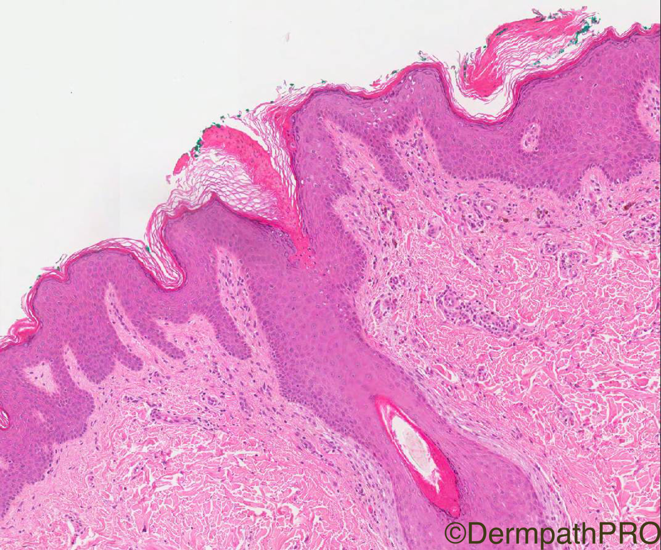
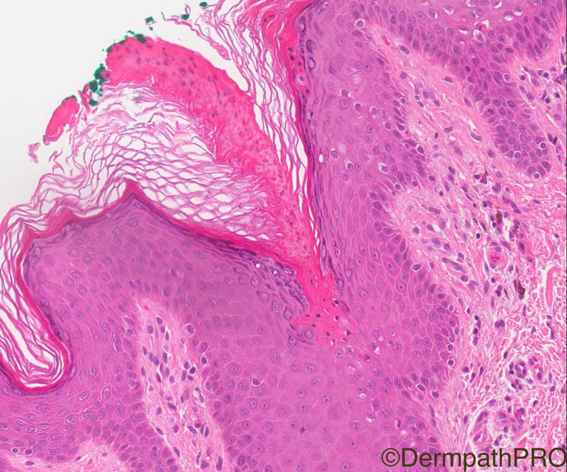
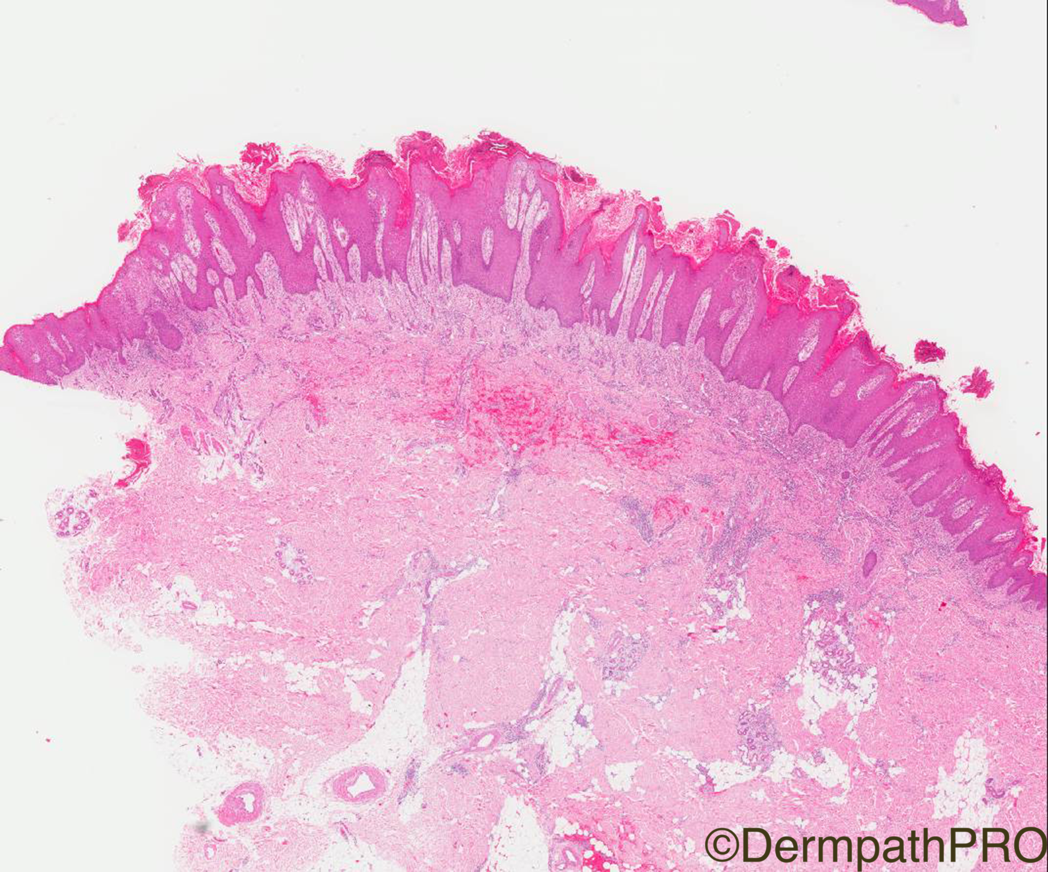
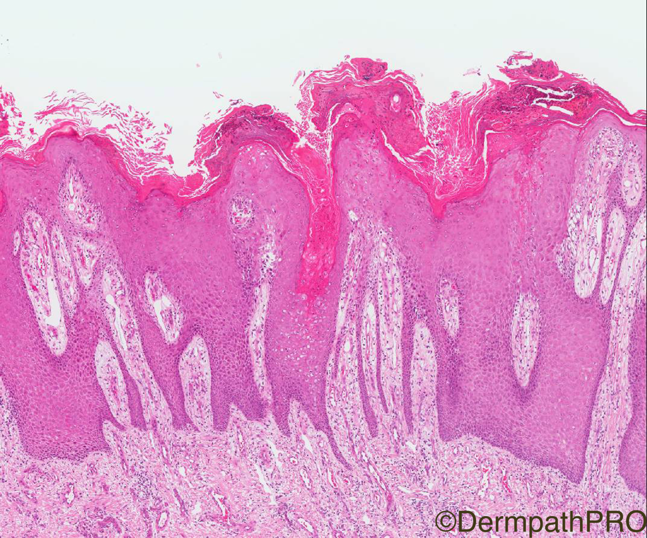
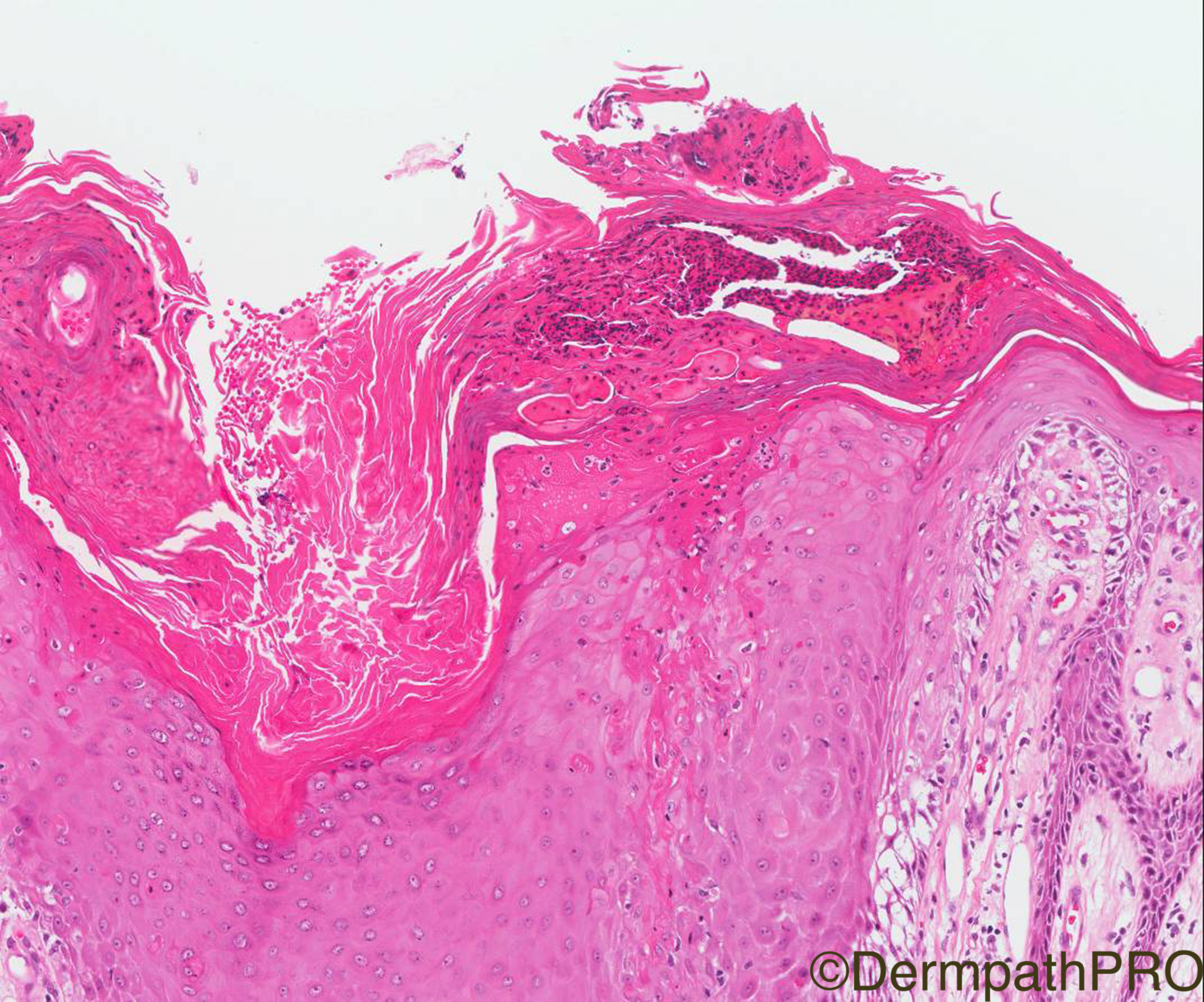
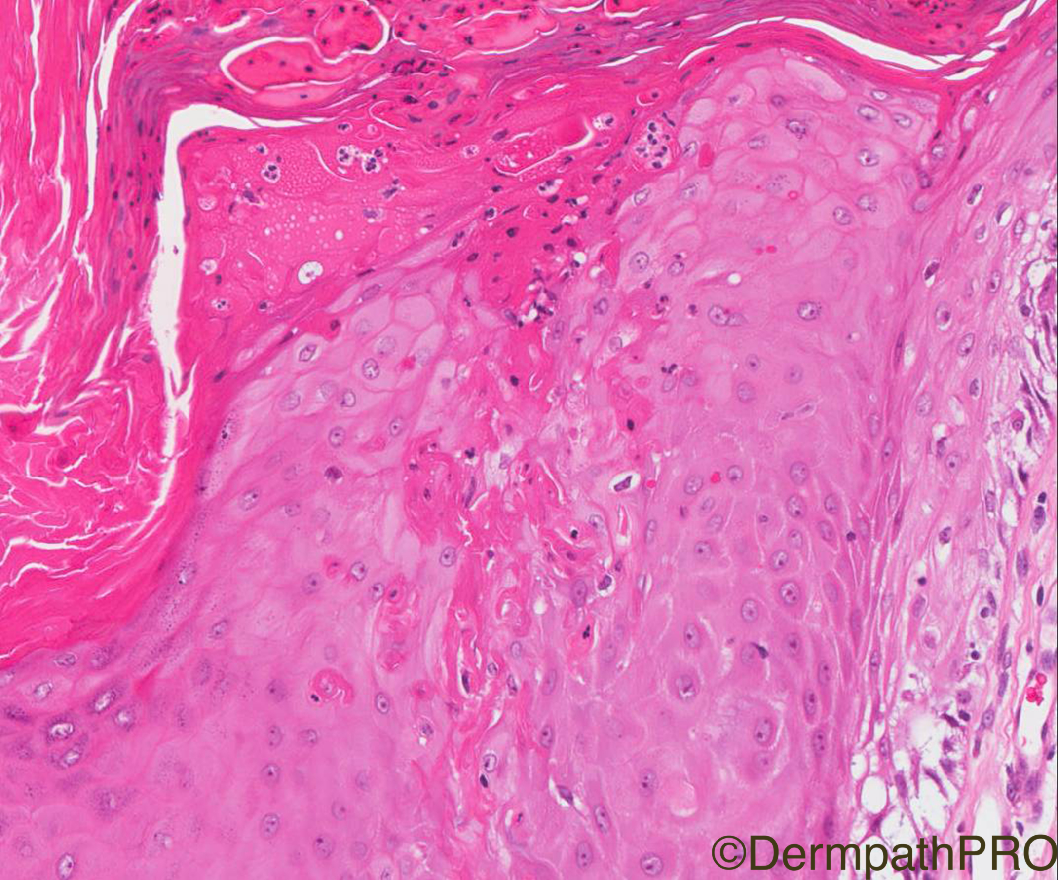
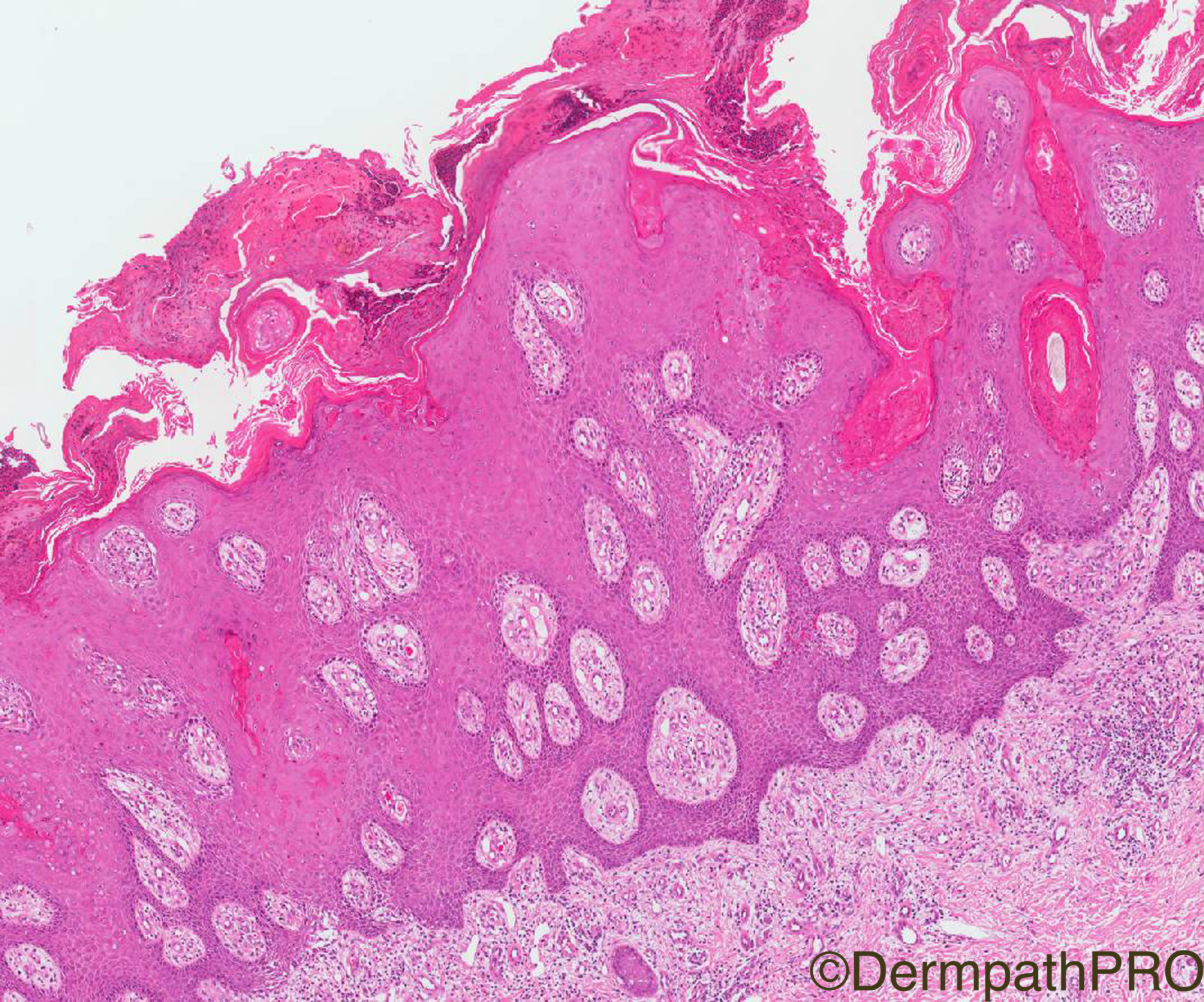
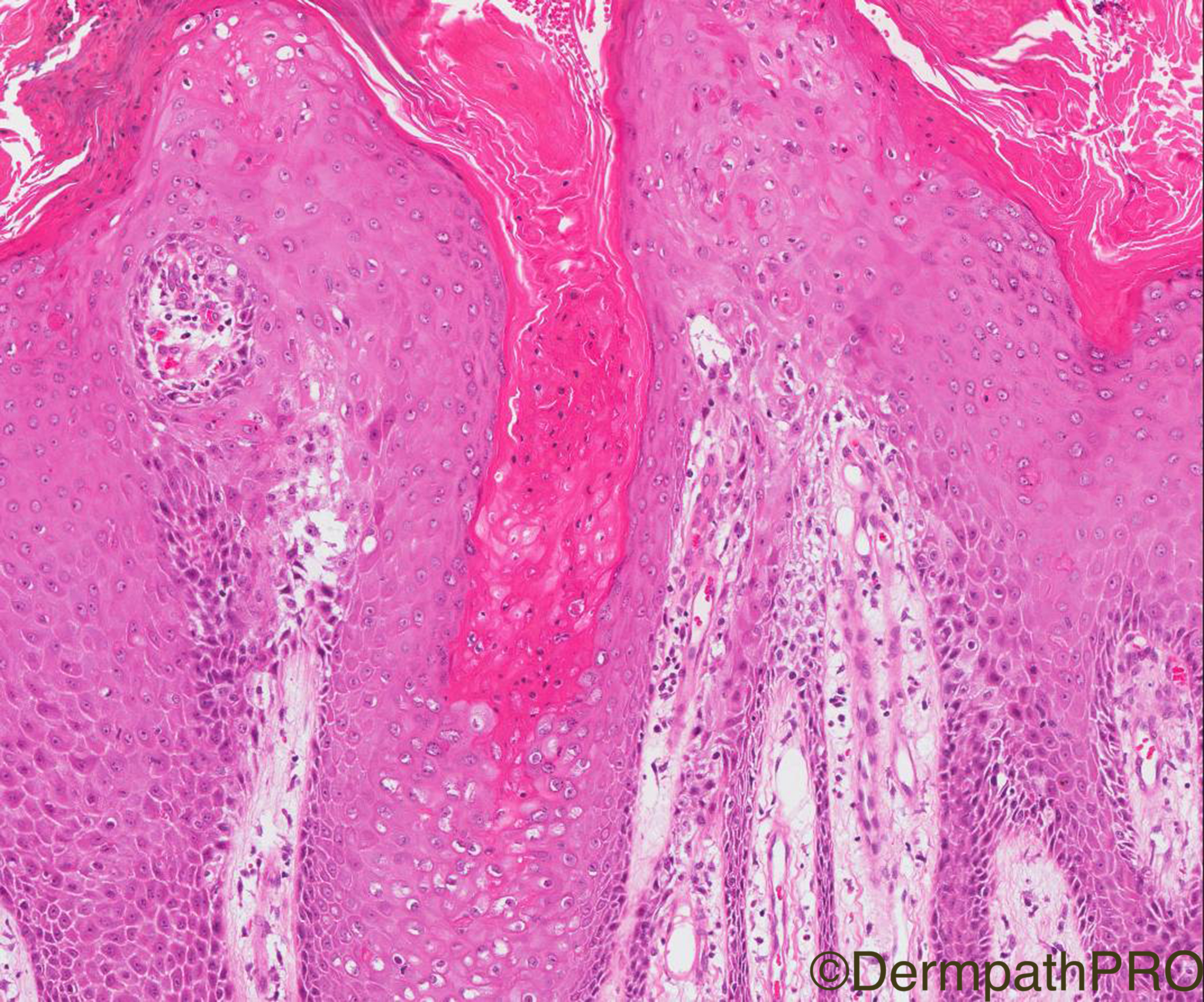
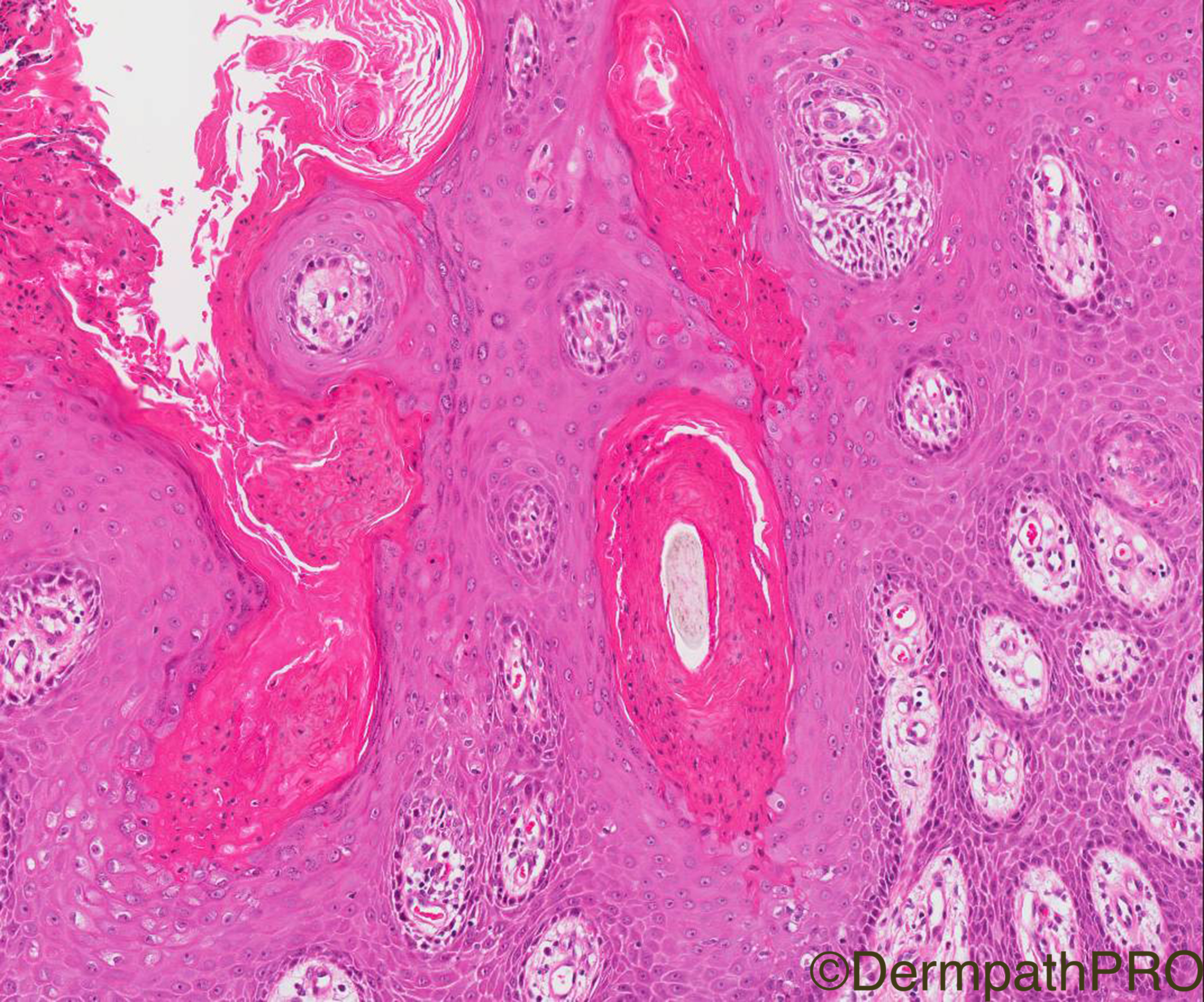
Clinical History: 54/M erythematous sharply demarcated scaly plaque buttocks extending into natal cleft, with satellite lesions. ?flexural psoriasis. 1st 7 images from main lesion in natal cleft, 9th and 10th image- Satellite lesion.

Case Posted by Dr Arti Bakshi

  Report Record

User Feedback


Admin_Dermpath

Posted

Sorry for slightly later than normal Daily Spot Posting, here we have a lovely case from Dr Arti Bakshi.


Cheers, Geoff Cross - DermpathPRO Projects

Share this comment


Link to comment
share_externally

Dr. Mona Abdel-Halim

Posted

Thinking of porokeratosis ptychotropica with possible secondary candidal superinfection. 

Share this comment


Link to comment
share_externally

Dr. Mona Abdel-Halim

Posted

The intracorneal neutrophils is what made me think of candidal superinfection as it can not be explained in the context of porokeratosis alone. 

Share this comment


Link to comment
share_externally

Robledo F. Rocha

Posted

Verrucous porokeratosis, a proposed terminology to encompass porokeratosis ptychotropica and other descriptive terms of the verrucous variant of porokeratosis involving the gluteal cleft.

Share this comment


Link to comment
share_externally

Raul Perret

Posted

Agree with porokeratosis either verrucous or ptychotropica as my colleagues suggested. I have read that the clinical appearance of the lesions is important to differentiate the entities (verrucous is always hyperkeratotic and can be located in other areas appart from the buttocks while ptychotropica is always located on the latter and is not hyperkeratotic). So based on these I favour verrucous in this case

Share this comment


Link to comment
share_externally

vincenzo polizzi

Posted

My first thought was of Porokeratosis, too. But after the first img I completely changed track...This because of my unknowledge of Porokeratosis Ptychotropica ( or Verrucous Porokeratosis ). Now I feel pretty sound what my Colleagues argue about this lesion. And agree with Mona that there is probably a Candida infection overlap to explain many neutrofils scale crusts. But I have a question to the experts: why not a flexural seborrheic dermatitis? 

Share this comment


Link to comment
share_externally

Dr. Mona Abdel-Halim

Posted

1 hour ago, vincenzo polizzi said:

My first thought was of Porokeratosis, too. But after the first img I completely changed track...This because of my unknowledge of Porokeratosis Ptychotropica ( or Verrucous Porokeratosis ). Now I feel pretty sound what my Colleagues argue about this lesion. And agree with Mona that there is probably a Candida infection overlap to explain many neutrofils scale crusts. But I have a question to the experts: why not a flexural seborrheic dermatitis? 

You will not see the typical cornoid lamelle in seb dermatitis Vincenzo, but u will c parakeratosis with neutrophil and exudate centered on follicular ostia. We have here typical cornoid lamellae with underlying agranulosis and dyskeratotic cells. 

Share this comment


Link to comment
share_externally

IgorSC

Posted (edited)

Nice case of Porokeratosis ptychotropica. It is important to recognize this entity, as some cases are clinically and even histologically misdiagnosed as HPV infection. I had 2 cases like this, one patient had 30 years of evolution without diagnosis and was treated as Lichen simplex chronicus!!

Edited by IgorSC
add some information at the end

Share this comment


Link to comment
share_externally

Arti Bakshi

Posted

Well done all!...this is a case of porokeratosis ptychotropica.

The images were posted in the wrong order. the 1st 2 are actually from the satellite lesion, which show typical cornoid lamellae without much inflammation. The rest of the images show significant inflammation (including neutrophilic collections in stratum corneum) and disruption of cornoid lamellae, which lead to an erroneous impression of psoriasis by the referring pathologist. PAS stain for fungal profiles was negative, so most likely the inflammatory changes seen are secondary to friction/trauma, given the site within the natal cleft. As Mona rightly pointed, the dyskeratotic cells are a clue to cornoid lamellae, even in the areas where typical tiers of parakeratosis are not seen. Completely agree with Igor, that these cases are easily missed.This case too has had previous biopsies reported as lichen simplex chronicus!

Share this comment


Link to comment
share_externally


Join the conversation

You can post now and register later. If you have an account, sign in now to post with your account.

Guest
Add a comment...

×   Pasted as rich text.   Paste as plain text instead

  Only 75 emoji are allowed.

×   Your link has been automatically embedded.   Display as a link instead

×   Your previous content has been restored.   Clear editor

×   You cannot paste images directly. Upload or insert images from URL.

Loading...

×
×
  • Create New...