Jump to content
In this section we have spot diagnoses posted on a daily basis since June 2010, now over 4000! You can review the archived cases and read the suggested diagnoses by users and the final comment by the contributors.
Case are uploaded each week day by 10 am UK time with the correct diagnosis will generally be posted at 8 pm UK time. Why not view the most recent spot diagnosis and proffer a diagnosis?

Case Number : Case 1735 - 20 January - Dr Richard A Carr Posted By: Guest

Please read the clinical history and view the images by clicking on them before you proffer your diagnosis.
Submitted Date :
   (0 reviews)

Clinical History: Spot Diagnosis

Case Posted by Dr Richard A Carr


  Report Record

User Feedback


Admin_Dermpath

Posted

A tidy case to wrap up the week by Dr Richard A Carr.


Geoff Cross - DermpathPRO Projects

Share this comment


Link to comment
share_externally

vincenzo polizzi

Posted

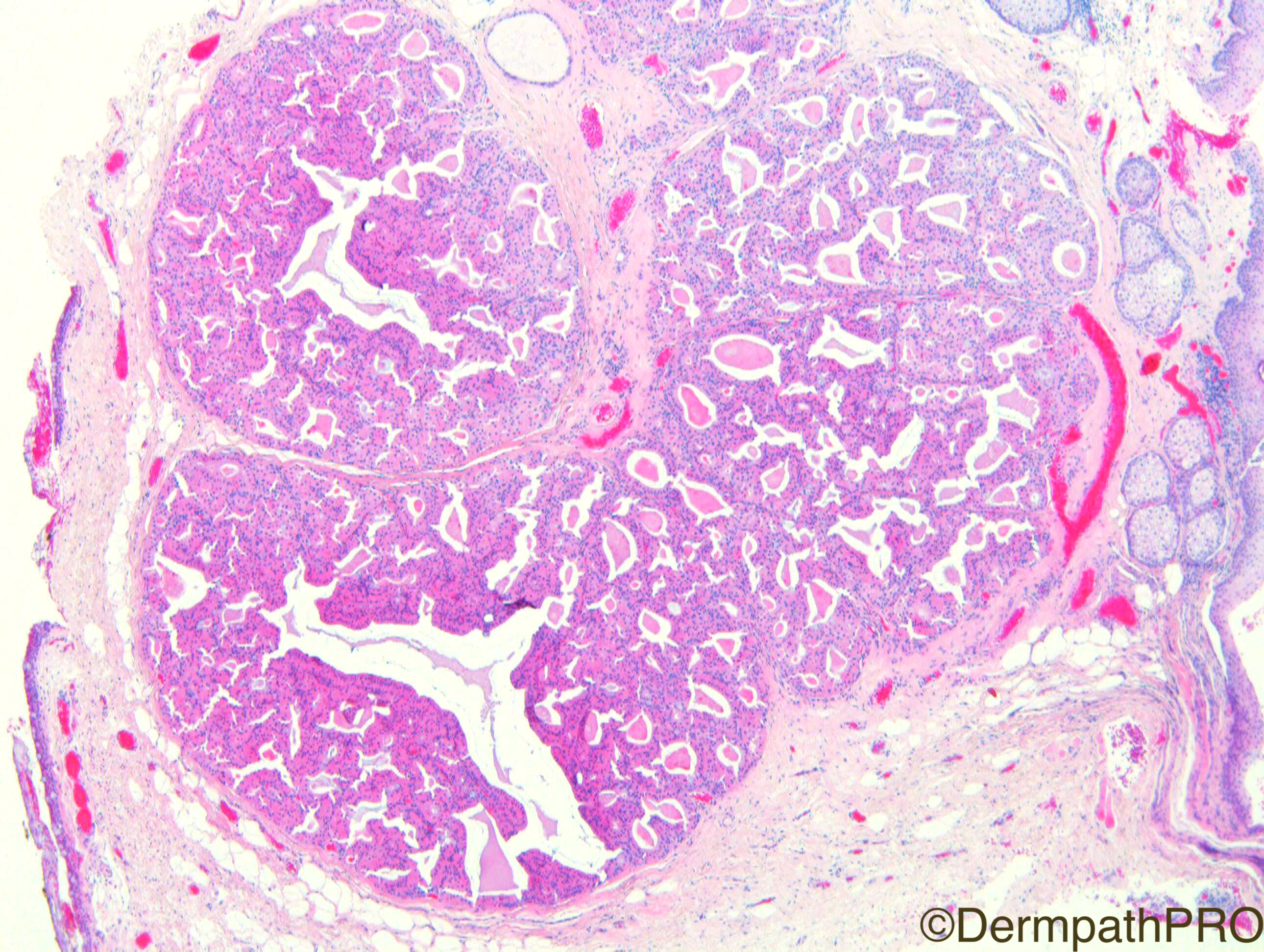
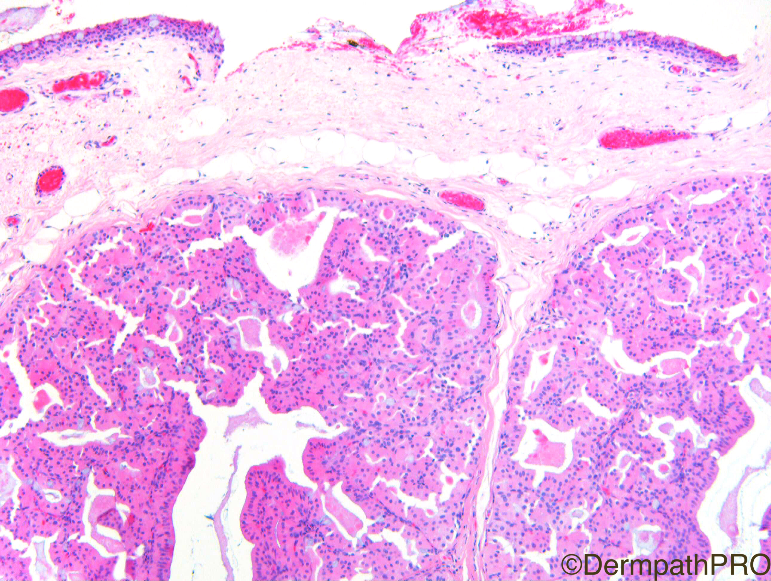
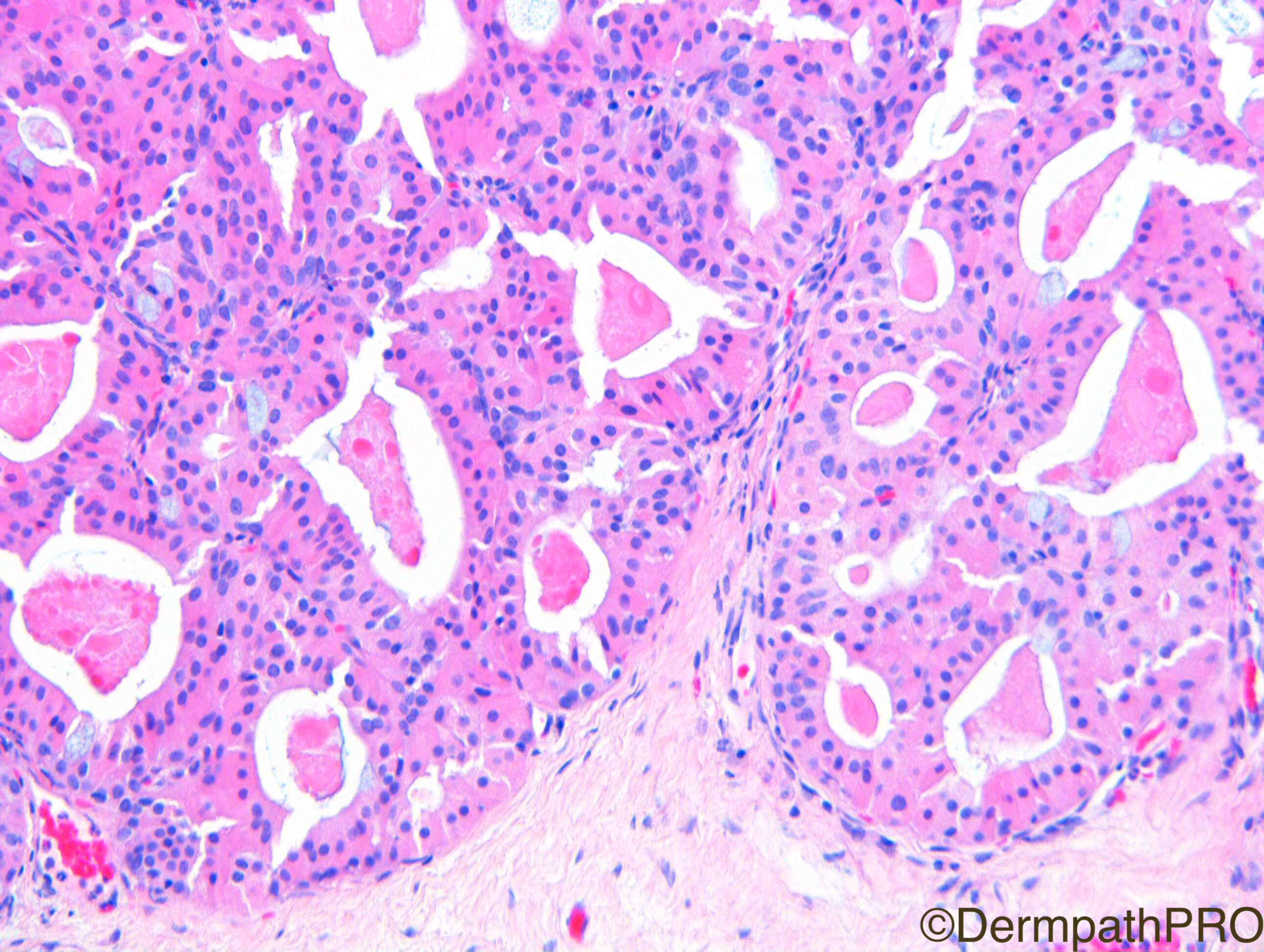
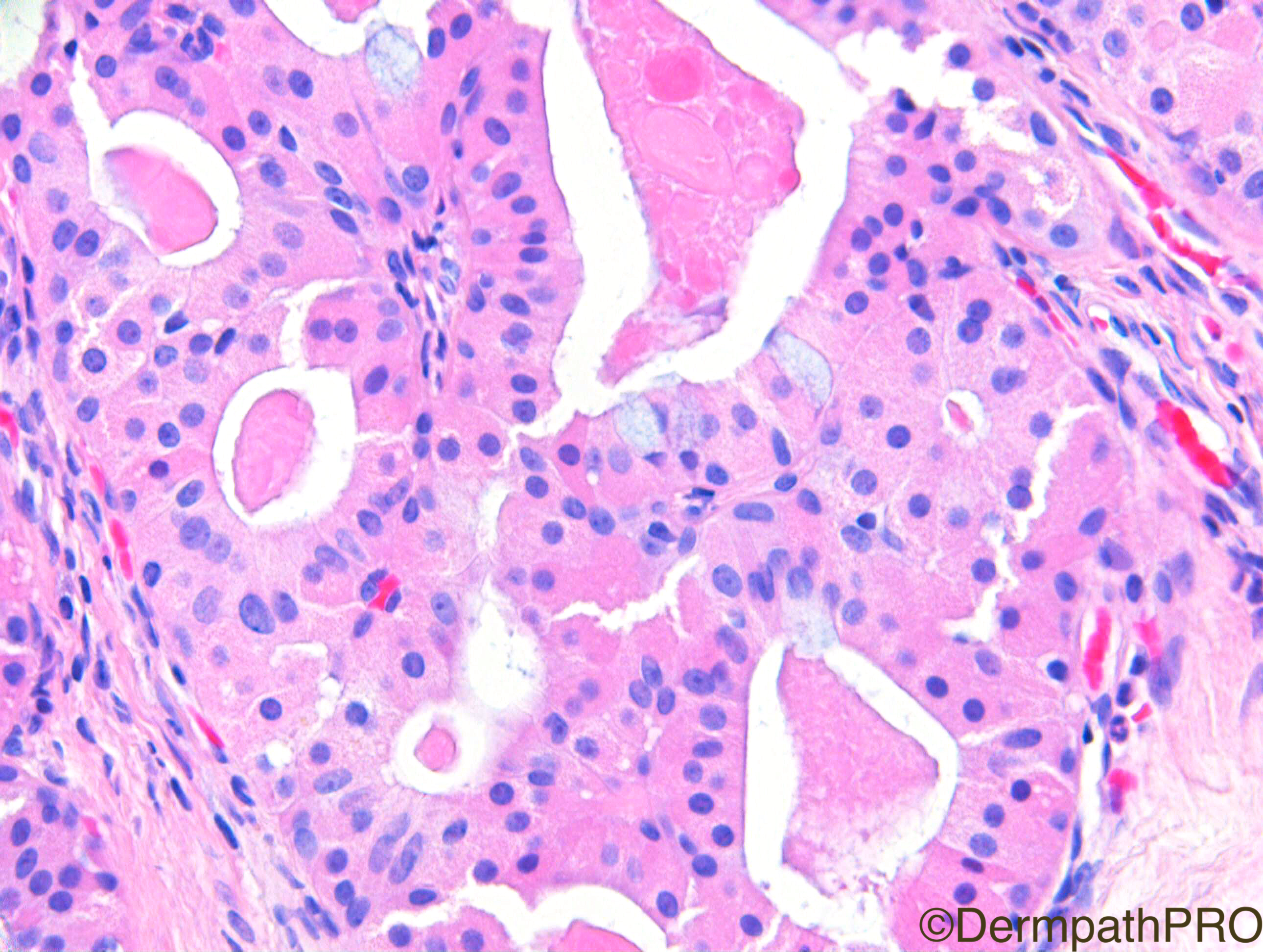
Looks like something with oncocytic differentiation, similar to, for example, a lymphocytic stroma poor Warthin tumor or a oncocytoma. May be I'm wrong...we don't know the topography of the lesion, but...I was thinking of an ocular adnexal oncocytoma.

Share this comment


Link to comment
share_externally

Dr. Mona Abdel-Halim

Posted

Agree with Vincenzo .. oncocytoma

Share this comment


Link to comment
share_externally

Arti Bakshi

Posted

Agree completely with Vincenzo! This is probably an eyelid lesion, as can see conjunctival epithelium on one surface. Interestingly, I was at the Royal Society of Medicine dermatology meeting in London yesterday and a rather similar case of primary cutaneous oncocytoma was presented in the CPC section.

Share this comment


Link to comment
share_externally

Robledo F. Rocha

Posted

Agree with Vicenzo. Solid-cystic lobulated oncocytic adenoma with some mucinous cells lining cystic spaces. Hidradenoma with predominant oxyphilic cellular population may be a point, but I cannot imagine a hidradenoma exclusively composed of this type of cells. Also, site favors oncocytic adenoma as at the periphery of the images one can find structures of the eyelid, including Meibomian sebaceous glands of the tarsal plate, and squamous stratified conjunctival epithelium with mucinous goblet cells.

Share this comment


Link to comment
share_externally

Nitin Khirwadkar

Posted

Agree with Vincenzo, oncoccytoma. Lovely case.

Share this comment


Link to comment
share_externally

Dr. Richard Carr

Posted

Well done this is an oncocytic adenoma of the conjunctival caruncle. A nice spot diagnosis.

Enjoy your weekends.

Share this comment


Link to comment
share_externally


Join the conversation

You can post now and register later. If you have an account, sign in now to post with your account.

Guest
Add a comment...

×   Pasted as rich text.   Paste as plain text instead

  Only 75 emoji are allowed.

×   Your link has been automatically embedded.   Display as a link instead

×   Your previous content has been restored.   Clear editor

×   You cannot paste images directly. Upload or insert images from URL.

Loading...

×
×
  • Create New...