Jump to content
In this section we have spot diagnoses posted on a daily basis since June 2010, now over 4000! You can review the archived cases and read the suggested diagnoses by users and the final comment by the contributors.
Case are uploaded each week day by 10 am UK time with the correct diagnosis will generally be posted at 8 pm UK time. Why not view the most recent spot diagnosis and proffer a diagnosis?

Case Number : Case 1741 - 30 January - Dr Limin Yu Posted By: Guest

Please read the clinical history and view the images by clicking on them before you proffer your diagnosis.
Submitted Date :
   (0 reviews)

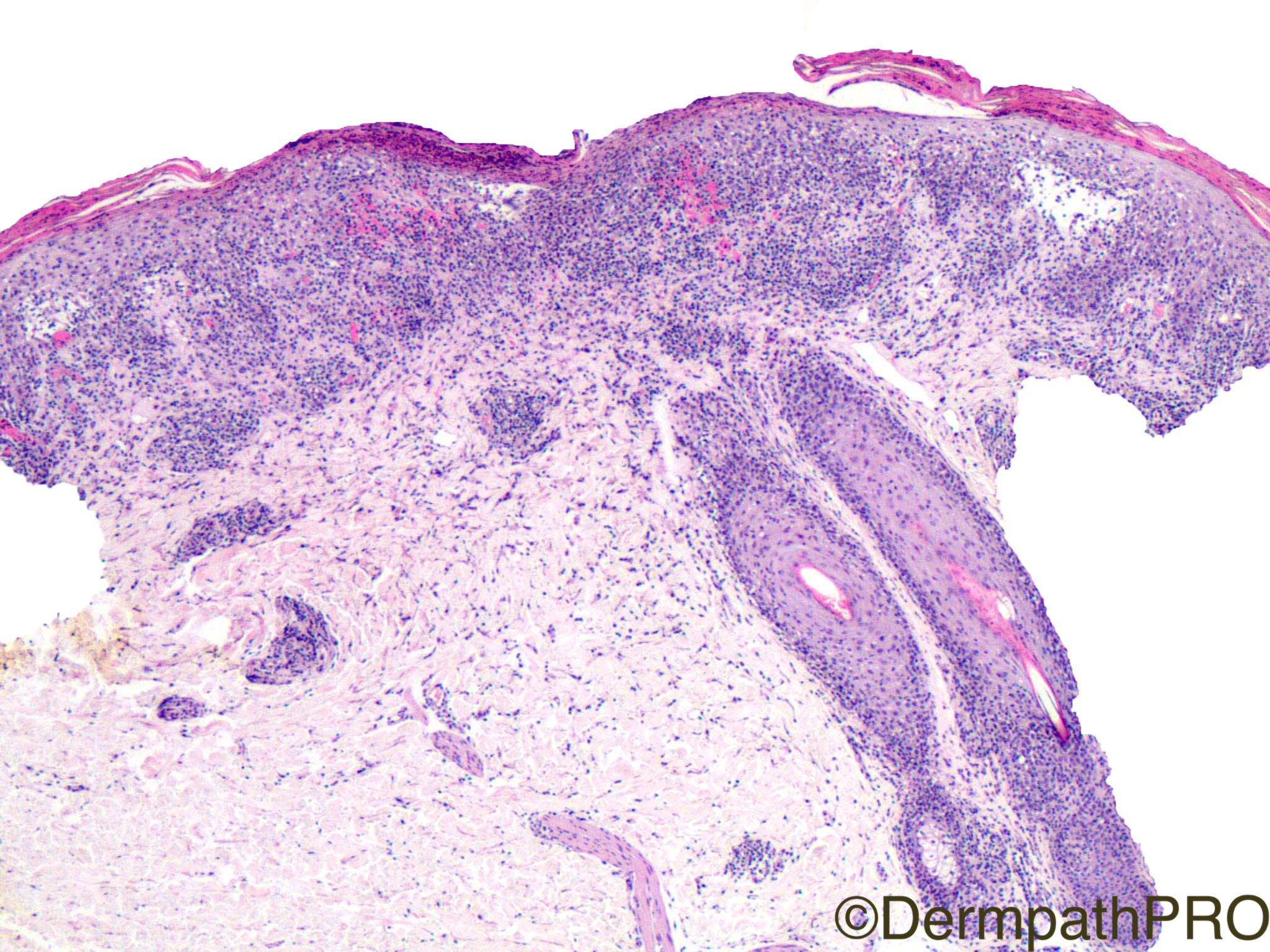
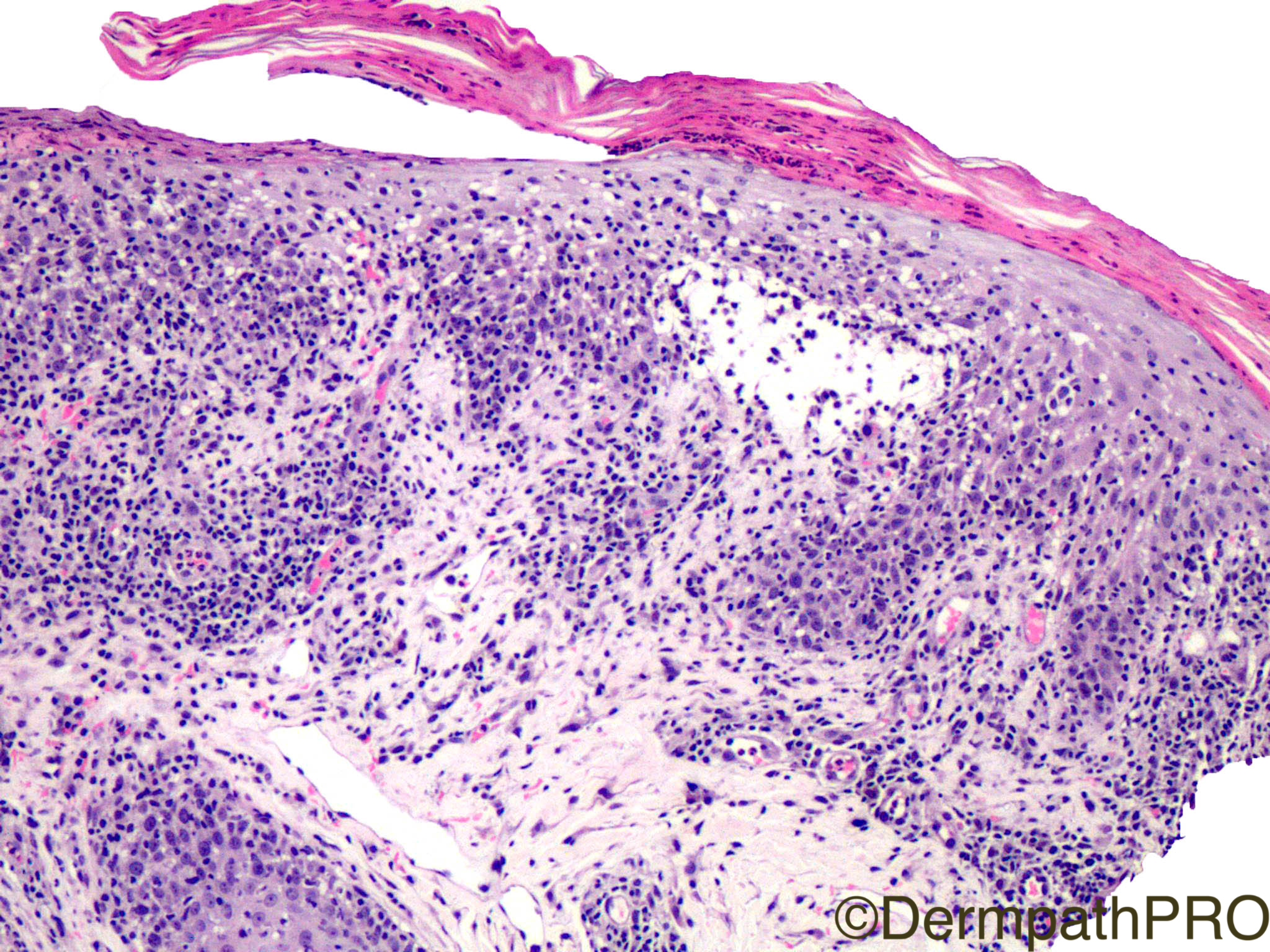
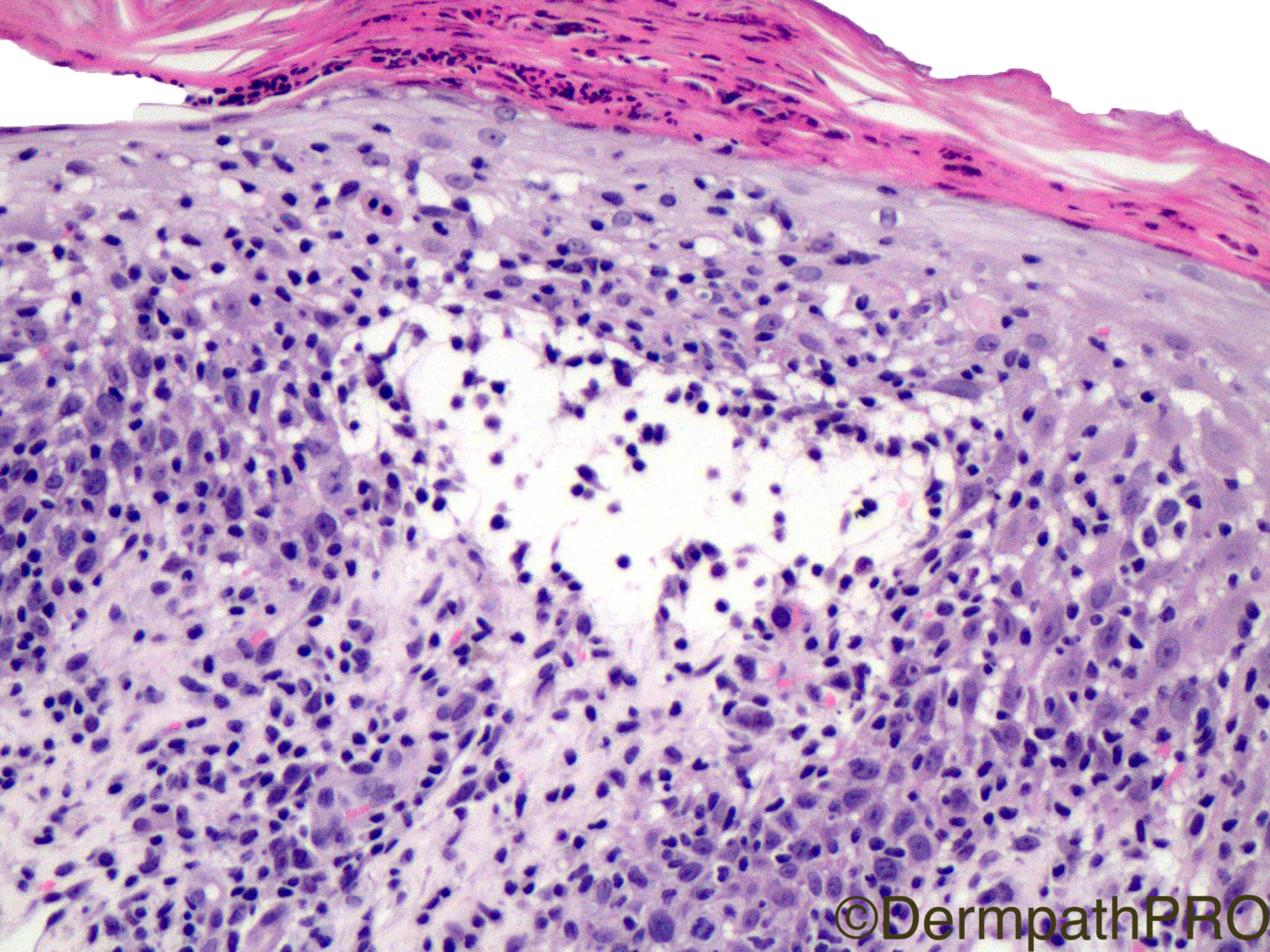
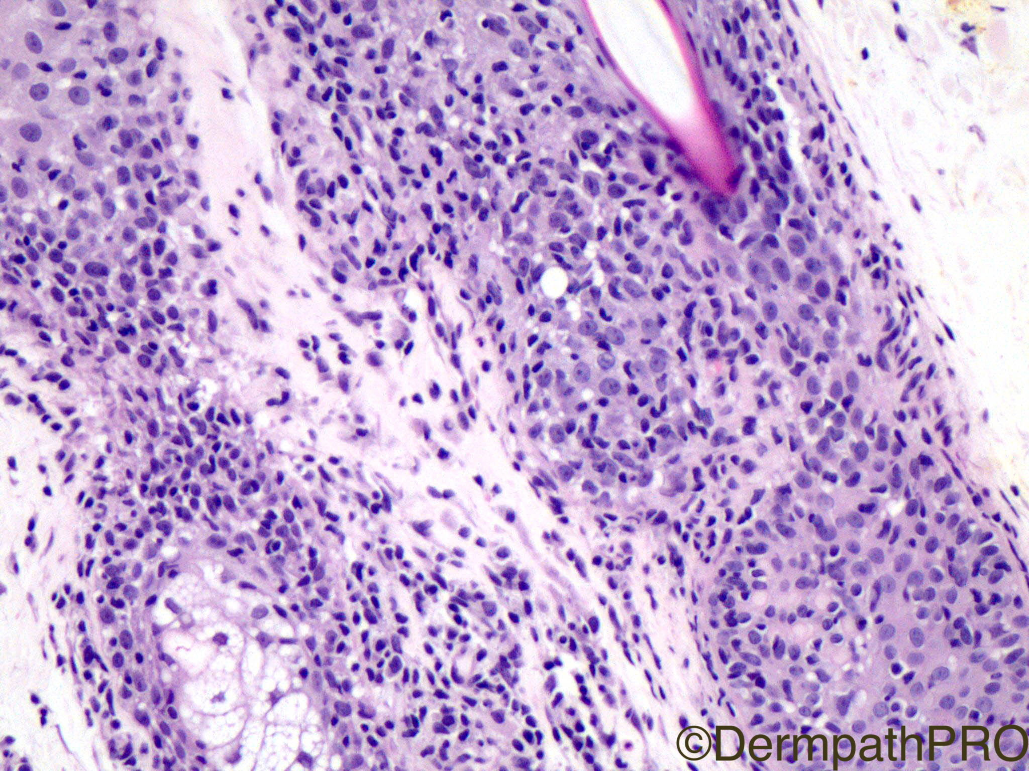
Clinical History: This is a 37 yo F with sudden onset of papules.

Case Posted by Dr Limin Yu


  Report Record

User Feedback


Admin_Dermpath

Posted

Start your week off with a neat case from Dr Limin Yu.


Geoff Cross - DermpathPRO Projects

Share this comment


Link to comment
share_externally

Dr. Mona Abdel-Halim

Posted

Pityriasis lichenoides spectrum. Sudden onset favors PLEVA if supported clinically by presence of necrotic lesions.

Share this comment


Link to comment
share_externally

vincenzo polizzi

Posted

Agree. I guess the large hypercromic lymphocytes are CD30 cells...

Share this comment


Link to comment
share_externally

Robledo F. Rocha

Posted

A hard case! Clinical history favors the acute form of pityriasis lichenoides, but caught my eyes the absence of extravasated erythrocytes and vacuolar change of the basal layer, and the presence of periadnexal lymphocytic infiltration. Were the lymphocytes abnormal and large, I’d go with lymphomatoid papulosis with 6p25.3 rearrangement due to the presence of, at low power magnification, pagetoid reticulosis-like epidermotropic lymphocytic infiltration and marked involvement of pilosebaceous units and eccrine glands.

Share this comment


Link to comment
share_externally

Arti Bakshi

Posted

Good suggestion by Robledo! Definitely need a CD30 in this case. However, PLEVA and LyP lie on a spectrum and one can get CD30+ cells in PLEVA. The clinical course of the disease would be important in the distinction.

Share this comment


Link to comment
share_externally

I think this is PLEVA, but I liked Robledo´s idea.

Share this comment


Link to comment
share_externally

Raul Perret

Posted

I thought of PLEVA in this case as all of you guys suggested. My impression was that we see some extravasated erythrocytes (although not super prominent)

Share this comment


Link to comment
share_externally

Nitin Khirwadkar

Posted

I am thinking of PLEVA. As Raul has commented, I too feel there is some RBC extravasation.

Share this comment


Link to comment
share_externally

I think the invading lymphocytes are atypical and there is ulceration. I would go for lymphomatoid papulosis but clinico path correlation required.

Share this comment


Link to comment
share_externally

Guest parasa gayatri devi

Posted

I am thinking of PLEVA.  Focal loss of stratum corneum and spongiosis favour the diagnosis of PLEVA

Share this comment


Link to comment
share_externally

You guys are awesome. Agree with respect!  This is a PLEVA. Your points are well taken!


Share this comment


Link to comment
share_externally


Join the conversation

You can post now and register later. If you have an account, sign in now to post with your account.

Guest
Add a comment...

×   Pasted as rich text.   Paste as plain text instead

  Only 75 emoji are allowed.

×   Your link has been automatically embedded.   Display as a link instead

×   Your previous content has been restored.   Clear editor

×   You cannot paste images directly. Upload or insert images from URL.

Loading...

×
×
  • Create New...