Jump to content
In this section we have spot diagnoses posted on a daily basis since June 2010, now over 4000! You can review the archived cases and read the suggested diagnoses by users and the final comment by the contributors.
Case are uploaded each week day by 10 am UK time with the correct diagnosis will generally be posted at 8 pm UK time. Why not view the most recent spot diagnosis and proffer a diagnosis?

Case Number : Case 1855 - 07 July - Dr Iskander Chaudhry (Invited) Posted By: Guest

Please read the clinical history and view the images by clicking on them before you proffer your diagnosis.
Submitted Date :
   (0 reviews)

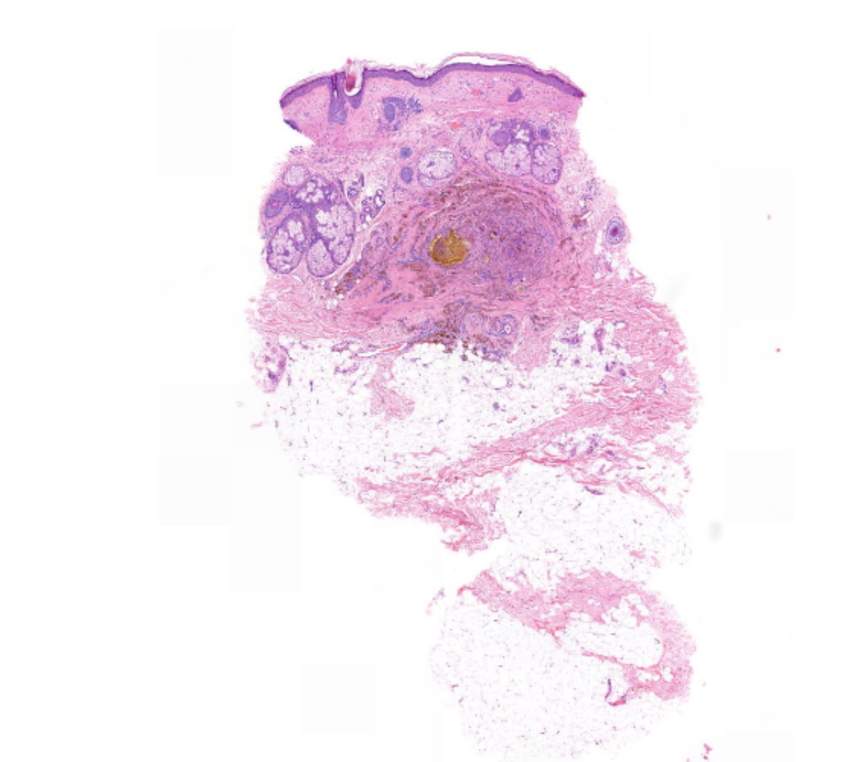
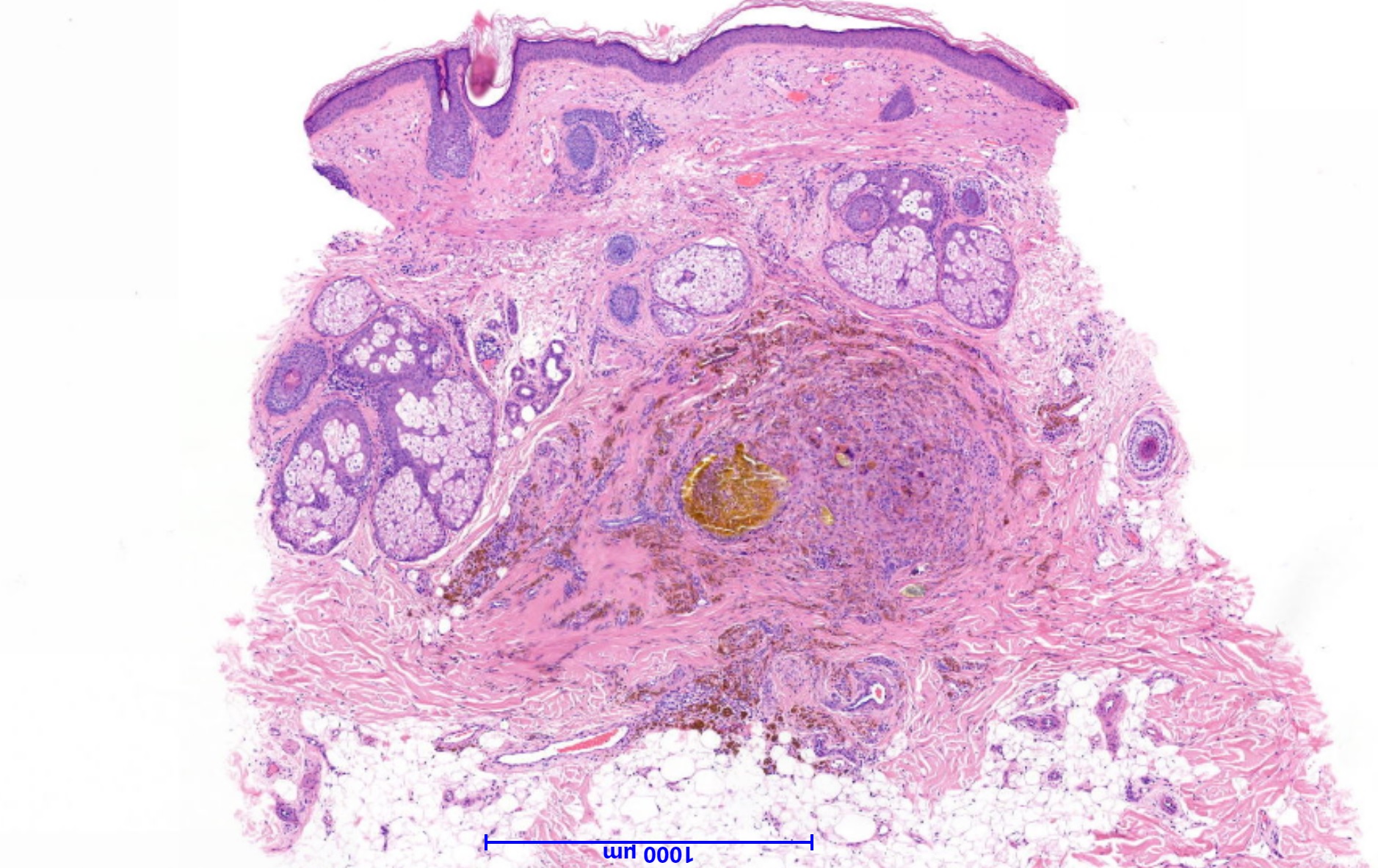
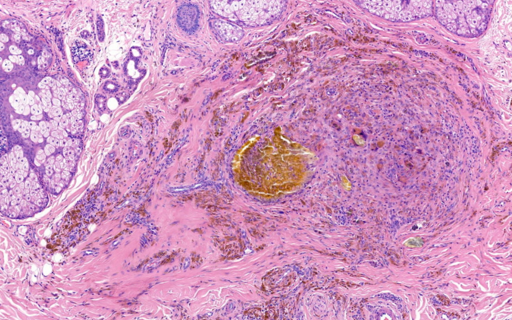
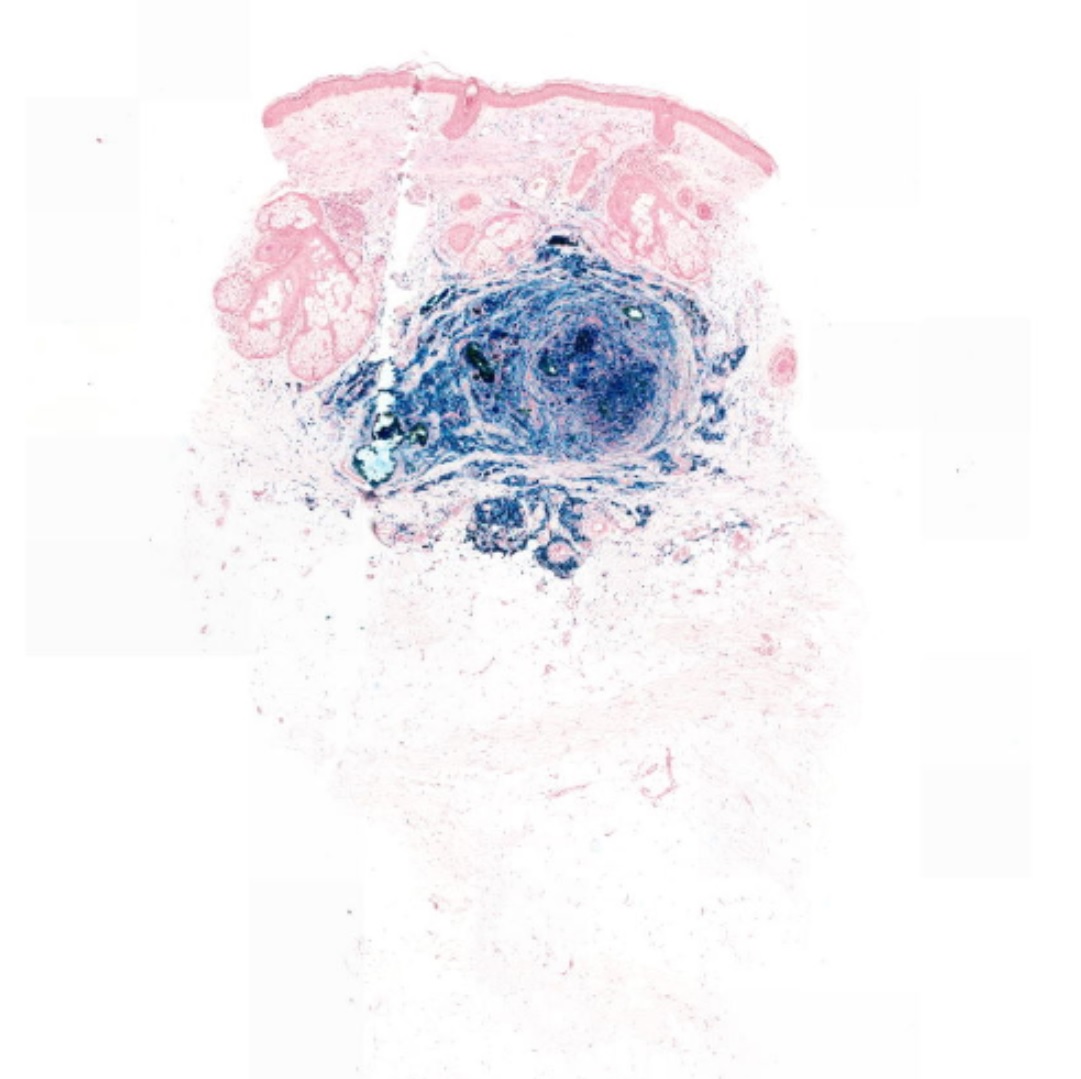
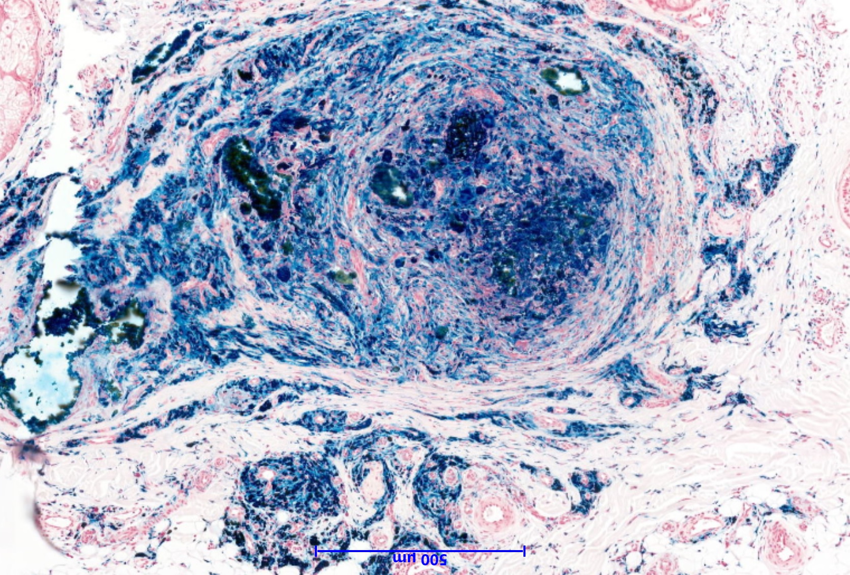
64 year old male, lesion on cheek ? blue naevus

Edited by Admin_Dermpath


  Report Record

User Feedback


Anil Patki

Posted

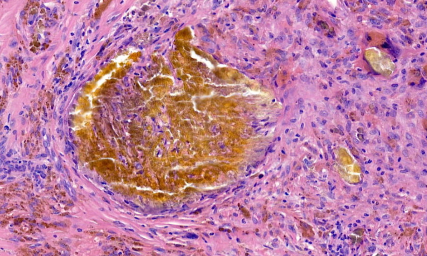
Botryomycosis / actinomycosis - grain with a granulomatous reaction

Share this comment


Link to comment
share_externally

Dr. Mona Abdel-Halim

Posted

Thought of aneurysmal fibrous histiocytoma. 

Share this comment


Link to comment
share_externally

Admin_Dermpath

Posted

Lots of views but not many responses :( 

Special stains were negative. 

Hope to get more responses  ......

Iskander

Share this comment


Link to comment
share_externally

Arti Bakshi

Posted

Looks like a foreign body reaction to extraneous matter...not sure about the nature of this extraneous brown material though!

Share this comment


Link to comment
share_externally

Robledo F. Rocha

Posted

Maybe this is a trauma-induced lesion. The golden-colored material resembles some pigment produced by the breakdown of hemoglobin, like hematoidin.

Share this comment


Link to comment
share_externally

Raul Perret

Posted

Looks like a localized form of hemosiderosis which can be caused by many entities, for example acne.

Share this comment


Link to comment
share_externally

vincenzo polizzi

Posted

An old organized trombus?

Share this comment


Link to comment
share_externally

Nitin Khirwadkar

Posted

Looks like hemosiderin within a thrombus. Certainly in the last image, the pigment is within a vessel.

Share this comment


Link to comment
share_externally

Admin_Dermpath

Posted

Many thanks everyone!


I have added the Perl's stain! 


Yes the diagnosis was localized form of hemosiderosis - as stated the causes are many. 


Iskander Chaudhry

Share this comment


Link to comment
share_externally


Join the conversation

You can post now and register later. If you have an account, sign in now to post with your account.

Guest
Add a comment...

×   Pasted as rich text.   Paste as plain text instead

  Only 75 emoji are allowed.

×   Your link has been automatically embedded.   Display as a link instead

×   Your previous content has been restored.   Clear editor

×   You cannot paste images directly. Upload or insert images from URL.

Loading...

×
×
  • Create New...