Jump to content
In this section we have spot diagnoses posted on a daily basis since June 2010, now over 4000! You can review the archived cases and read the suggested diagnoses by users and the final comment by the contributors.
Case are uploaded each week day by 10 am UK time with the correct diagnosis will generally be posted at 8 pm UK time. Why not view the most recent spot diagnosis and proffer a diagnosis?

Case Number : Case 1860 - 14 July - Dr Richard Carr Posted By: Guest

Please read the clinical history and view the images by clicking on them before you proffer your diagnosis.
Submitted Date :
   (0 reviews)

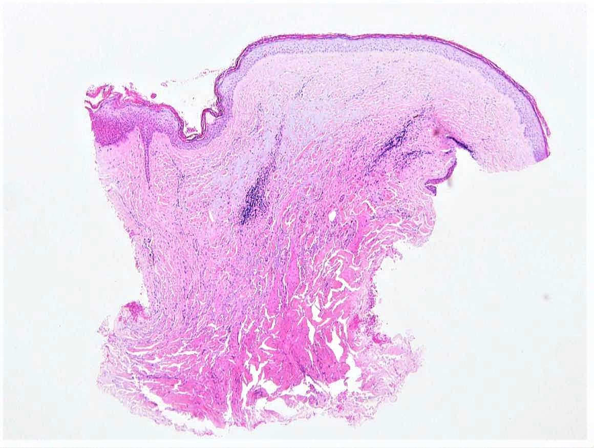
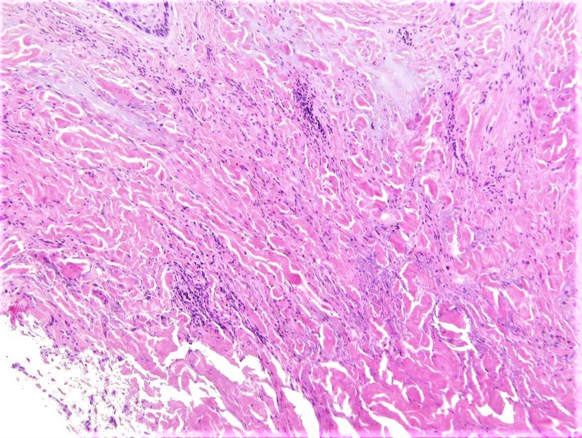
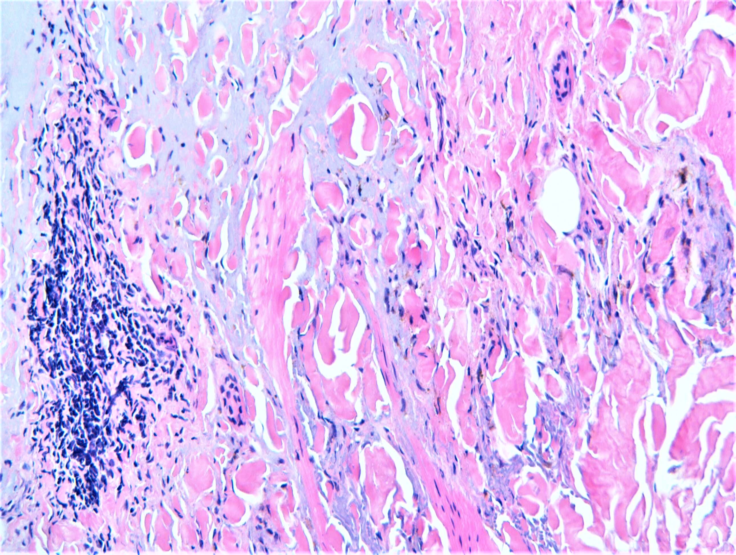
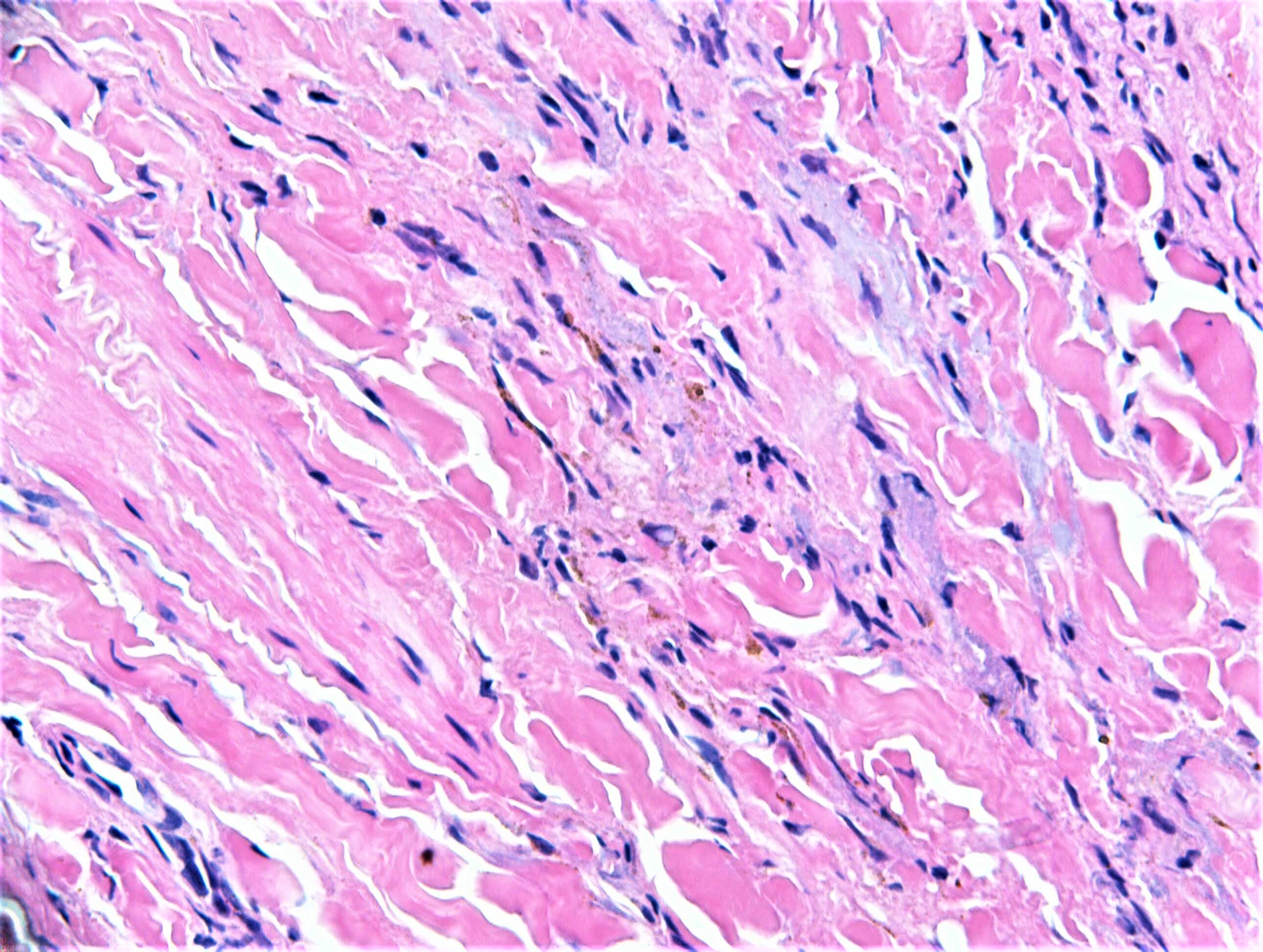
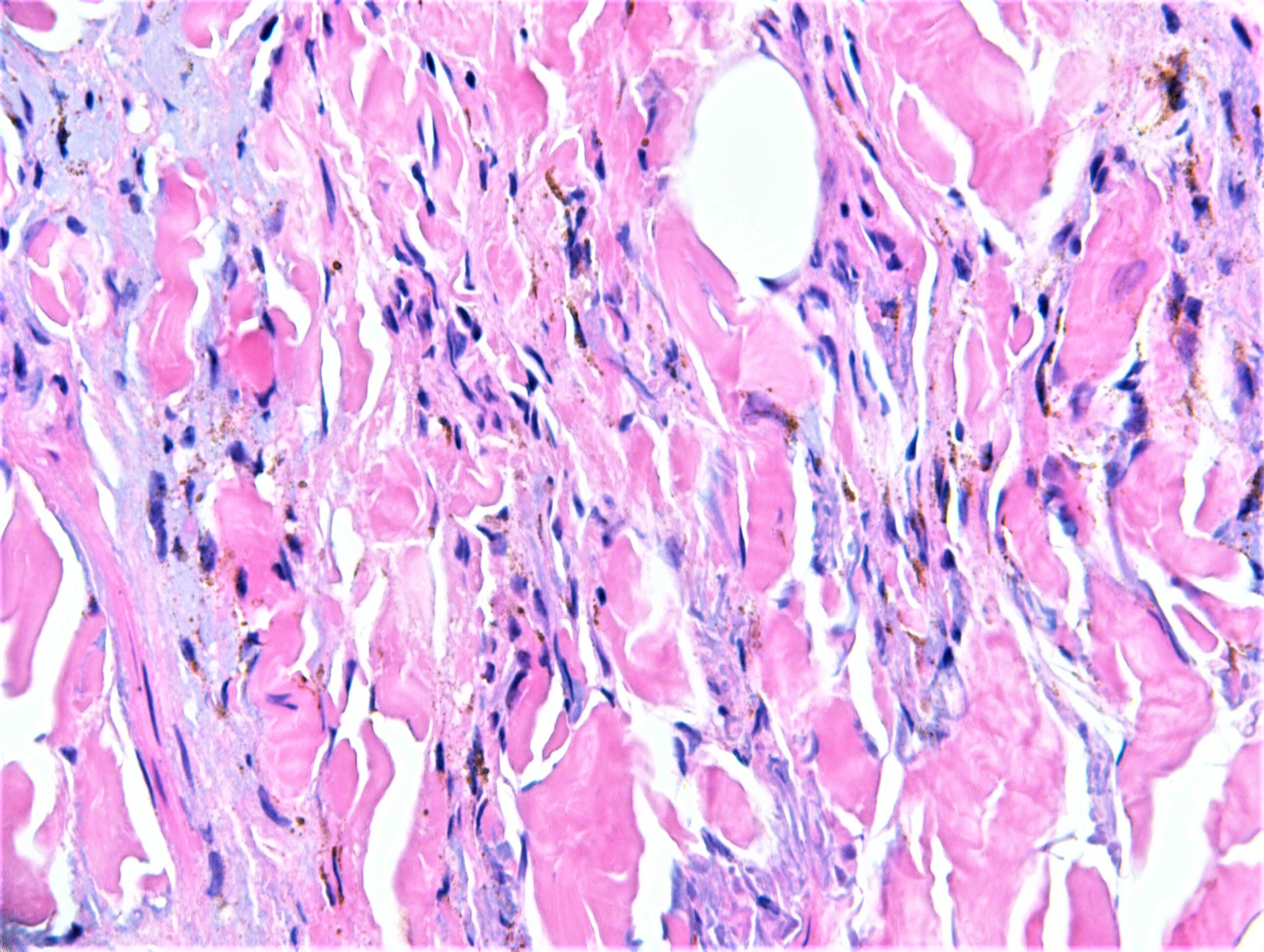
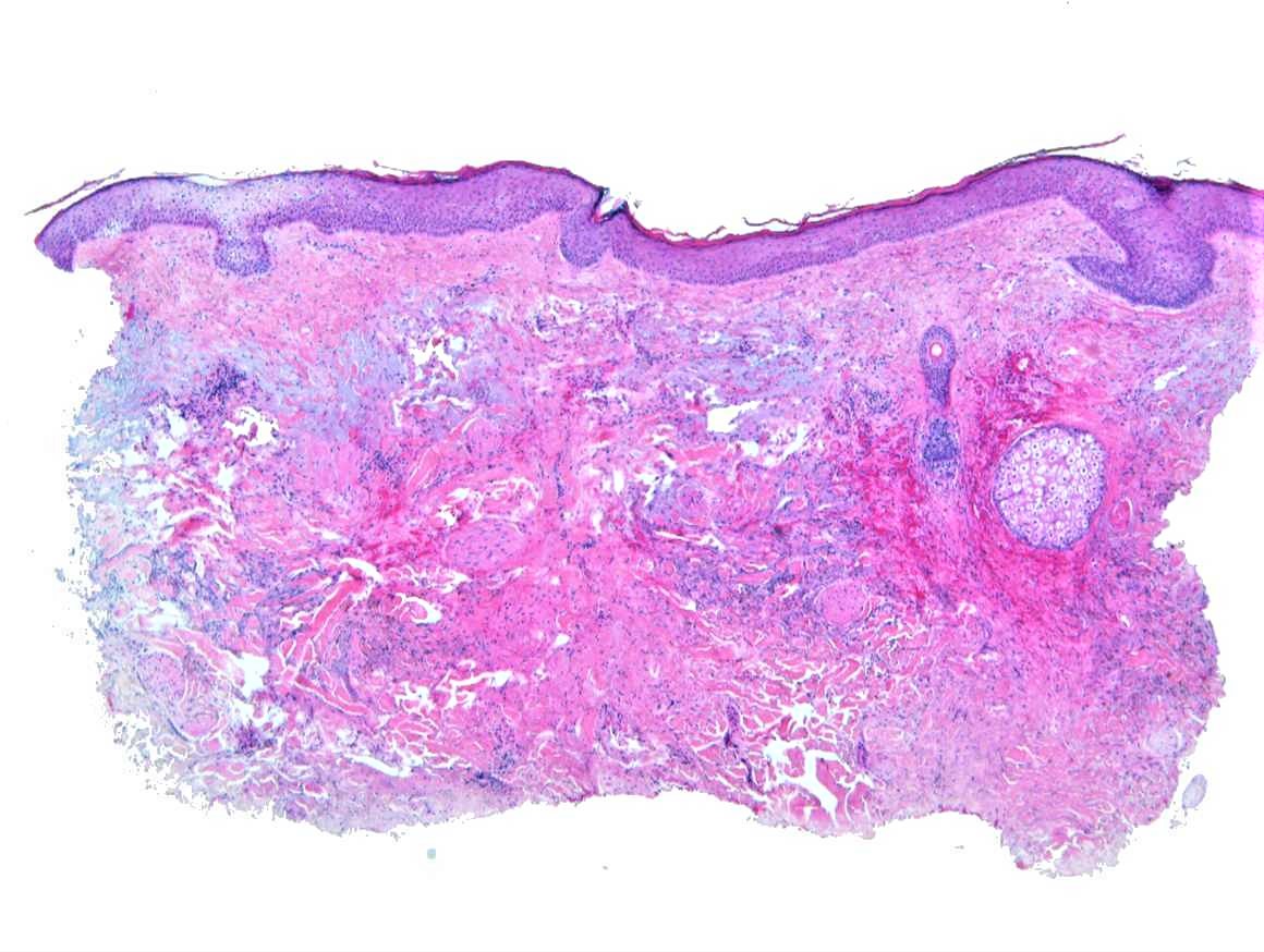
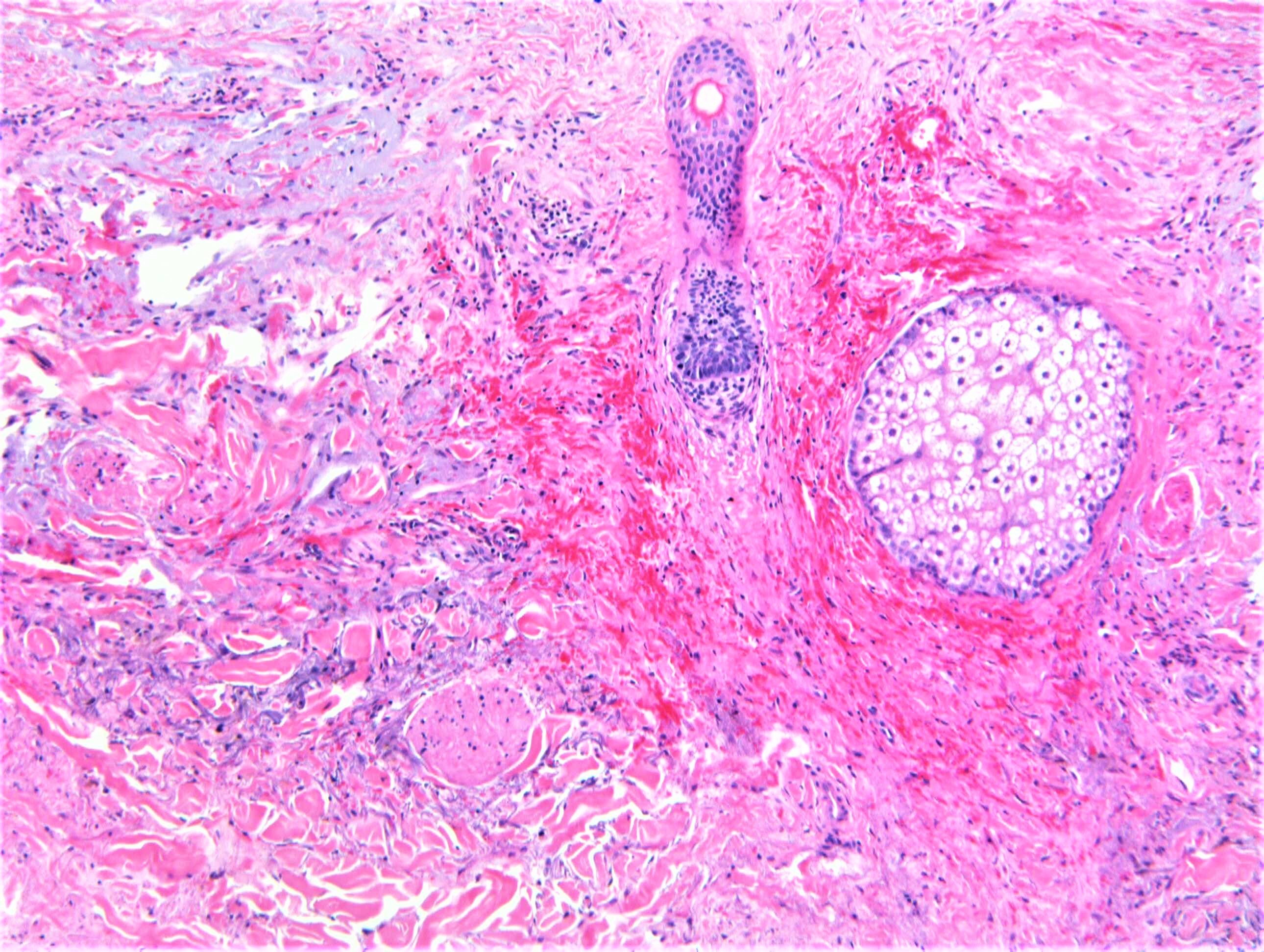
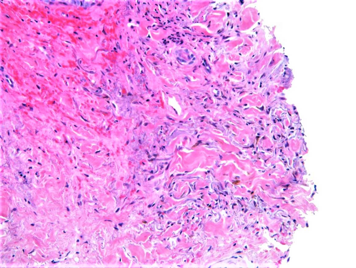
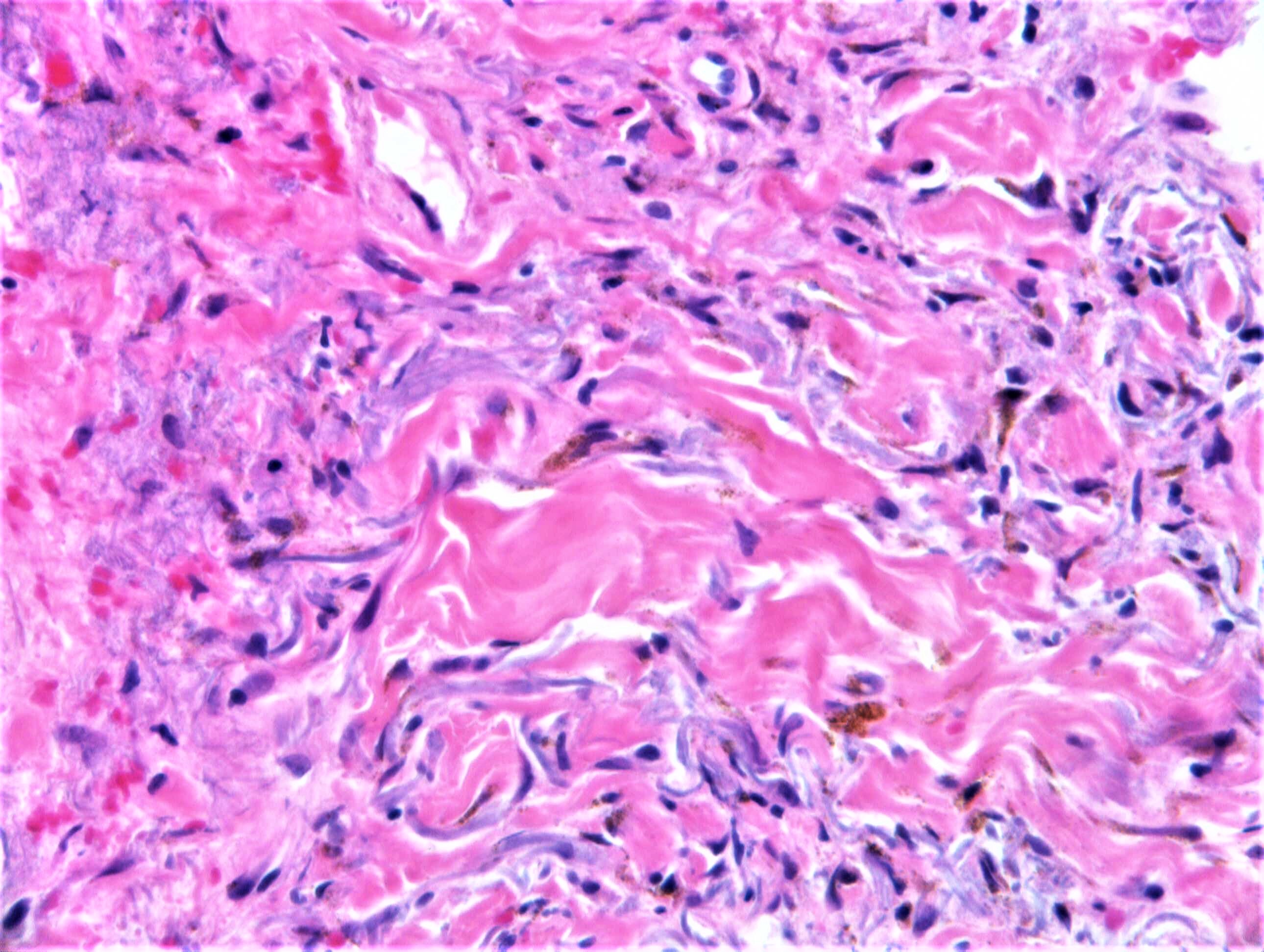
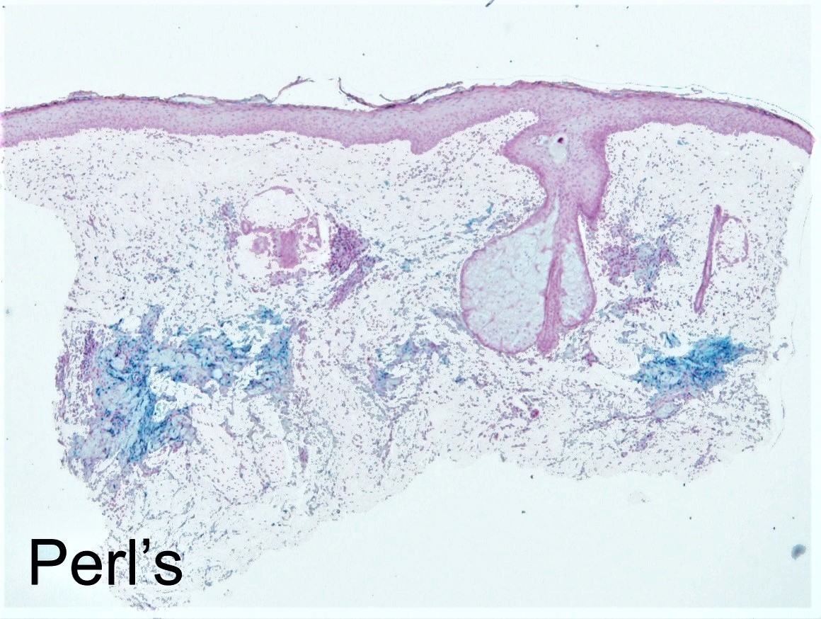
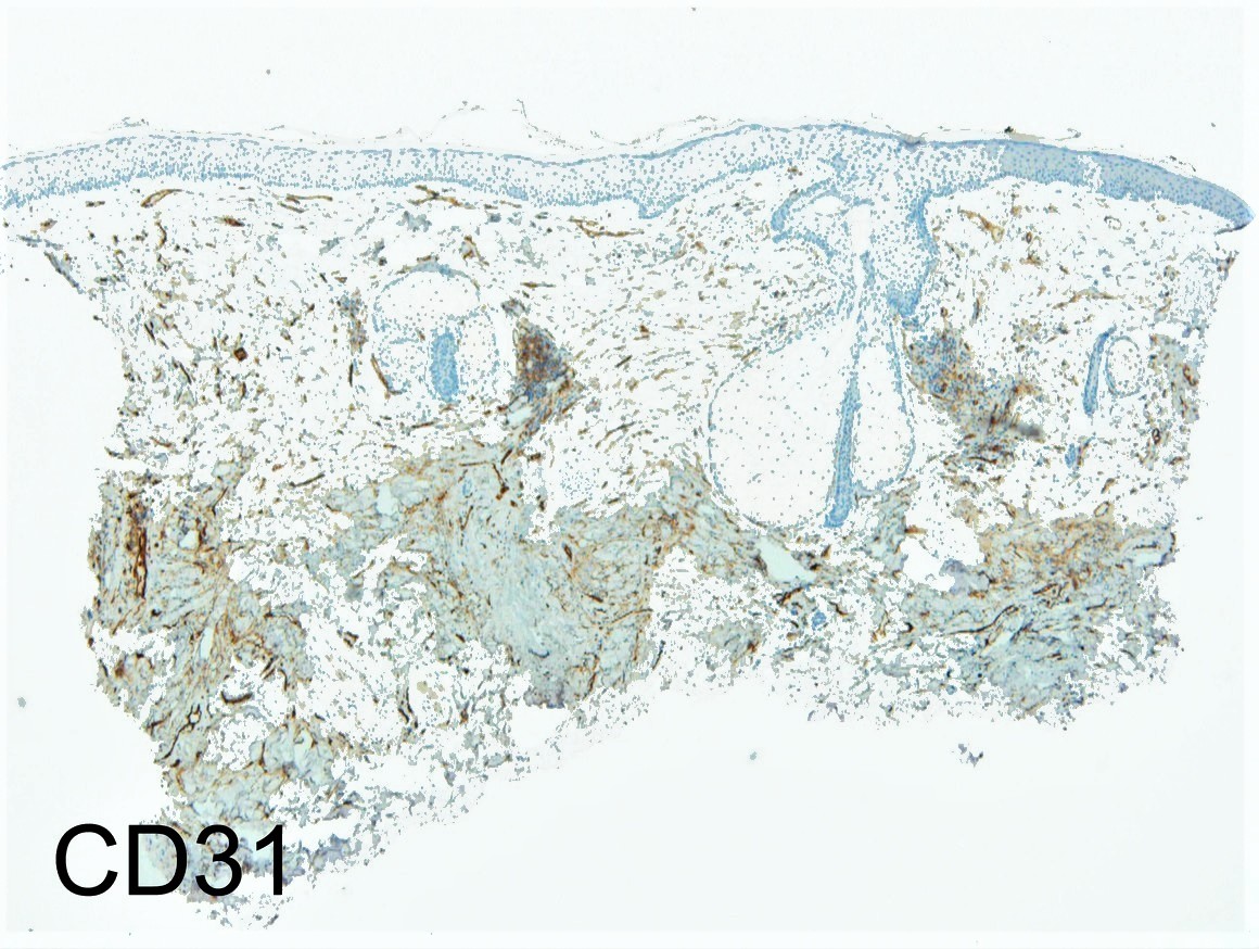
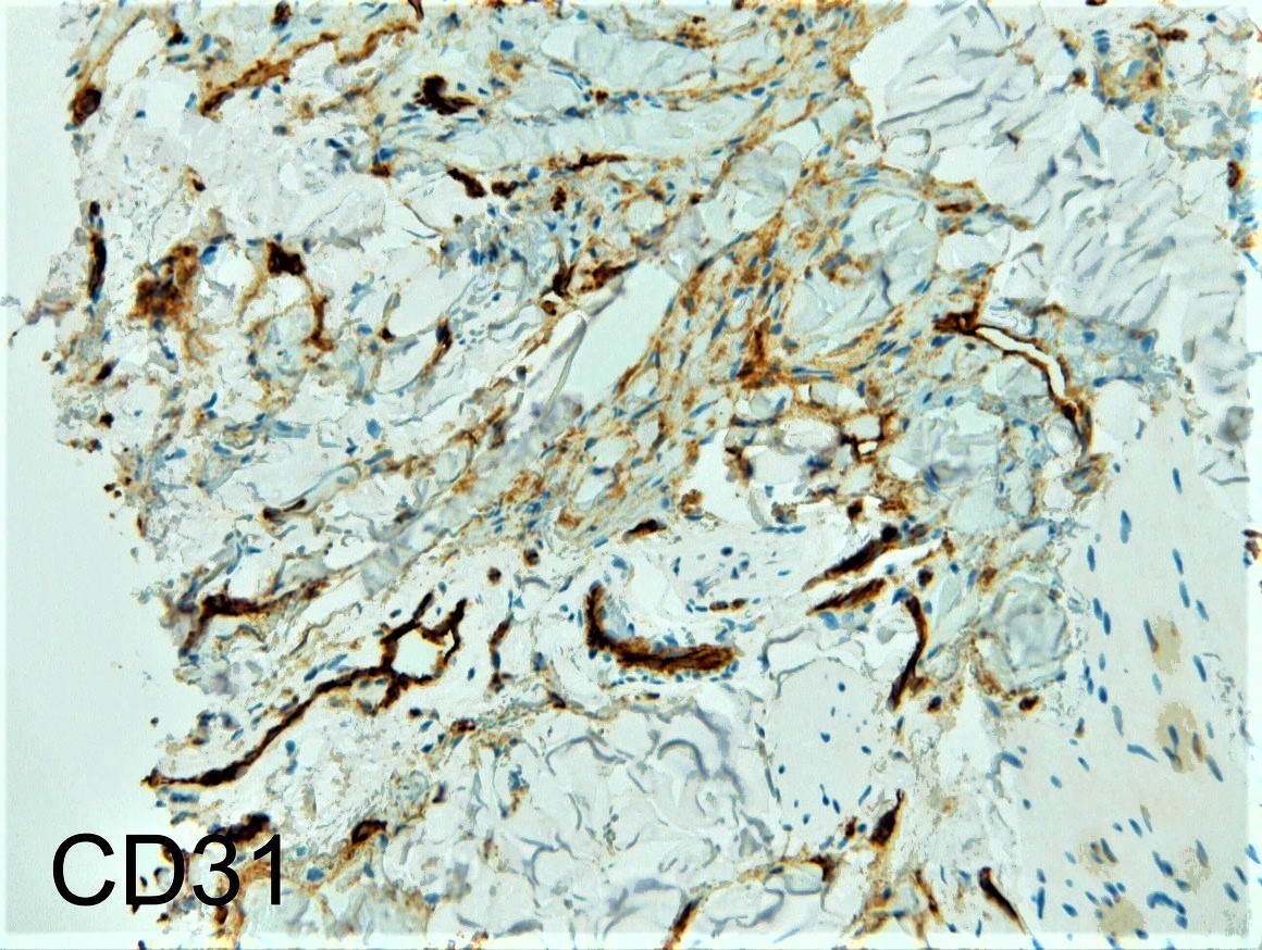
M80. Three scalp lesions. ?All actinic keratoses, ?lichenoid keratoses, ?actinic granuloma. Representative images from 2 of the biopsies but all showed similar features.

Edited by Admin_Dermpath


  Report Record

User Feedback


Dr. Mona Abdel-Halim

Posted

Worried about blue nevus like met melanoma. At some point I was thinking early angiosarc but the pigmented spindled and dendritic cells made me favor a melanocytic lesion.

Share this comment


Link to comment
share_externally

urmilapandey

Posted

wondered if this could be a desmoplastic melanoma with stromal myxoid change


Share this comment


Link to comment
share_externally

vincenzo polizzi

Posted

Desmoplastic Melanoma was my first thought on my iPhone, too...but after read the Mona's comment I deferred the diagnosis to the study with the computer, and now Angiosarcoma is my favorite spot.

Share this comment


Link to comment
share_externally

Dr. Mona Abdel-Halim

Posted

1 hour ago, vincenzo polizzi said:

Desmoplastic Melanoma was my first thought on my iPhone, too...but after read the Mona's comment I deferred the diagnosis to the study with the computer, and now Angiosarcoma is my favorite spot.

But why the pigmented spindled and dendritic cells Vincenzo?

Share this comment


Link to comment
share_externally

vincenzo polizzi

Posted

I can’t answer to this question, Mona. I’m thinking of angipsarcoma because of many hypercromatic cells stuck to collagen bands, reminiscent of a dissecans pattern...but this is a difficult case...of course!!!

Share this comment


Link to comment
share_externally

Nitin Khirwadkar

Posted

Difficult case. Agree with the differentials. Favour an angiosarcoma ( a rather sneaky one) especially with the last two images. Case requires IHC.

Share this comment


Link to comment
share_externally

Raul Perret

Posted

To be honest the picture that struck me the most was the first picture of the second biopsy (the one at low power) there are many suspicious empty spaces/clefts something that we usually see en angiosarc, are those spaces real or artefactual?

Share this comment


Link to comment
share_externally

Raul Perret

Posted

P.S. For me the pigment is hemosiderin due to haemorrhage

Share this comment


Link to comment
share_externally

vincenzo polizzi

Posted

Yes, agree. Hemosiderin. I’d like to see other images of this intriguing case...

Share this comment


Link to comment
share_externally

Dr. Mona Abdel-Halim

Posted

So the first impression was correct!

One should stick to his first impression!

Angiosarc it is!

Very nice case Dr Carr, Thanks for sharing..

Share this comment


Link to comment
share_externally

urmilapandey

Posted

just as well we don't report (at least some) cases without immunohistochemistry! Great case Richard, thanks for sharing

Share this comment


Link to comment
share_externally

vincenzo polizzi

Posted

Great case. Thanks Richard. And kudos to Mona. 

Share this comment


Link to comment
share_externally

Dr. Richard Carr

Posted

I think this is a very sneaky case but I gave you fully representative images and the clinical information exactly as provided. I don't think we definitely have the neoplastic vessels in these biopsies. I think however you have to look at the clinical and if there is any suspicion for angiosarcoma request larger biopsies. The clinical in fact was suspicious for angiosarcoma (when reviewed but that suspicion had not be stated on the request form) and only one of 3 repeat generous incision biopsies included clear-cut, well differentiated, rather superficial, vasoformative angiosarcoma. Also in the intervening few months the lesions had become much more purple and concerning clinically for angiosarcoma.

Share this comment


Link to comment
share_externally


Join the conversation

You can post now and register later. If you have an account, sign in now to post with your account.

Guest
Add a comment...

×   Pasted as rich text.   Paste as plain text instead

  Only 75 emoji are allowed.

×   Your link has been automatically embedded.   Display as a link instead

×   Your previous content has been restored.   Clear editor

×   You cannot paste images directly. Upload or insert images from URL.

Loading...

×
×
  • Create New...