Jump to content
In this section we have spot diagnoses posted on a daily basis since June 2010, now over 4000! You can review the archived cases and read the suggested diagnoses by users and the final comment by the contributors.
Case are uploaded each week day by 10 am UK time with the correct diagnosis will generally be posted at 8 pm UK time. Why not view the most recent spot diagnosis and proffer a diagnosis?

Case Number : Case 1862 - 18 July - Dr Uma Sundram Posted By: Guest

Please read the clinical history and view the images by clicking on them before you proffer your diagnosis.
Submitted Date :
   (0 reviews)

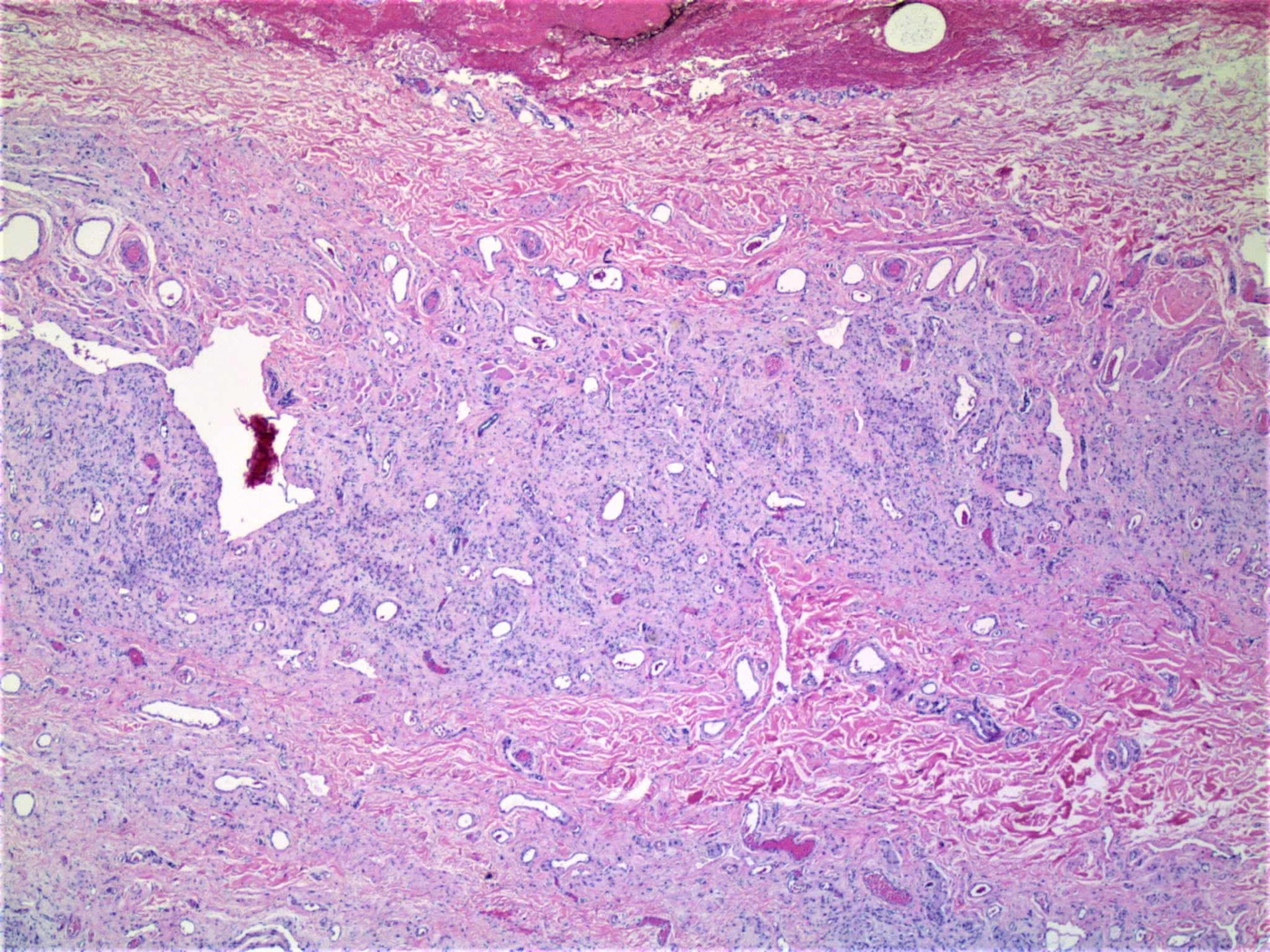
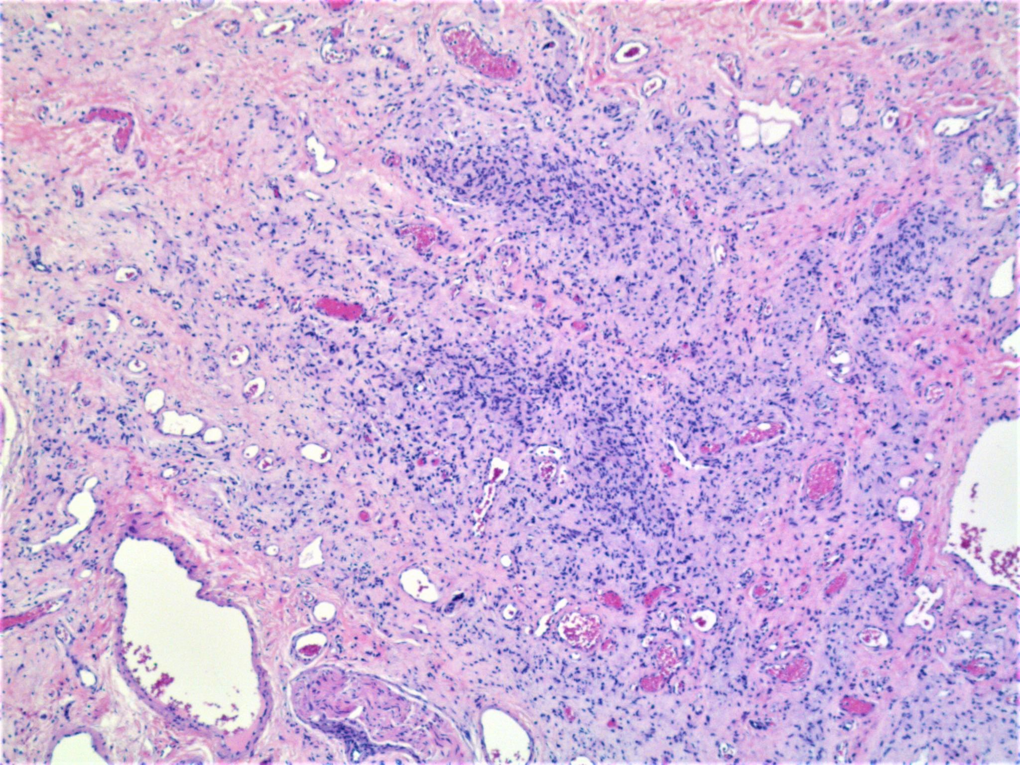
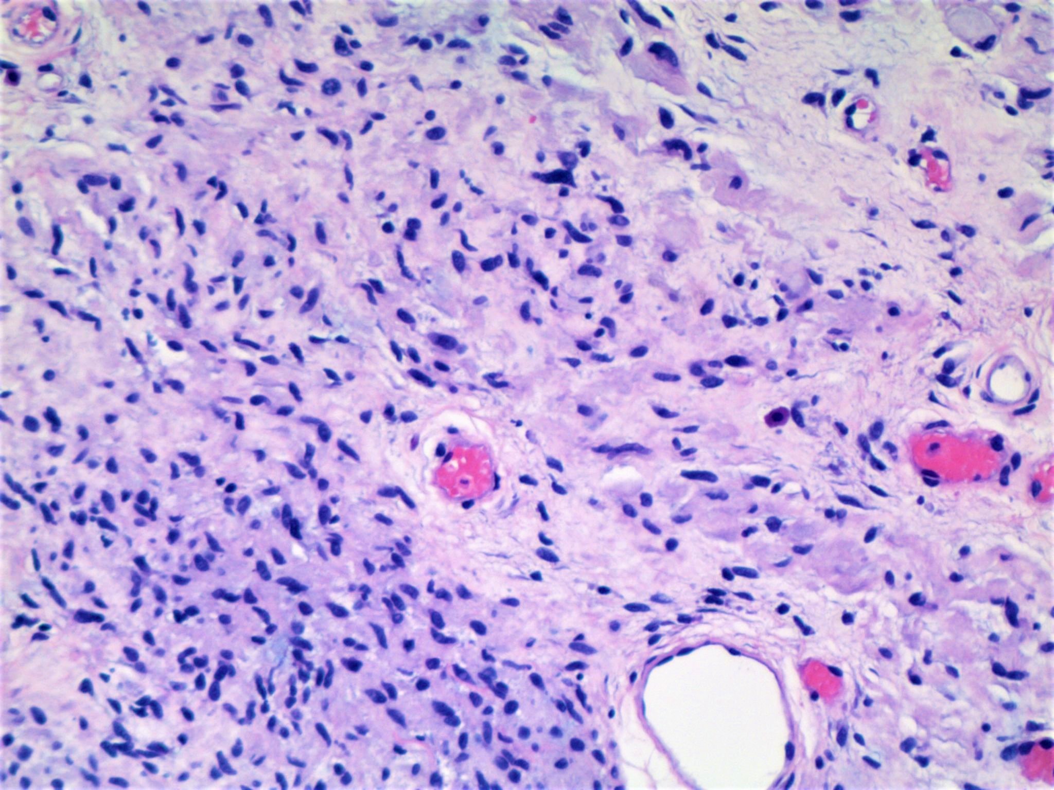
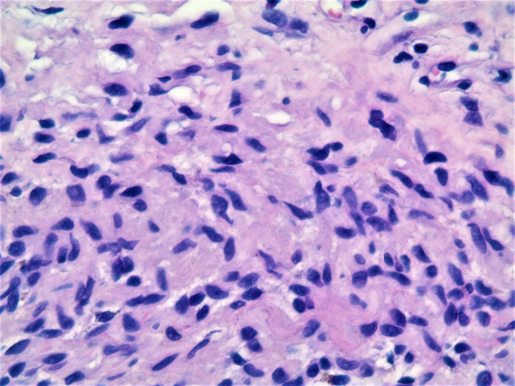
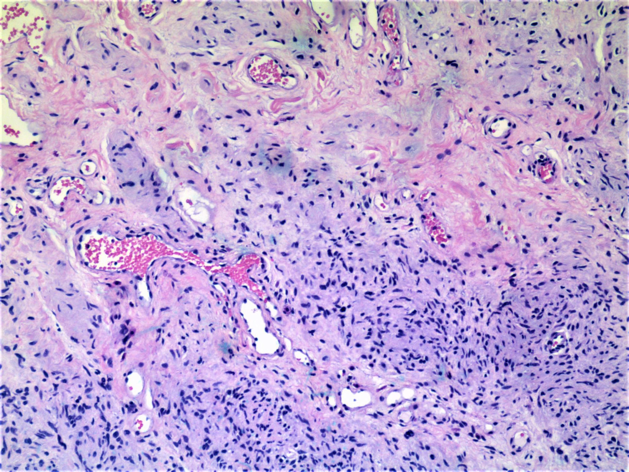
18 year old male with ‘lipoma’ right temporal scalp.

Edited by Admin_Dermpath


  Report Record

User Feedback


Anil Patki

Posted

Angiofibroma aka fibrous papule of face

Share this comment


Link to comment
share_externally

urmilapandey

Posted (edited)

nuclear features, mast cells and myxoid stromal change make me think of a neural lesion: neurofibroma, solitary circumscribed neuroma and even perineurioma. would have done S100, EMA, GFAP.some degenerate changes reminiscent of schwannoma but no capsule.

Edited by urmilapandey

Share this comment


Link to comment
share_externally

Raul Perret

Posted

I think this is one of those cases where the immunos clear easily the pathway. Would perform S-100, Desmin, CD34 and CD31. Morphologically I see a prominence of vessels that strikes me, some even with a staghorn morphology, there are also some areas that look myoid. For me this is Myopericytoma/Myofibroma vs Neurofibroma to confirm with immunos

Share this comment


Link to comment
share_externally

Nitin Khirwadkar

Posted

Agree with the comments made by Raul. Needs IHC. More for a myopericytoma.

Share this comment


Link to comment
share_externally

vincenzo polizzi

Posted

The silhouette is one of a plaque in medium deep dermis. The spindle cells lie side by side in bands parallels to epidermis and show abundant esosinophilic cytoplasm. There is often a contiguity relationship with the angiectatic vessels, angioleiomyoma-like. 

For all these reasons my favorite spot is Dermatomyofibroma.

Odd site, I know. And actina and desmin are many times negative in dermatomyofibroma. 

Share this comment


Link to comment
share_externally

Robledo F. Rocha

Posted

I favor a nerve sheath tumor, particularly diffuse neurofibroma. The short fusiform cells display wavy hyperchromatic nuclei and permeative pattern of growth. There are sparse mast cells interspersed among them. In the last image, some elongated structures are reminiscent of Wagner-Meissner corpuscles to my eyes.

Share this comment


Link to comment
share_externally

Dr. Mona Abdel-Halim

Posted

My impression is also neural; neurofibroma may be with some ancient changes. S100 can easily confirm. 

Share this comment


Link to comment
share_externally

Uma Sundram

Posted

Great DDX and I always love these discussions! The analysis of a series of photomicrographs with a DDX based on morphology is a lost art (at least, unfortunately, in the US). This is a young man with a long standing nevus on the scalpe where the clinician neglected to give us the superficial aspects of the lesion; the nested appearance of the superficial dermal component would have clinched the diagnosis. S100 and MART1 (Melan A) were both positive in lesional cells. Also interesting to note various features often associated with 'soft tissue tumors' such as staghorn vessels.

Share this comment


Link to comment
share_externally

anjali shinde

Posted

So, final diagnosis is long staving nevus?


Share this comment


Link to comment
share_externally


Join the conversation

You can post now and register later. If you have an account, sign in now to post with your account.

Guest
Add a comment...

×   Pasted as rich text.   Paste as plain text instead

  Only 75 emoji are allowed.

×   Your link has been automatically embedded.   Display as a link instead

×   Your previous content has been restored.   Clear editor

×   You cannot paste images directly. Upload or insert images from URL.

Loading...

×
×
  • Create New...