Jump to content
In this section we have spot diagnoses posted on a daily basis since June 2010, now over 4000! You can review the archived cases and read the suggested diagnoses by users and the final comment by the contributors.
Case are uploaded each week day by 10 am UK time with the correct diagnosis will generally be posted at 8 pm UK time. Why not view the most recent spot diagnosis and proffer a diagnosis?

Case Number : Case 1870 - 28 July - Dr Richard Carr Posted By: Guest

Please read the clinical history and view the images by clicking on them before you proffer your diagnosis.
Submitted Date :
   (0 reviews)

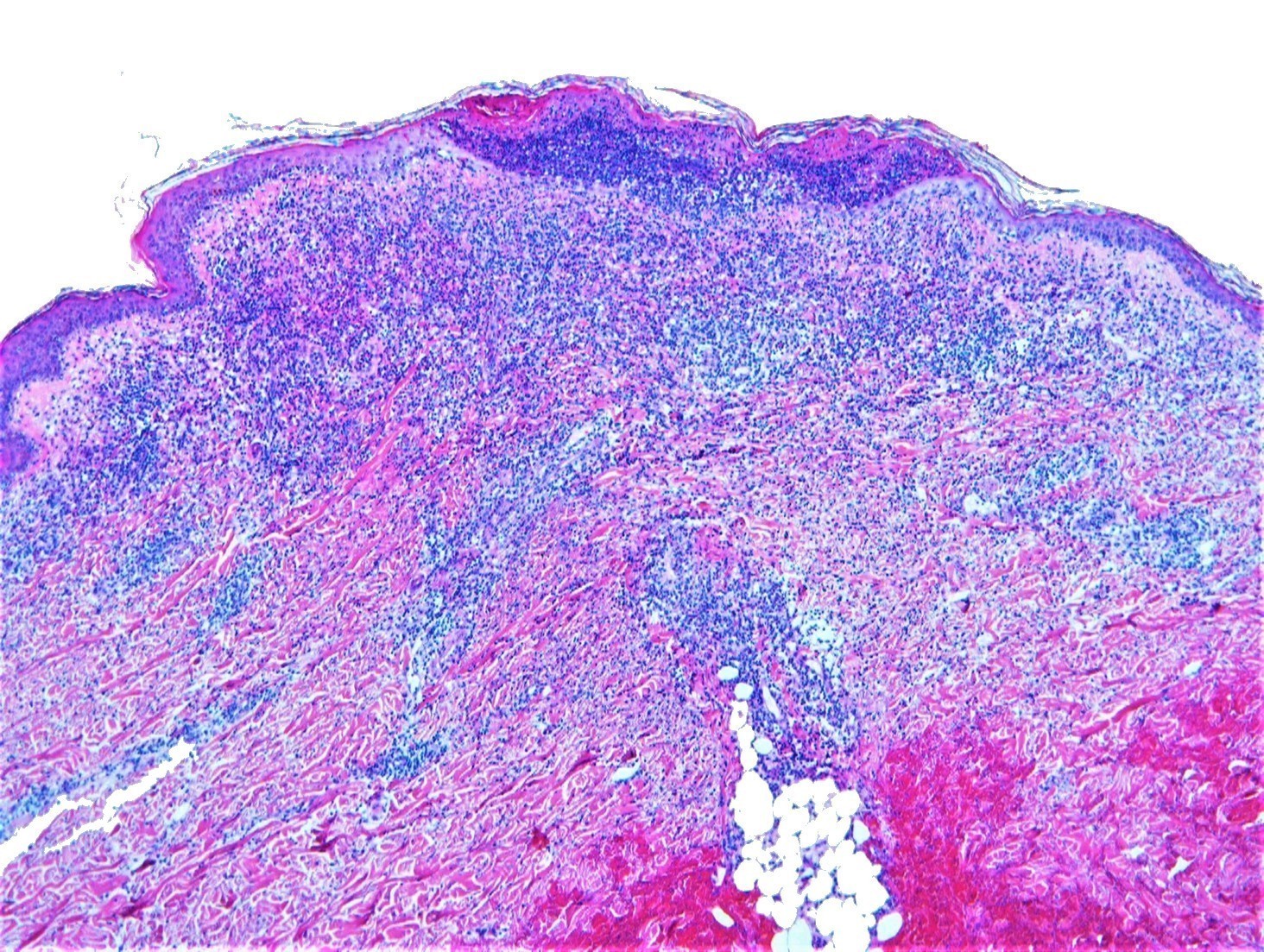
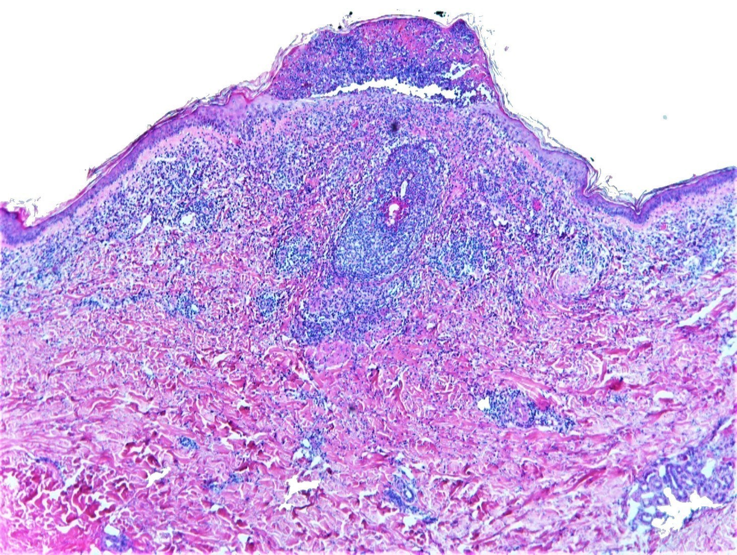
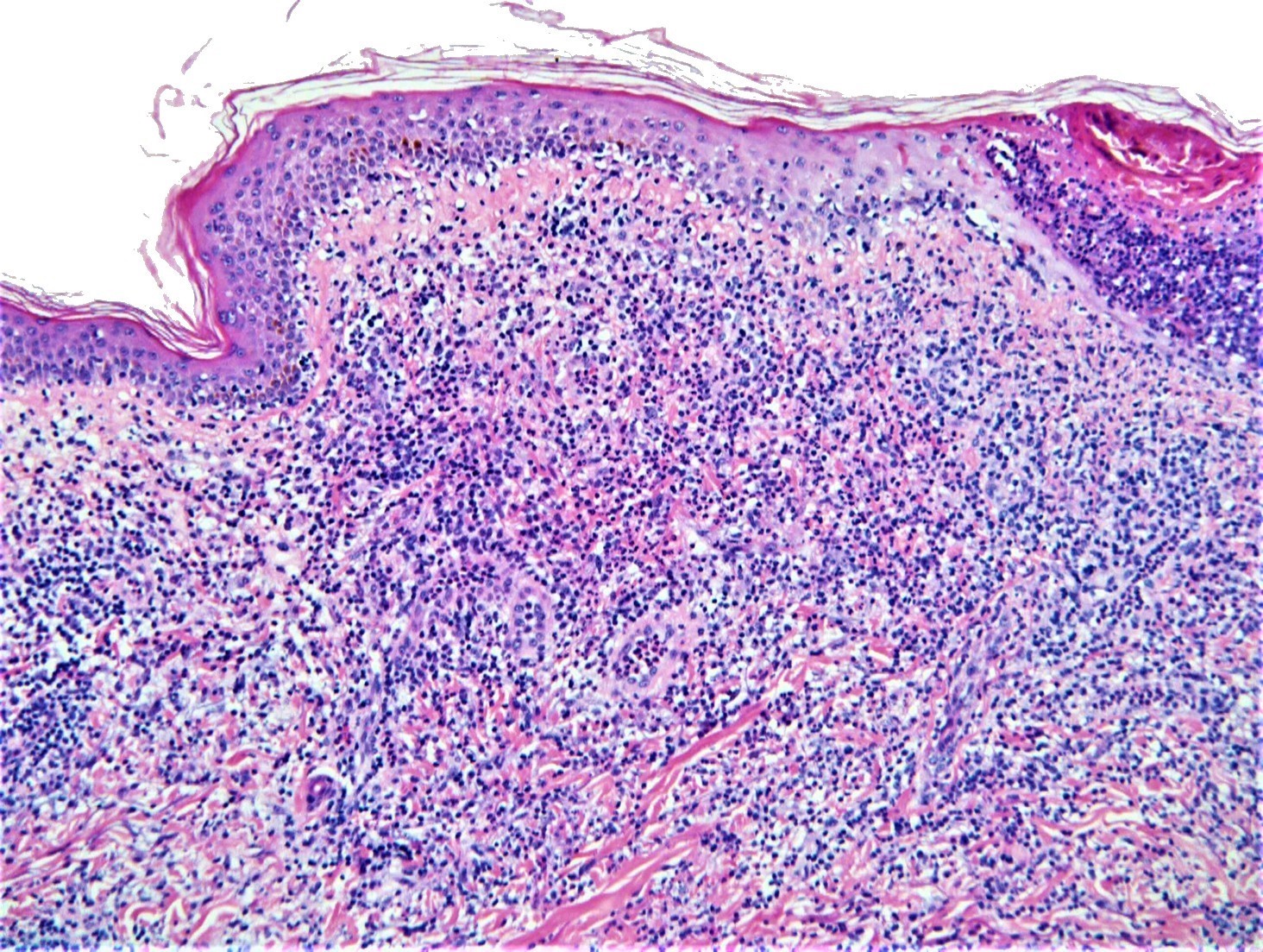
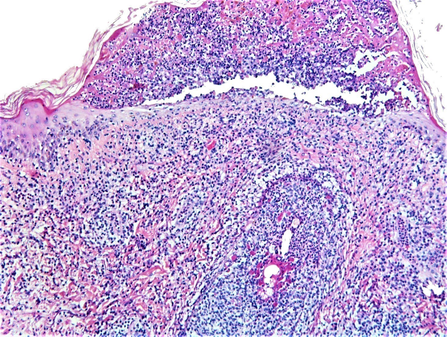
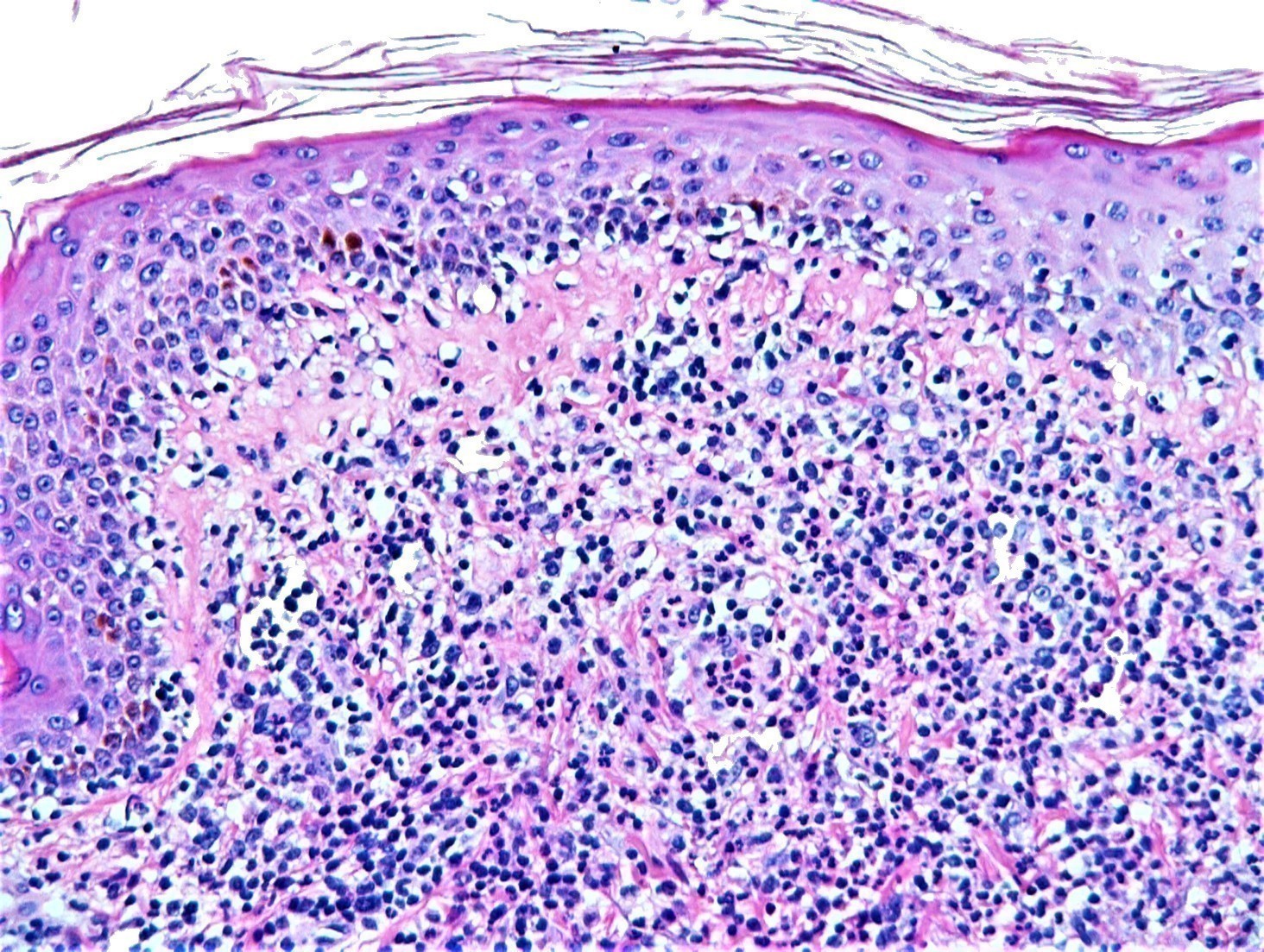
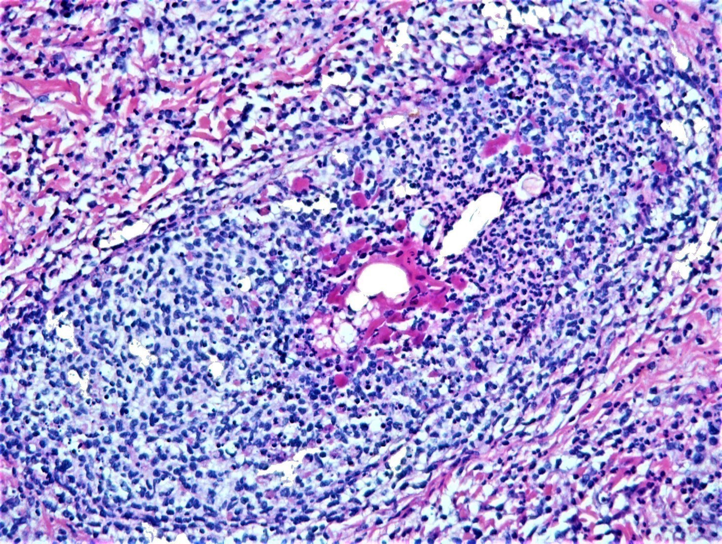
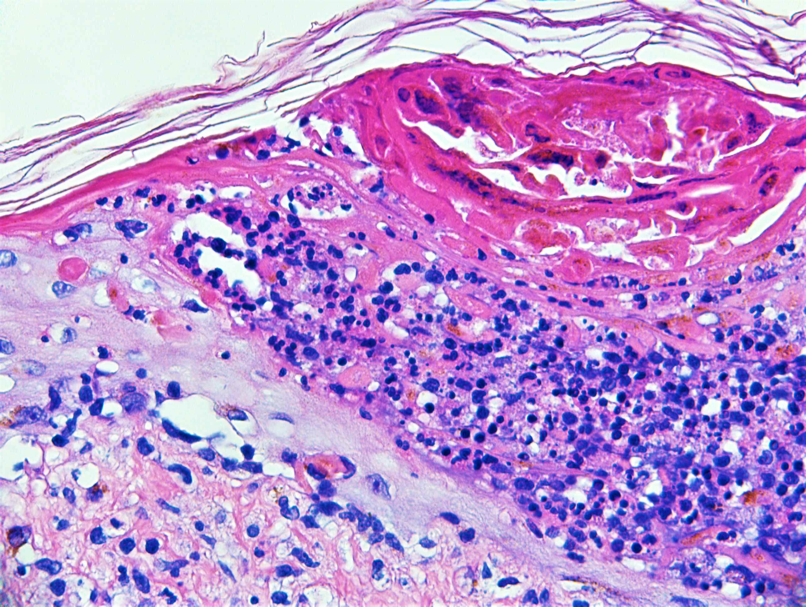
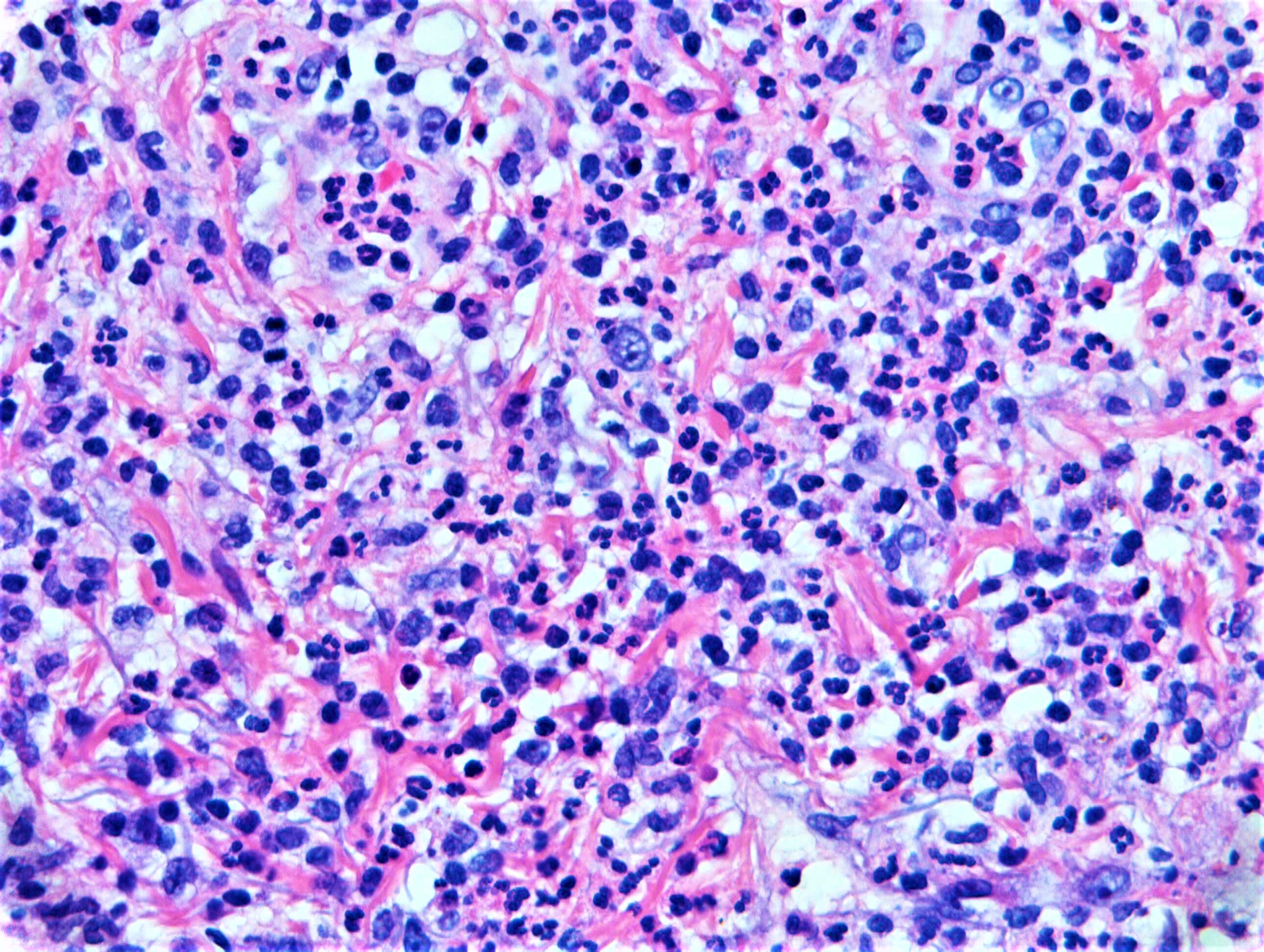
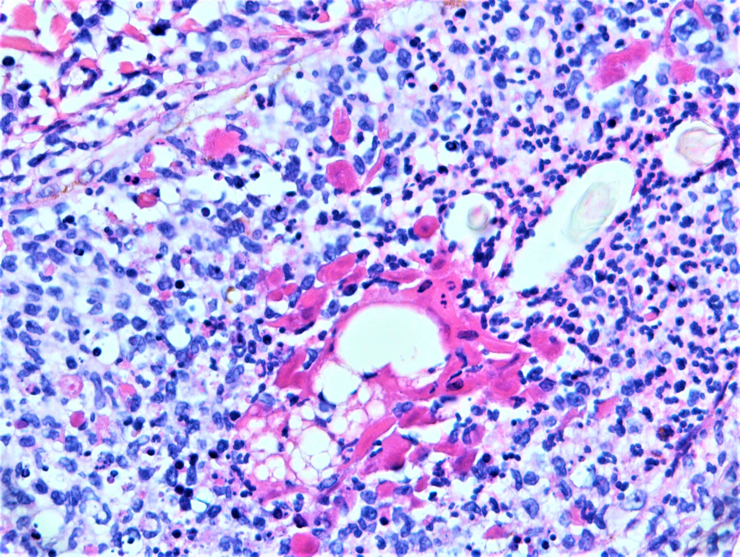
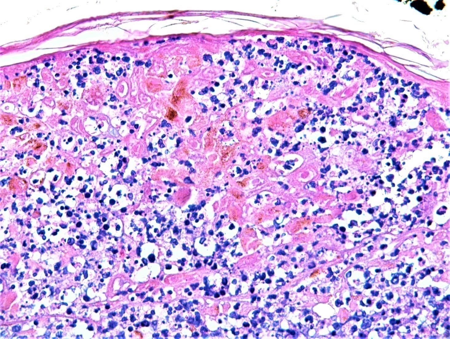
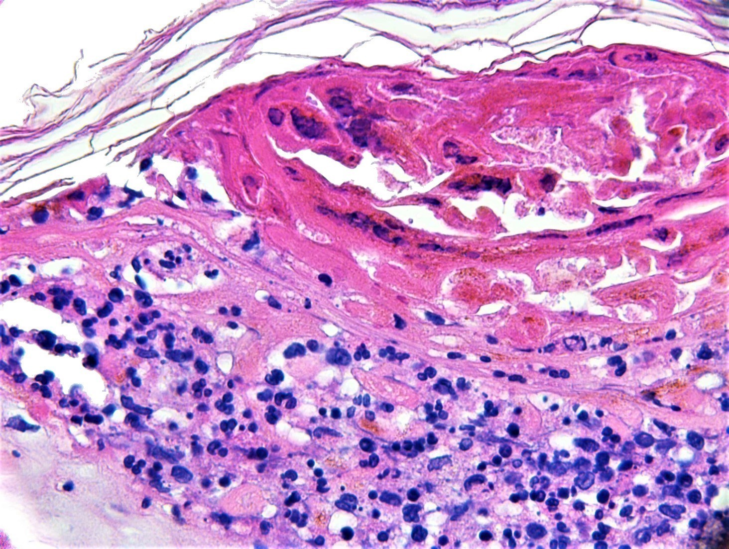
F65. Pervious history of low grade NHL with high grade transformation. Plaque on cheek.

Edited by Admin_Dermpath


  Report Record

User Feedback


urmilapandey

Posted

wondering if there is vasculitis, don't know if the dense pink stuff is fibrin. patient may be immunocompromised (given the history) so stains for bugs; ? reaction to drugs. doesn't look like lymphoma to me but then i have little expertise in this area.

Share this comment


Link to comment
share_externally

Raul Perret

Posted

My feeling was that the main pattern is that of an acute folliculitis with pustular formation and some lichenoid infiltrate. I would perform some serial sections, special stains for microorganisms, and of course check for herpes and other viruses. The lichenoid infiltrate puzzles me, that is why I think is wise to remark that a drug reaction cannot be discarded. There was this term used in the past: follicular toxic pustuloderma for some drug reactions but they are actually cases of AGEP, and as we know this is generalized and not localized like in this case.

Share this comment


Link to comment
share_externally

vincenzo polizzi

Posted

What about an arthropod bite reaction in patient with Richter syndrome ( cll in DLBCL transformation )?

Share this comment


Link to comment
share_externally

Dr. Mona Abdel-Halim

Posted

Looks folliculitis, may be viral? 

Share this comment


Link to comment
share_externally

Nitin Khirwadkar

Posted

Definitely acute folliculitis. To rule out bugs. In addition there is almost an interface in images 3 & 5. Drug induced folliculitis?

Share this comment


Link to comment
share_externally

Admin_Dermpath

Posted


Dear All 

Images 7- 11 added at the request of Dr Carr. 

DermpathPRO Admin

Happy Weekend!

Share this comment


Link to comment
share_externally

Saman Fatah

Posted

Intra-epidermal collection of mixed inflammatory cell + necrotic keratinocytes overlying a clear folliculitis. At DEJ, the individual lymphocytes seems to be surrounded by a clear halo "lymph in holes" they are aligned along DEJ, I couldn't see convincing apoptosis at this level. 

If colleagues agree that this is interface/lichenoid, would appreciate if they elaborate on why? 

Infective cause is crucial to exclude especially herpes folliculitis in the current context. Aware IHC for HSV/VZV is an option but from Clinician point of view, viral swab from the plaque for PCR is  a sensitive and extremely easy to do with results in 1-2 days. 

Herpes infection is one of the well known lymphoma mimicker which may fit well with some of the changes in this biopsy. 

Share this comment


Link to comment
share_externally

Saman Fatah

Posted

Just for record, I have seen the first 6 photomicrographs only when posted the earlier comment and my screen was no refreshed whilst busy with typing so was not really aware of them! 

Share this comment


Link to comment
share_externally

vincenzo polizzi

Posted

Yes, agree with HSV/VZV

nice case. 

Share this comment


Link to comment
share_externally

Admin_Dermpath

Posted

Dr Richard Carr

Final Diagnosis: Herpes zoster folliculitis.

Share this comment


Link to comment
share_externally

Dr. Richard Carr

Posted

Thanks Admin for posting the diagnosis (I was on leave). I thought this was a nice example of prominent necrotic follicular keratinocytes with sparse multinucleate cytopathic changes in keeping with VZV folliculitis. In fact the clinical was of numerous pustules erupting on the chest and face on an erythematous base with a large vesicular plaque on the cheek ?varicella zoster. This case demonstrates the pseudolymphomatous pattern (presumably exaggerated here due to immune suppression) and interface reaction pattern that can be seen and if you miss the follicular involvement or subtle cytopathic changes (both absent in re-cut sections) you may not make the diagnosis. Well done to those for thinking of zoster on the first set of images.

Share this comment


Link to comment
share_externally


Join the conversation

You can post now and register later. If you have an account, sign in now to post with your account.

Guest
Add a comment...

×   Pasted as rich text.   Paste as plain text instead

  Only 75 emoji are allowed.

×   Your link has been automatically embedded.   Display as a link instead

×   Your previous content has been restored.   Clear editor

×   You cannot paste images directly. Upload or insert images from URL.

Loading...

×
×
  • Create New...