Jump to content
In this section we have spot diagnoses posted on a daily basis since June 2010, now over 4000! You can review the archived cases and read the suggested diagnoses by users and the final comment by the contributors.
Case are uploaded each week day by 10 am UK time with the correct diagnosis will generally be posted at 8 pm UK time. Why not view the most recent spot diagnosis and proffer a diagnosis?

Case Number : Case 1809 - 04 May - Dr Arti Bakshi Posted By: Guest

Please read the clinical history and view the images by clicking on them before you proffer your diagnosis.
Submitted Date :
   (0 reviews)

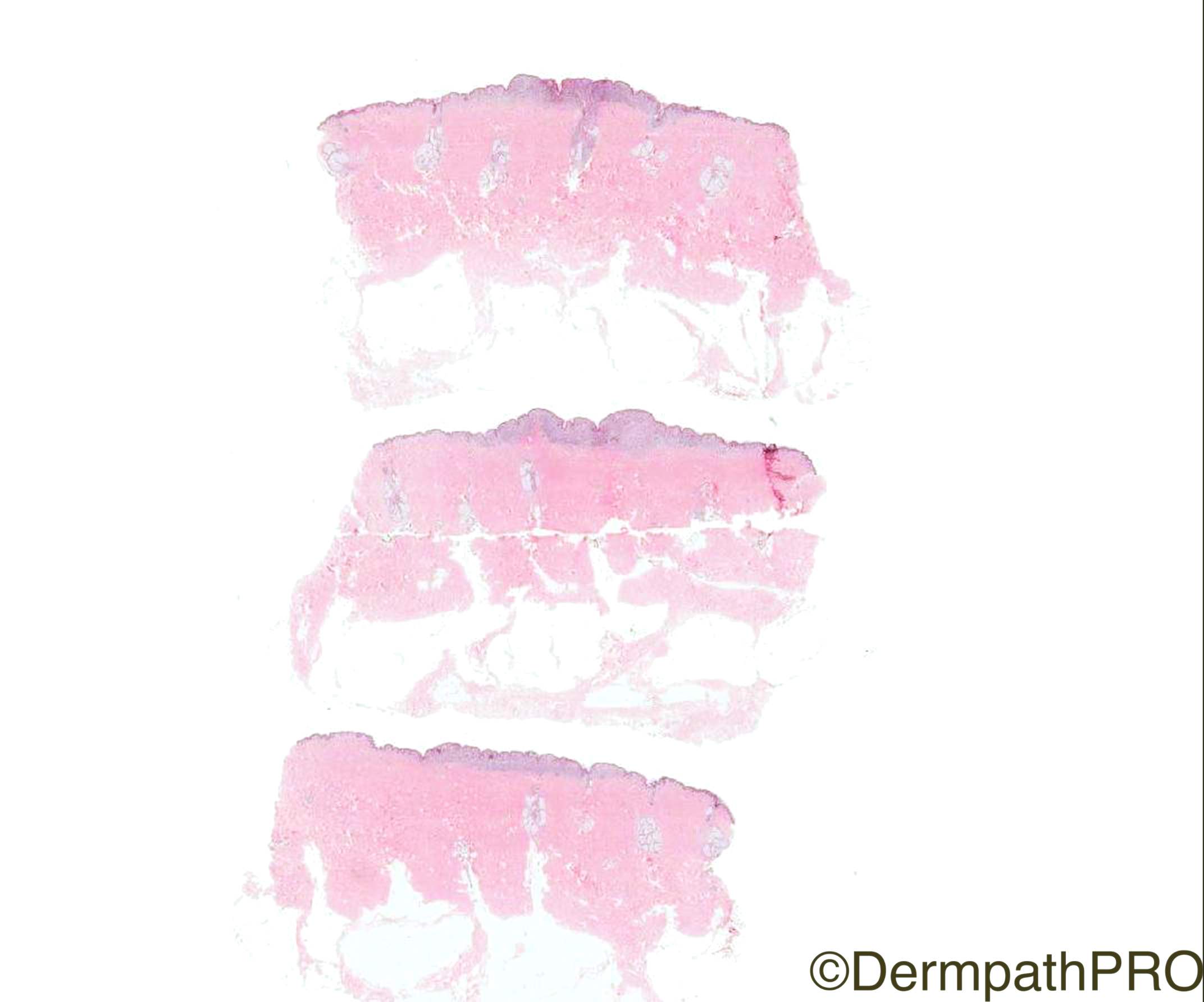
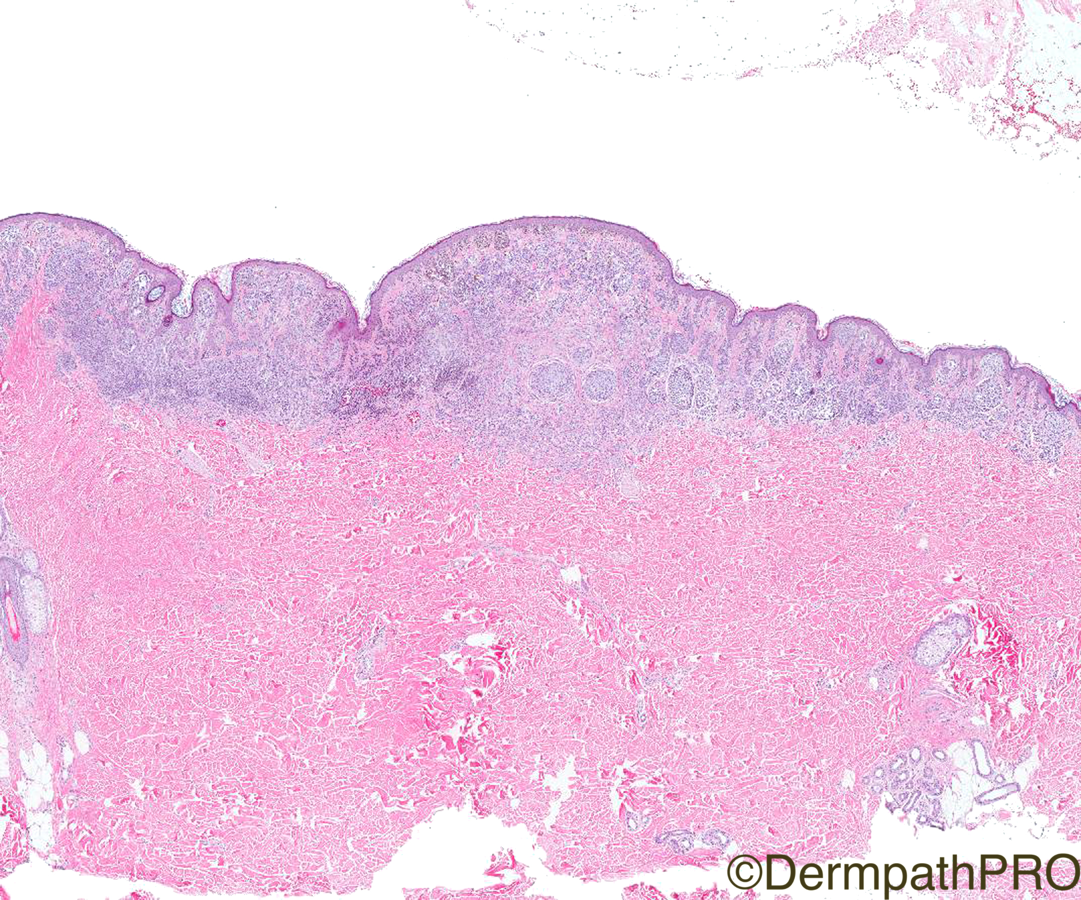
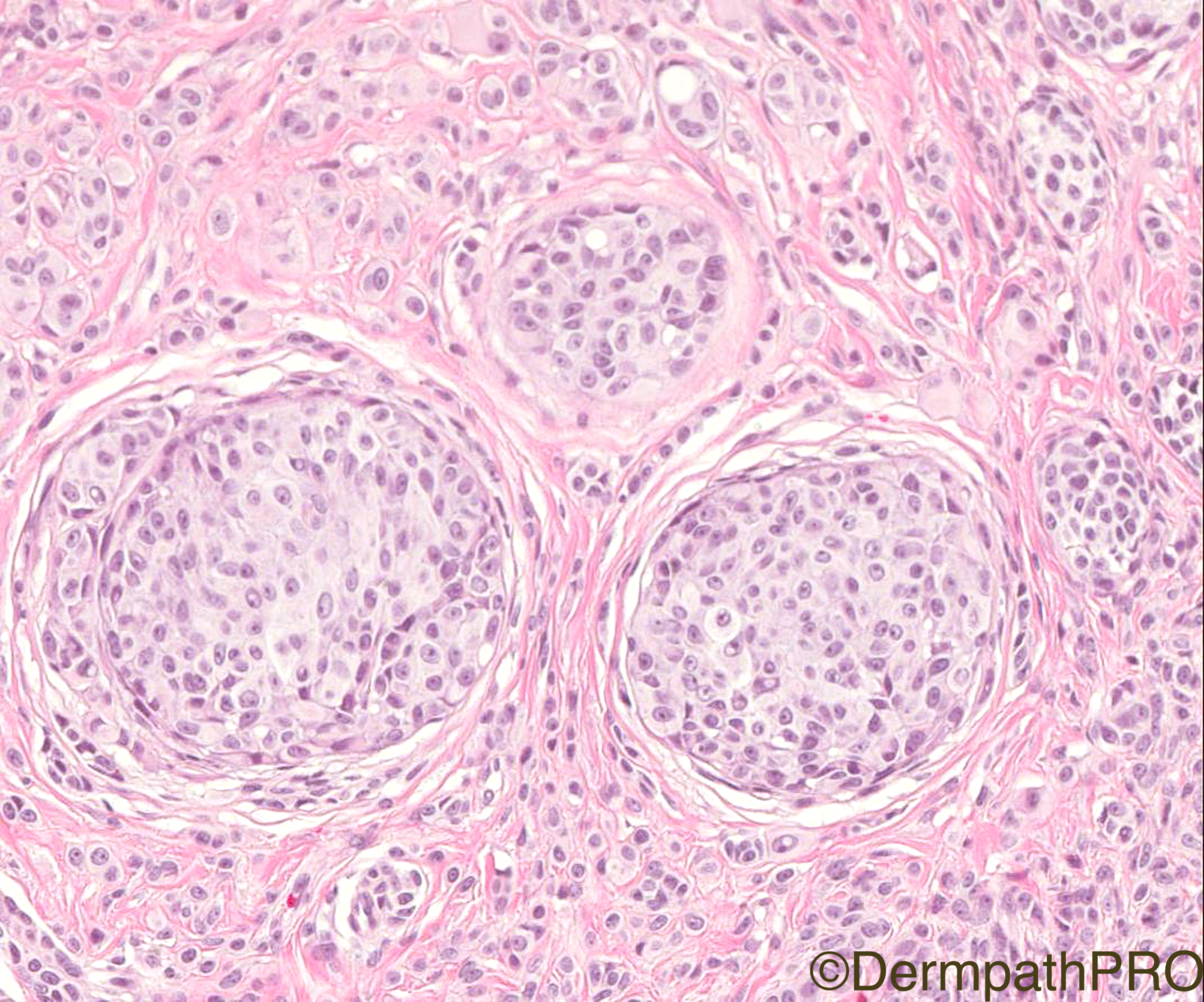
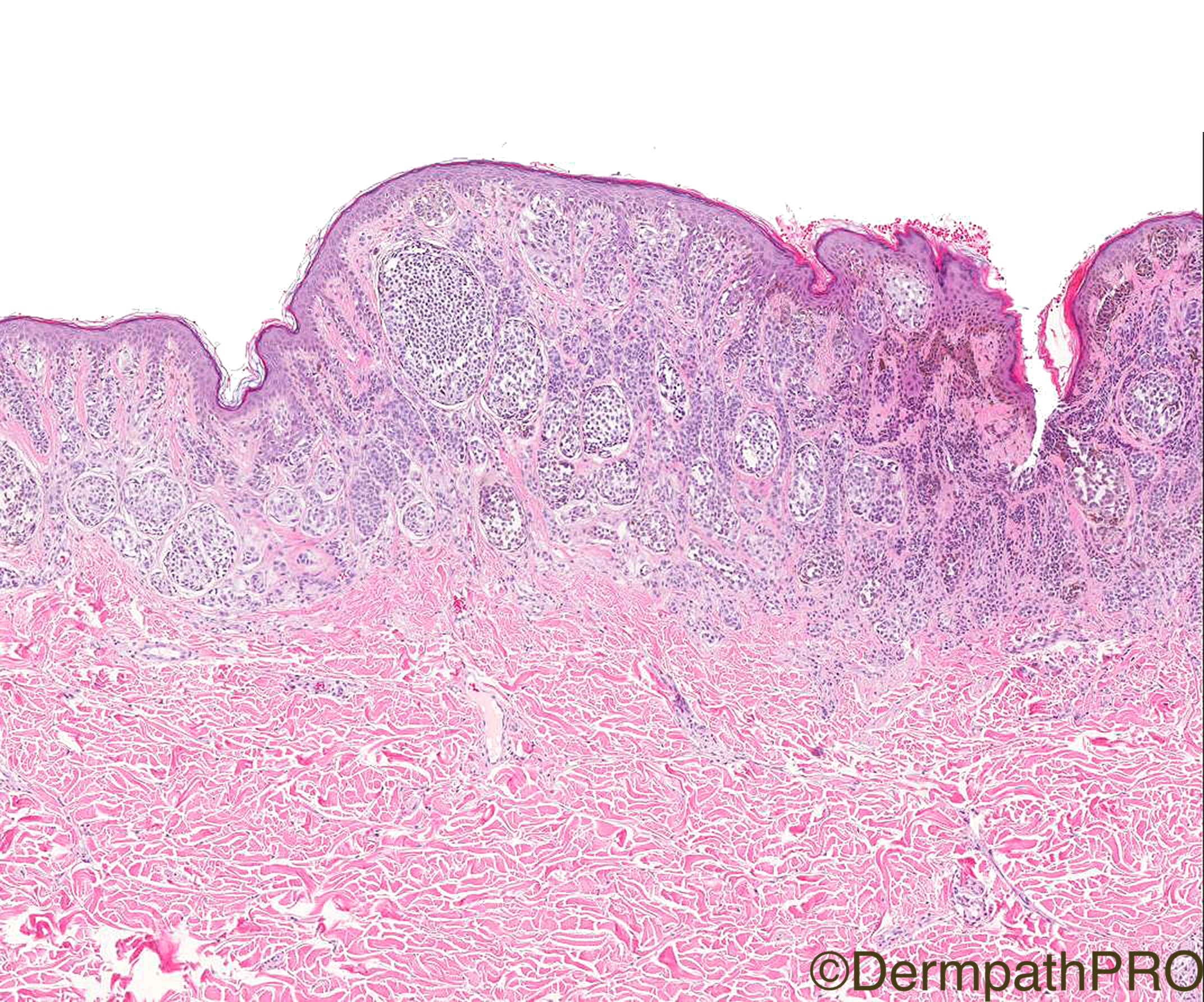
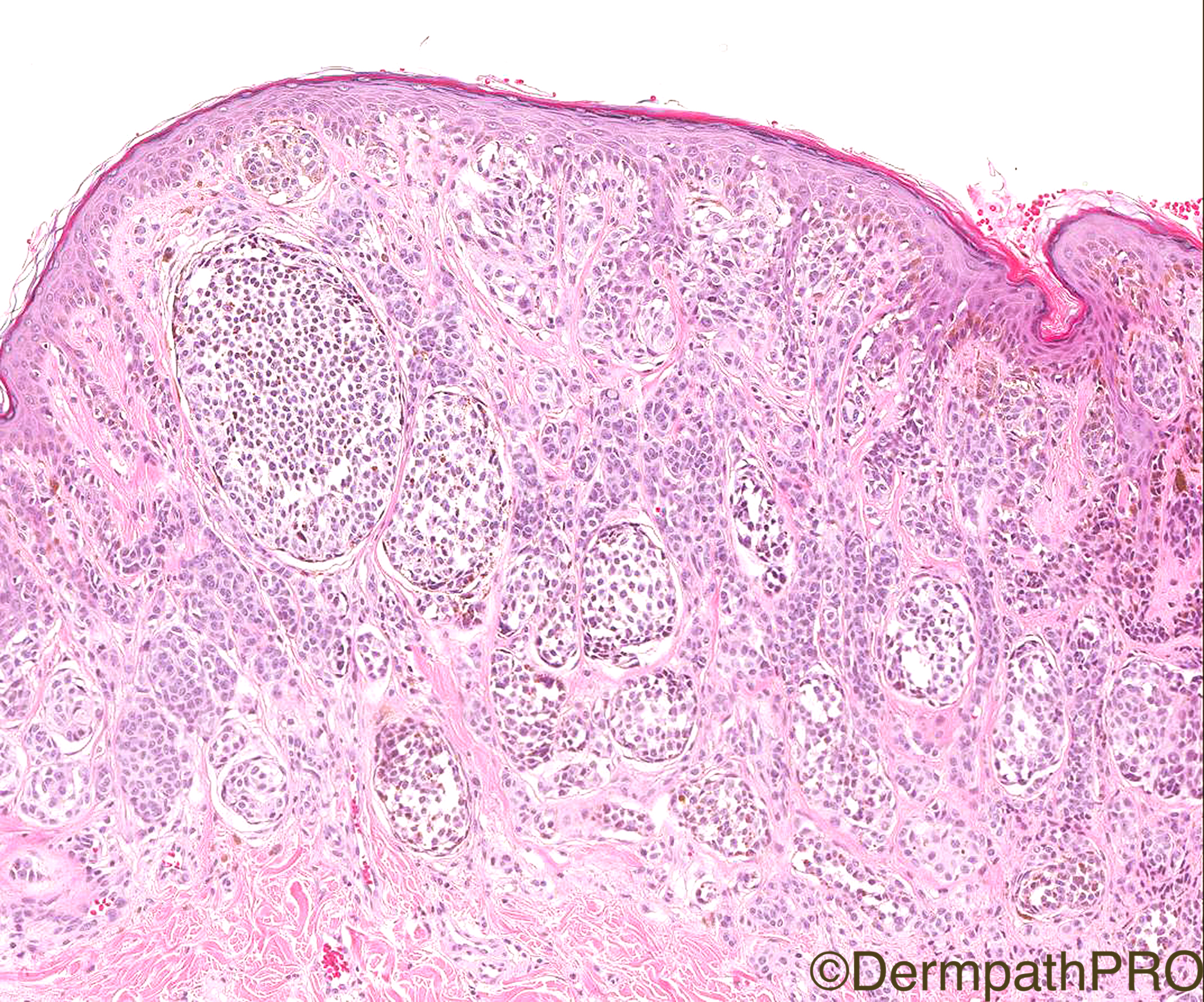
Clinical History: 30/F, changing mole right upper back, case c/o Dr Naveen Sharma.

Case Posted by Dr Arti Bakshi


  Report Record

User Feedback


Raul Perret

Posted

I am away for an event so I can only see the case on my phone, but I thought of MELTUMP in this case, we should definetely try to do some inmunos  including BAP-1 (even if the lesion has a junctional component). To be honest my gut tells me  that this lesion is biologically benign

Share this comment


Link to comment
share_externally

Mariantonieta Tirado

Posted

I favor benign, I agree with the previous comment and do BAP. Another consideration would be nevoid melanoma. Not sure if the epidermis shows some scatter. Didn't see dermal mitoses. I would also add MelA, HMB45 and maybe p16

Share this comment


Link to comment
share_externally

Robledo F. Rocha

Posted

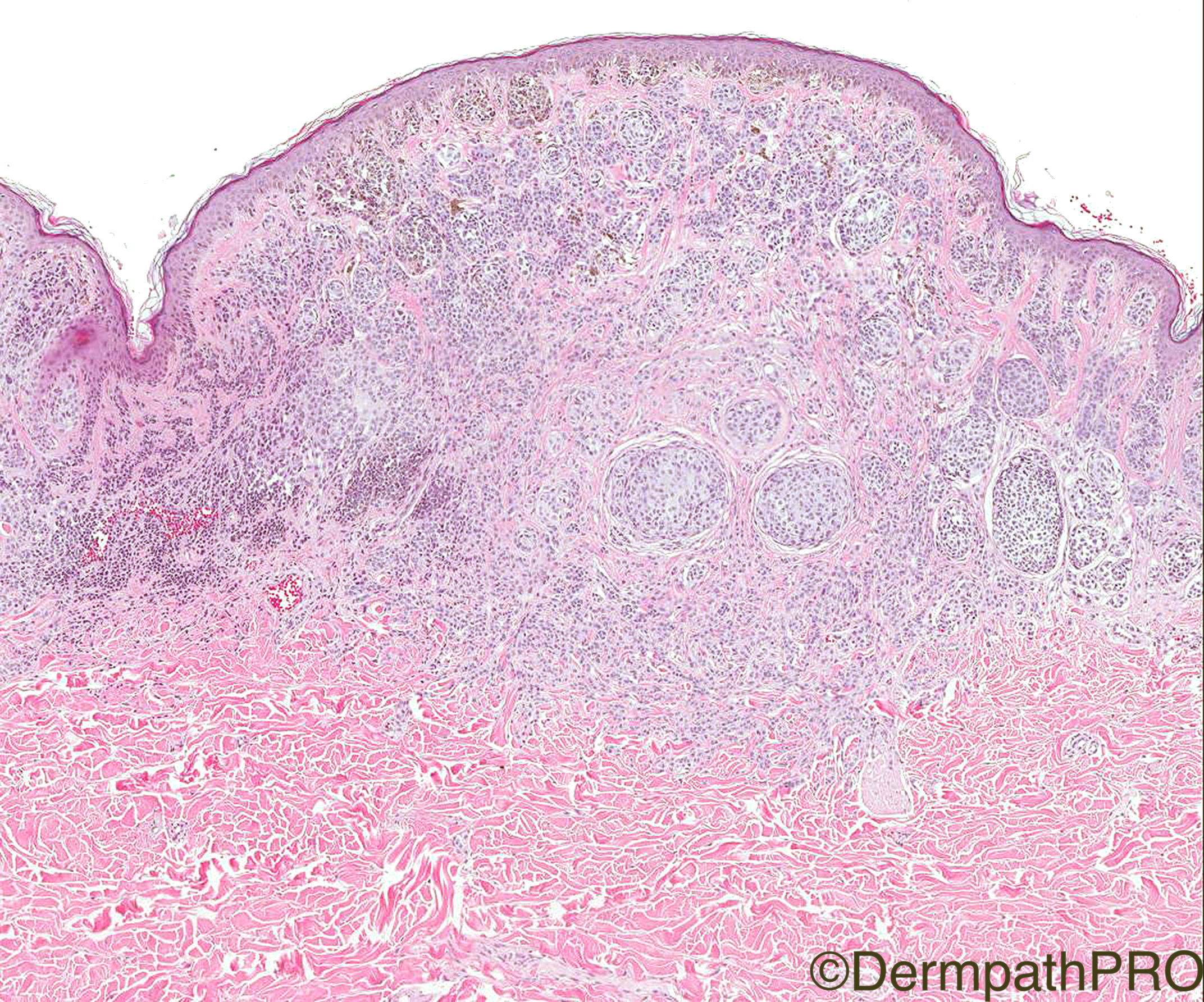
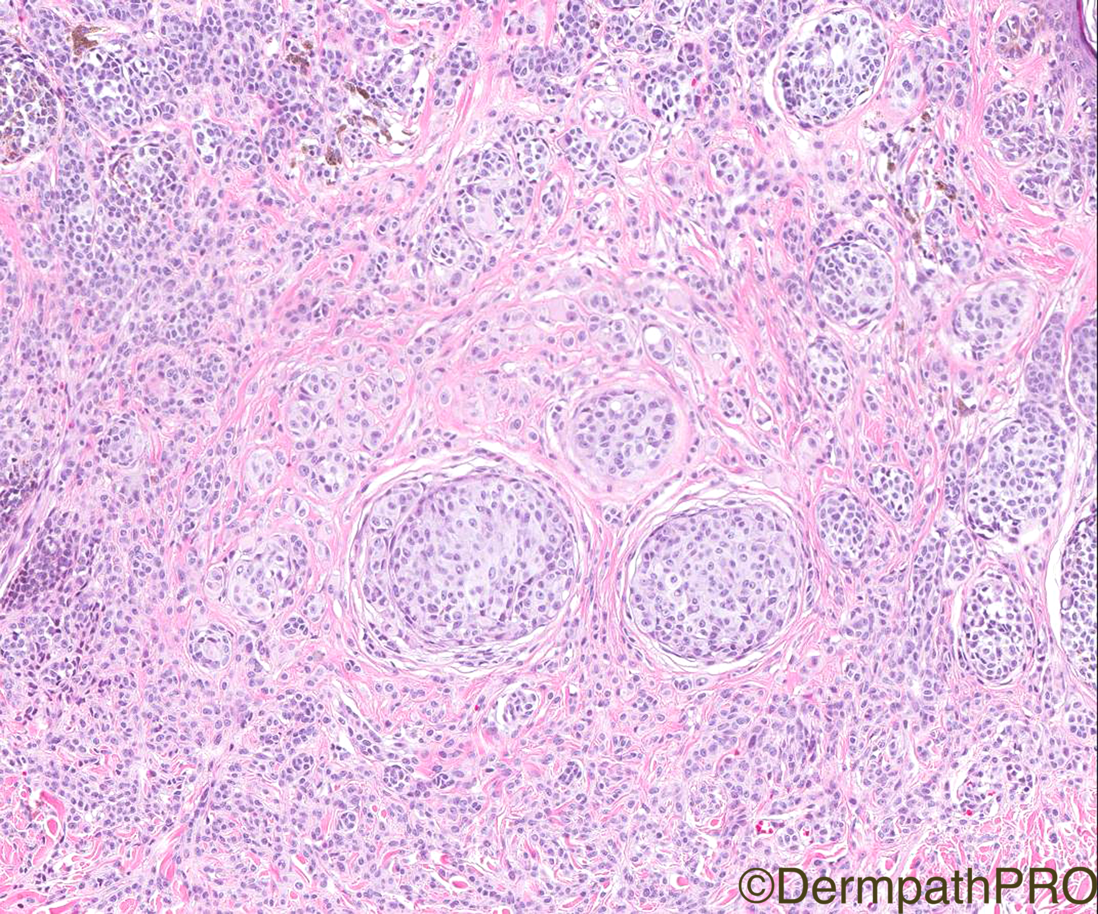
A banal melanocytic nevus showing some intradermal nests of type A melanocytes in the deeper reaches of the lesion, and those intradermal nests are surrounded by type B melanocytes. Inverted type A nevus, clonal nevus, and nevus with atypical dermal nodules are some names, but multiple levels may show this lesion represents a combined nevus or a deep penetrating nevus.

Share this comment


Link to comment
share_externally

Fernando Cabo

Posted

Nevoid melanoma in the diferential 

Share this comment


Link to comment
share_externally

Dr. Mona Abdel-Halim

Posted

Worried about nevoid melanoma.

Share this comment


Link to comment
share_externally

Neil Catterall

Posted

Agree with Robledo - benign with some clinal nests. Would like to see mitoses before considering naevoid melanoma. There is good maturation of nuclei even in areas of colonial nests.

Share this comment


Link to comment
share_externally

vincenzo polizzi

Posted

I'm away with only my iPhone, but  

not sure about benignity.  Agree with Mona and Fernando. 

Share this comment


Link to comment
share_externally

Arti Bakshi

Posted

I went for benign. I called it a 'proliferative nodule- like lesion' within a benign compound naevus (a clonal naevus is equally appropriate).  Although the dermal nests are expansile, they merge with the surrounding naevocytes, are not particularly atypical cytologically and are mitotically inactive. (there were no mitoses at all in the dermal component in the levels.).

For what its worth, Ki67 fraction was low and p16 was retained throughout the lesion. I did not do a BAP1, I have to admit, mainly because there was a florid junctional component. But i think we have had the discussion before on this forum that in some series, BAPOMAS can be compound lesions, so maybe worth a try!

Share this comment


Link to comment
share_externally

Sasi Attili

Posted

Extremely difficult case.  Initially though, just a proliferation nodule. But am not so sure after looking at it again. The nests are a bit large even at the deep dermis. I guess this is one I would ensure complete excision and recommend long term follow-up (i.e. treat as a MELTUMP). . 

Share this comment


Link to comment
share_externally


Join the conversation

You can post now and register later. If you have an account, sign in now to post with your account.

Guest
Add a comment...

×   Pasted as rich text.   Paste as plain text instead

  Only 75 emoji are allowed.

×   Your link has been automatically embedded.   Display as a link instead

×   Your previous content has been restored.   Clear editor

×   You cannot paste images directly. Upload or insert images from URL.

Loading...

×
×
  • Create New...