Jump to content
In this section we have spot diagnoses posted on a daily basis since June 2010, now over 4000! You can review the archived cases and read the suggested diagnoses by users and the final comment by the contributors.
Case are uploaded each week day by 10 am UK time with the correct diagnosis will generally be posted at 8 pm UK time. Why not view the most recent spot diagnosis and proffer a diagnosis?

Case Number : Case 1811 - 08 May - Dr Limin Yu Posted By: Guest

Please read the clinical history and view the images by clicking on them before you proffer your diagnosis.
Submitted Date :
   (0 reviews)

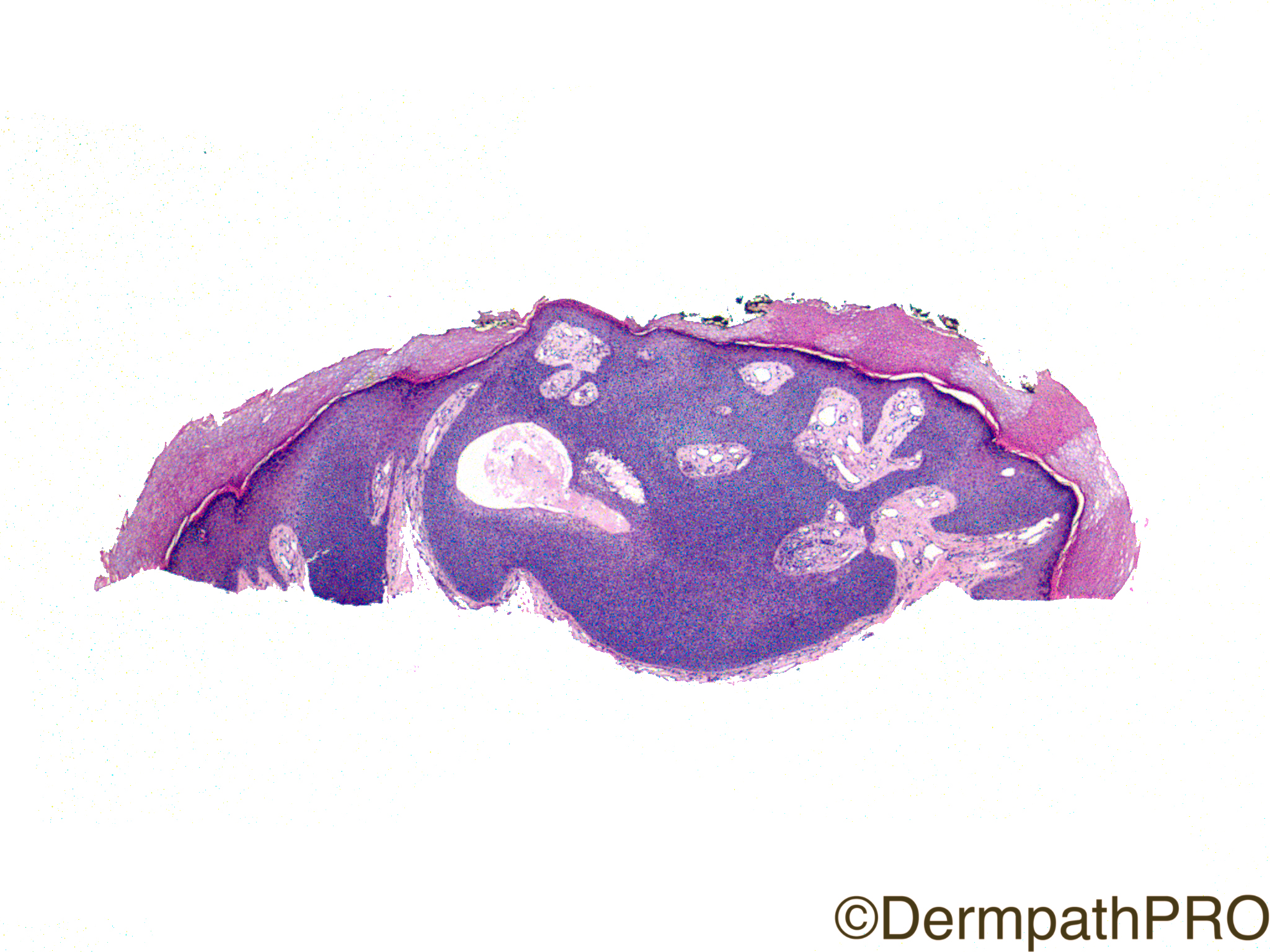
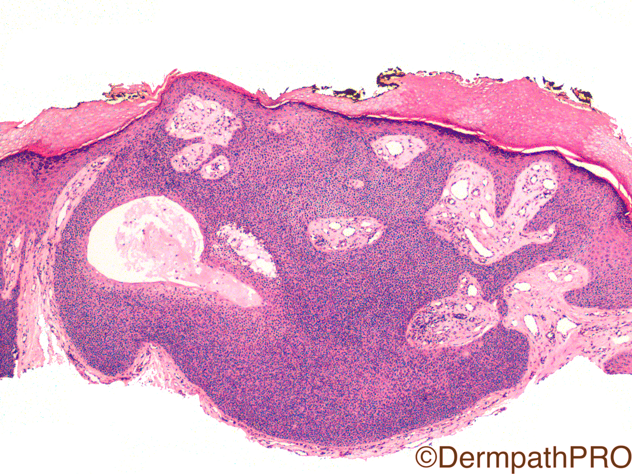
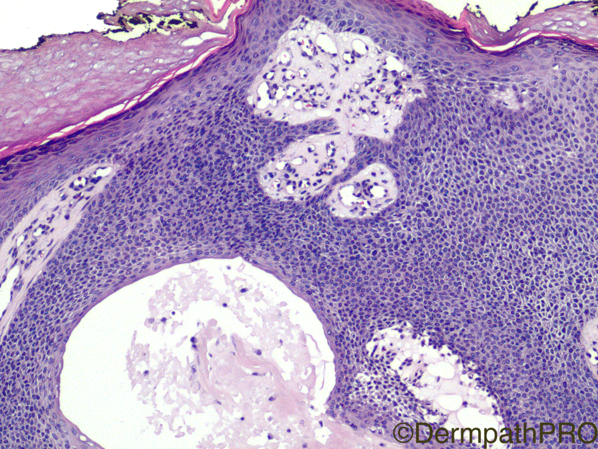
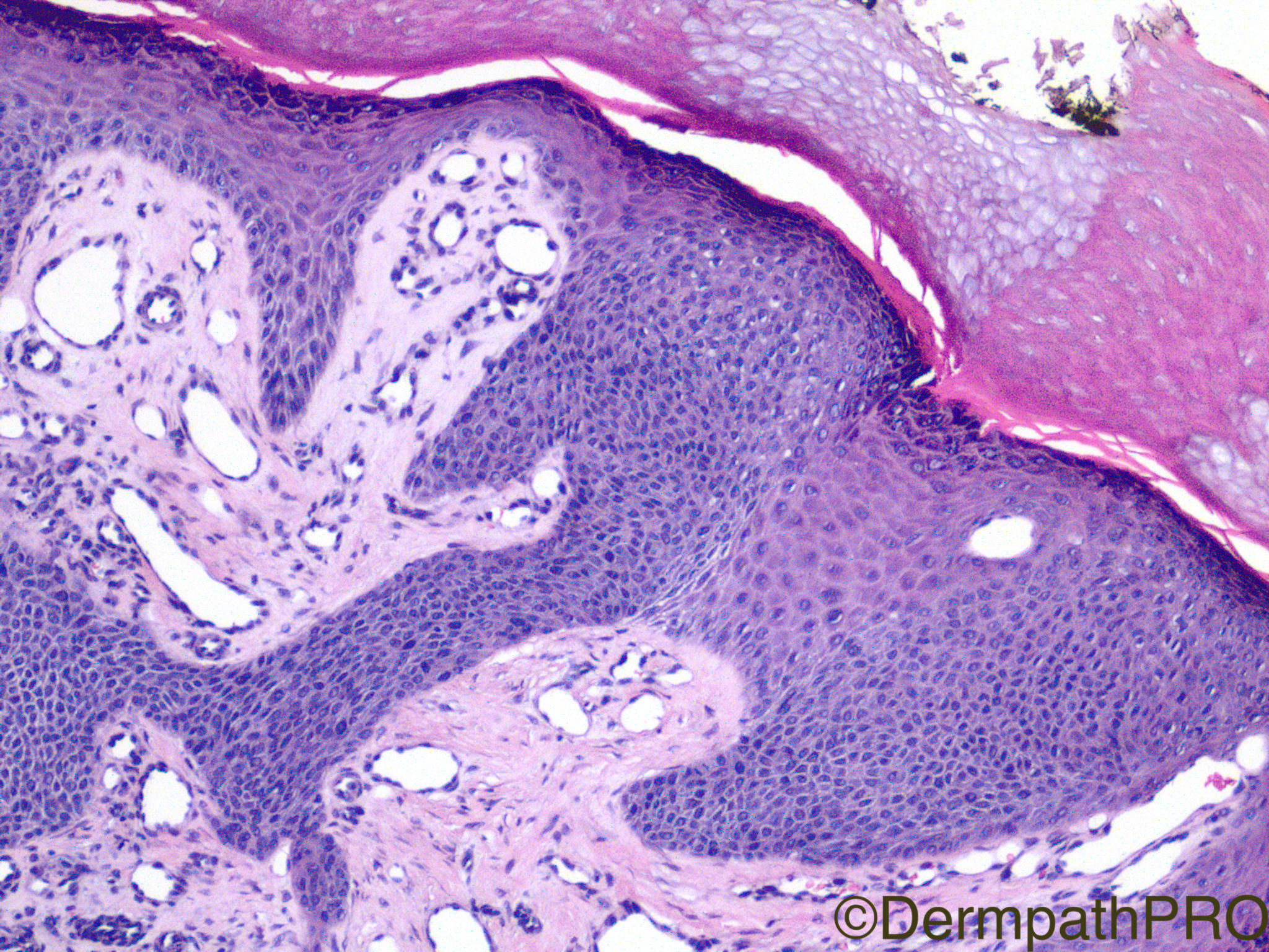
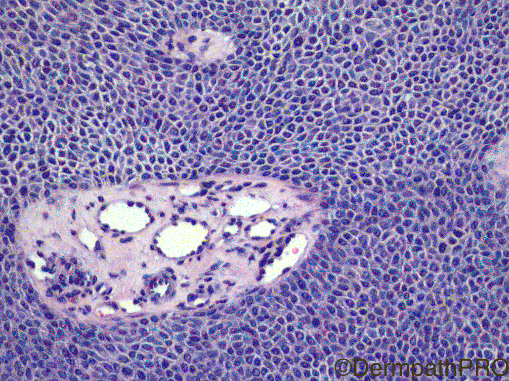
Clinical History: 23 year old woman, lesion on the left foot.

Case Posted by Dr Limin Yu


  Report Record

User Feedback


Admin_Dermpath

Posted

A tidy case from Dr Limin Yu to get your week off to a great start.

Geoff Cross - DermpathPRO Projects


The New York Dermatopathology Review Course

9th & 10th June 2017


Running in its second year, with excellent feedback from delegates!

A two day practical course in Dermatopathology designed for dermatologists, pathologists, residents, fellows and faculty.  Delivered by highly distinguished specialists from across the USA.


•  First course using PathPresenter Digital Pathology integration of cases into presentations.

•  First course ever with Live Streaming of the Course and FULL access to case materials one month prior and after.

•  ALL sessions recorded in HD and then available using FREE three-month DermpathPRO full membership subscription.

•  ALL sessions will be streamed live using GoToWebinar for FULL remote course participation (same course fee).

Register Now!!

Practical Dermatopathology.png

Share this comment


Link to comment
share_externally

urmilapandey

Posted

hidroacanthoma simplex


Share this comment


Link to comment
share_externally

Mariantonieta Tirado

Posted

They look like cuticular cells, though the pictures don't show ductal structures. A lesion from the poroma group.

Share this comment


Link to comment
share_externally

vincenzo polizzi

Posted

A typical dome shaped eccrine poroma, with typical mixed location (epidermal and dermal), and typical hyaline vascular stroma...in a typical site. 

Share this comment


Link to comment
share_externally

Dr. Mona Abdel-Halim

Posted

Yes, lovely 

Share this comment


Link to comment
share_externally

Dr. Mona Abdel-Halim

Posted

Yes, lovely 

Share this comment


Link to comment
share_externally

Fernando Cabo

Posted (edited)

Clasic Poroma . Poroid cells, cuticle cell with dúctal dif. at the botton of 3th pic and necrosis 

Edited by Fernando Cabo

Share this comment


Link to comment
share_externally

Robledo F. Rocha

Posted

The cellular composition is common to all four tumors of poroma group, but the architectural pattern (epithelial broad columns emanating from the undersurface of the epidermis and extending into the dermis) and the site (lower extremity rather than trunk) is of classic poroma.

Share this comment


Link to comment
share_externally

Nitin Khirwadkar

Posted

Agree, classic poroma.

Share this comment


Link to comment
share_externally

Limin Yu

Posted

Thank all for the discussion. I agree with everyone -- this is a pretty good example of poroma

Share this comment


Link to comment
share_externally


Join the conversation

You can post now and register later. If you have an account, sign in now to post with your account.

Guest
Add a comment...

×   Pasted as rich text.   Paste as plain text instead

  Only 75 emoji are allowed.

×   Your link has been automatically embedded.   Display as a link instead

×   Your previous content has been restored.   Clear editor

×   You cannot paste images directly. Upload or insert images from URL.

Loading...

×
×
  • Create New...