Jump to content
In this section we have spot diagnoses posted on a daily basis since June 2010, now over 4000! You can review the archived cases and read the suggested diagnoses by users and the final comment by the contributors.
Case are uploaded each week day by 10 am UK time with the correct diagnosis will generally be posted at 8 pm UK time. Why not view the most recent spot diagnosis and proffer a diagnosis?

Case Number : Case 1819 - 18 May - Dr Arti Bakshi Posted By: Guest

Please read the clinical history and view the images by clicking on them before you proffer your diagnosis.
Submitted Date :
   (0 reviews)

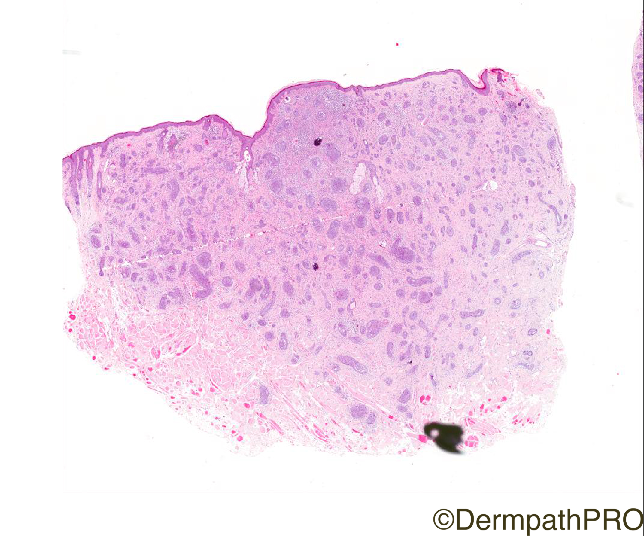
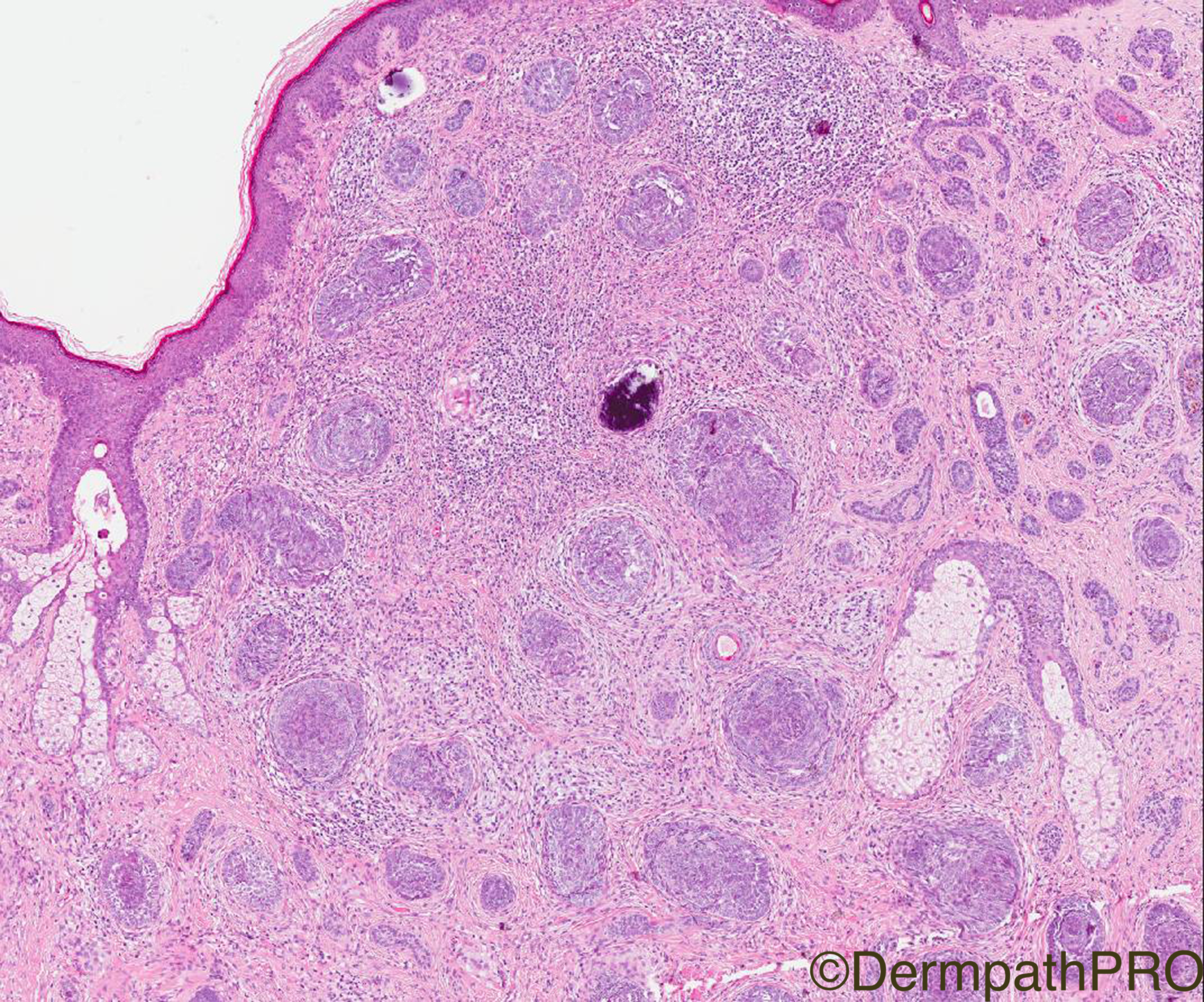
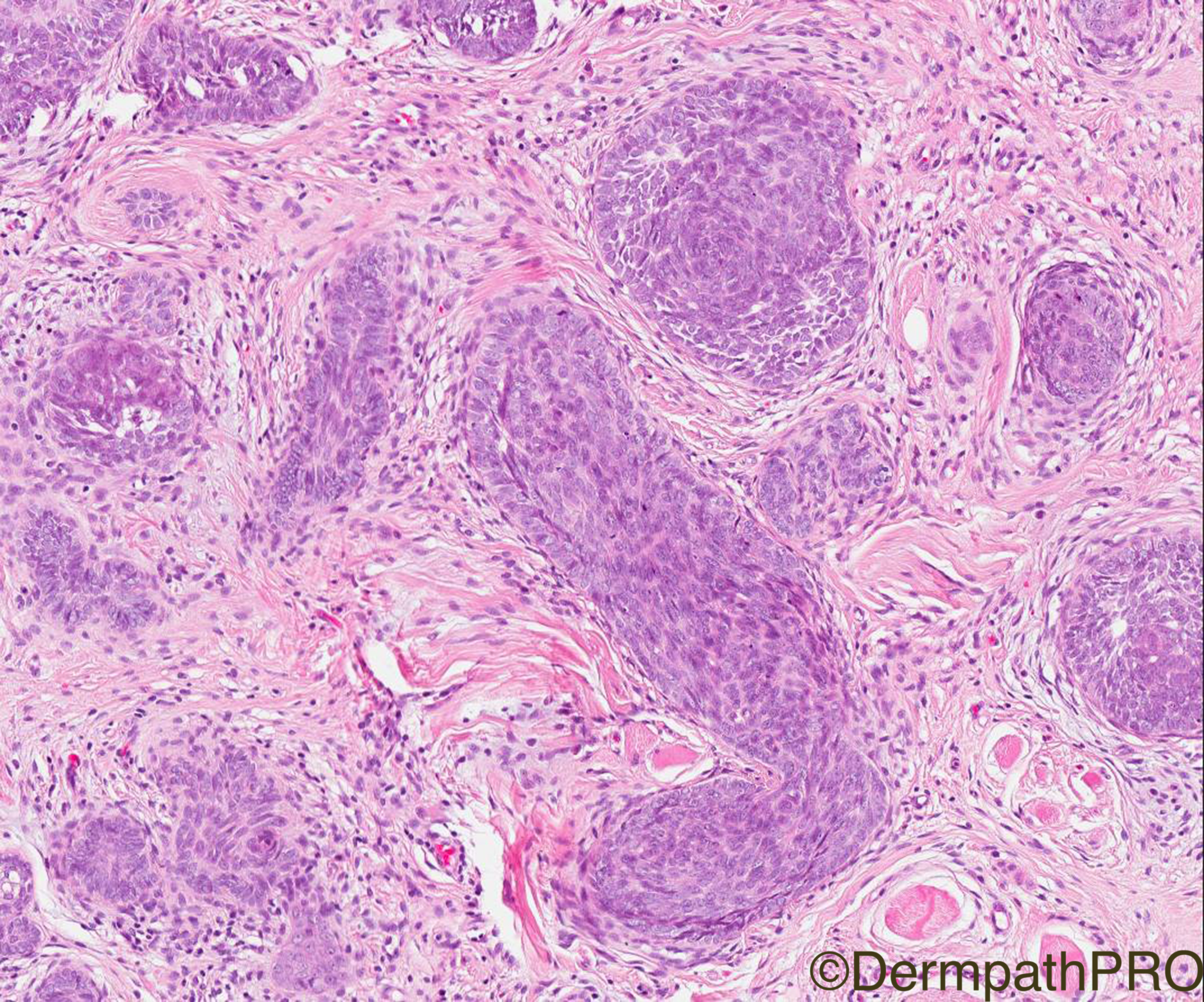
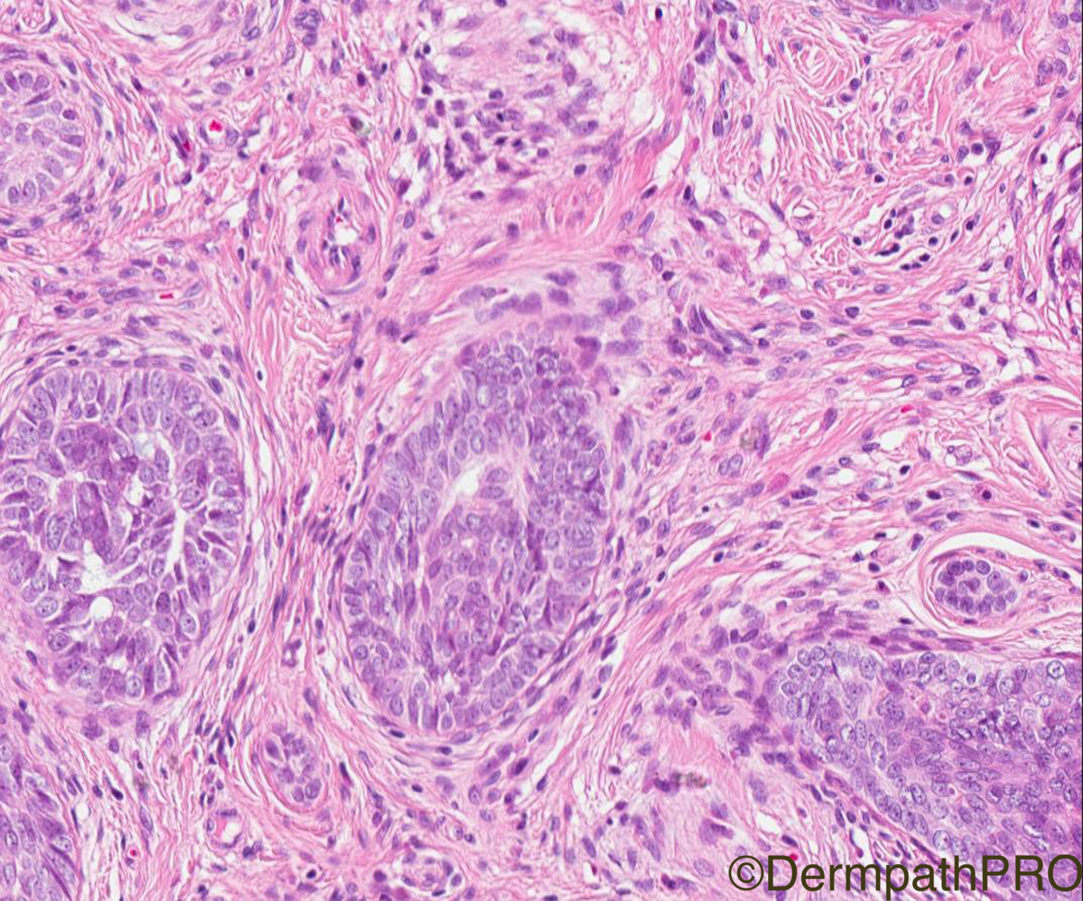
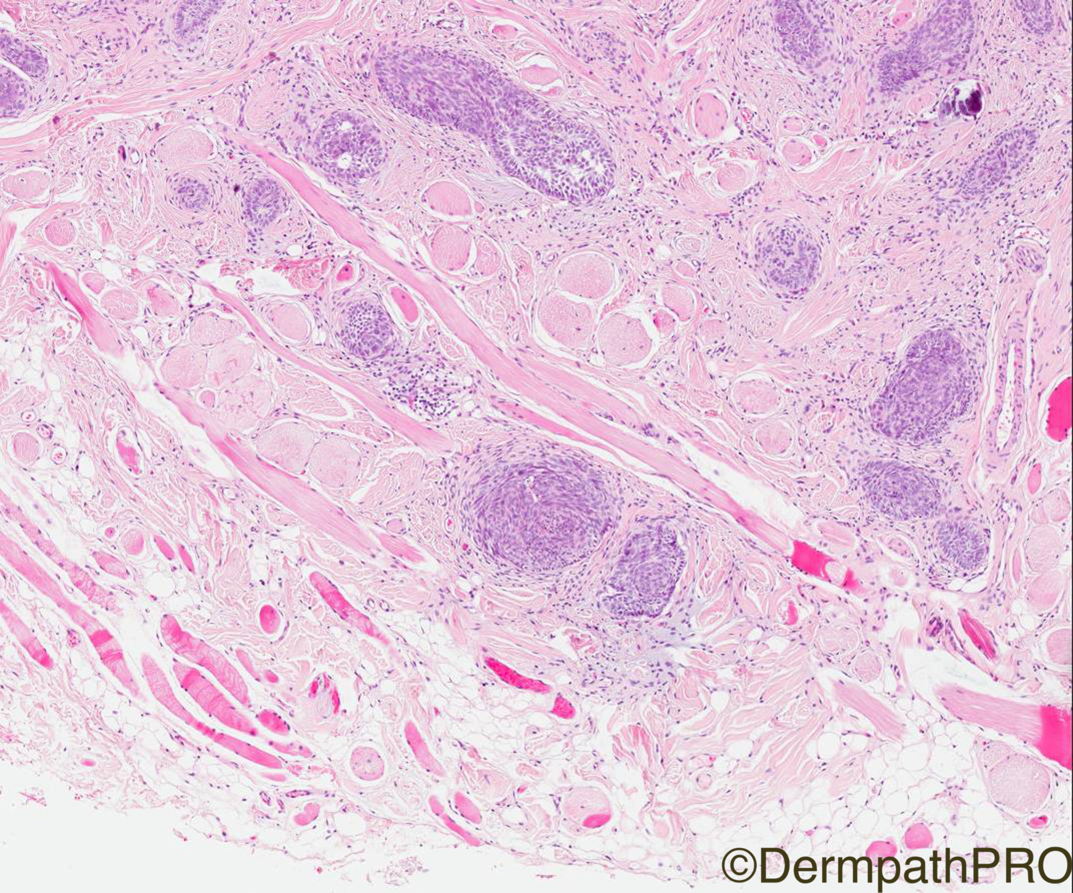
Clinical History: 45 year-old Male ?BCC lesion cheek.

Case Posted by Dr Arti Bakshi


  • Like 1
  Report Record

User Feedback


Admin_Dermpath

Posted

Dr Arti Bakshi was presenting at last week's London Dermatopathology Symposium so was not able to post her case as scheduled. No worries, here it is, and tomorrow you will get her regular case. Enjoy.

Geoff Cross - DermpathPRO Projects

Share this comment


Link to comment
share_externally

vincenzo polizzi

Posted

A multi lobular/nodular intradermal growth of basaloid cells surrounded by abundant specialized fibrous stroma ( PMB-like ), with subtle stroma-stroma retraction effect ( fig3 ) makes me thinking of Trichoblastoma. 

There is a peculiar excess of specialized stroma in this case, so it could be defined trichoblastic fibroma, but I'm a practical pathologist and favour the easier definition of trichoblastoma.

Share this comment


Link to comment
share_externally

Mariantonieta Tirado

Posted

Possibly Trichoblastoma. Can't see if there is any connection to the epidermis. Moderately cellular stroma. Didn't see obvious germ and papillae structures. No retraction between tumor and stroma

Share this comment


Link to comment
share_externally

Dr. Richard Carr

Posted

I think (on quick look) a nice example of "small nodular" trichoblastoma.

Moh's can be a good treatment for this particular subtype at this location.

Some authors consider this subtype a low grade carcinoma (not my personal view).

I'm pasting comments from our 2007 review:

Although trichoblastoma are benign tumours, a few cases are accompanied by foci indistinguishable

from conventional BCC.17 BCC have rarely been reported in association with trichoepithelioma78

and in patients with multiple trichoepithelioma, 79–81 but it was uncertain if BCC arose directly

from trichoepithelioma in the latter cases.80 Locally recurrent trichoblastoma is exceedingly

rare but a peri-anal giant solitary trichoepithelioma did recur some 17 years after initial excision,74 and

another peri-anal trichoblastoma transformed to a frank carcinoma after many years of quiescence.82

There have been several reports of tumours showing features of trichoblastoma but with asymmetrical,

plaque-like or deeply infiltrative growth on the central face, and the authors have considered

these tumours to represent indolent, locally invasive carcinomas.83–85 There are other

isolated reports of malignant transformation in familal trichoepithelioma86 and solitary trichoblastoma87;

the latter giving rise to disseminated metastases resulting in death. In view of these rare

instances of local recurrence and malignant transformation, complete excision, with narrow margins,

may be the preferred treatment for any mitotically active trichoblastoma.

Share this comment


Link to comment
share_externally

Arti Bakshi

Posted

Thanks Richard for your very educational comment. I think you have answered all the questions associated with this case!

I reported it as a trichoblastoma too and asked for a complete excision in view of the deep infiltration and presence of tumour at margin. 


Share this comment


Link to comment
share_externally


Join the conversation

You can post now and register later. If you have an account, sign in now to post with your account.

Guest
Add a comment...

×   Pasted as rich text.   Paste as plain text instead

  Only 75 emoji are allowed.

×   Your link has been automatically embedded.   Display as a link instead

×   Your previous content has been restored.   Clear editor

×   You cannot paste images directly. Upload or insert images from URL.

Loading...

×
×
  • Create New...