Jump to content
In this section we have spot diagnoses posted on a daily basis since June 2010, now over 4000! You can review the archived cases and read the suggested diagnoses by users and the final comment by the contributors.
Case are uploaded each week day by 10 am UK time with the correct diagnosis will generally be posted at 8 pm UK time. Why not view the most recent spot diagnosis and proffer a diagnosis?

Case Number : Case 1822 - 23 May - Dr Uma Sundram Posted By: Guest

Please read the clinical history and view the images by clicking on them before you proffer your diagnosis.
Submitted Date :
   (0 reviews)

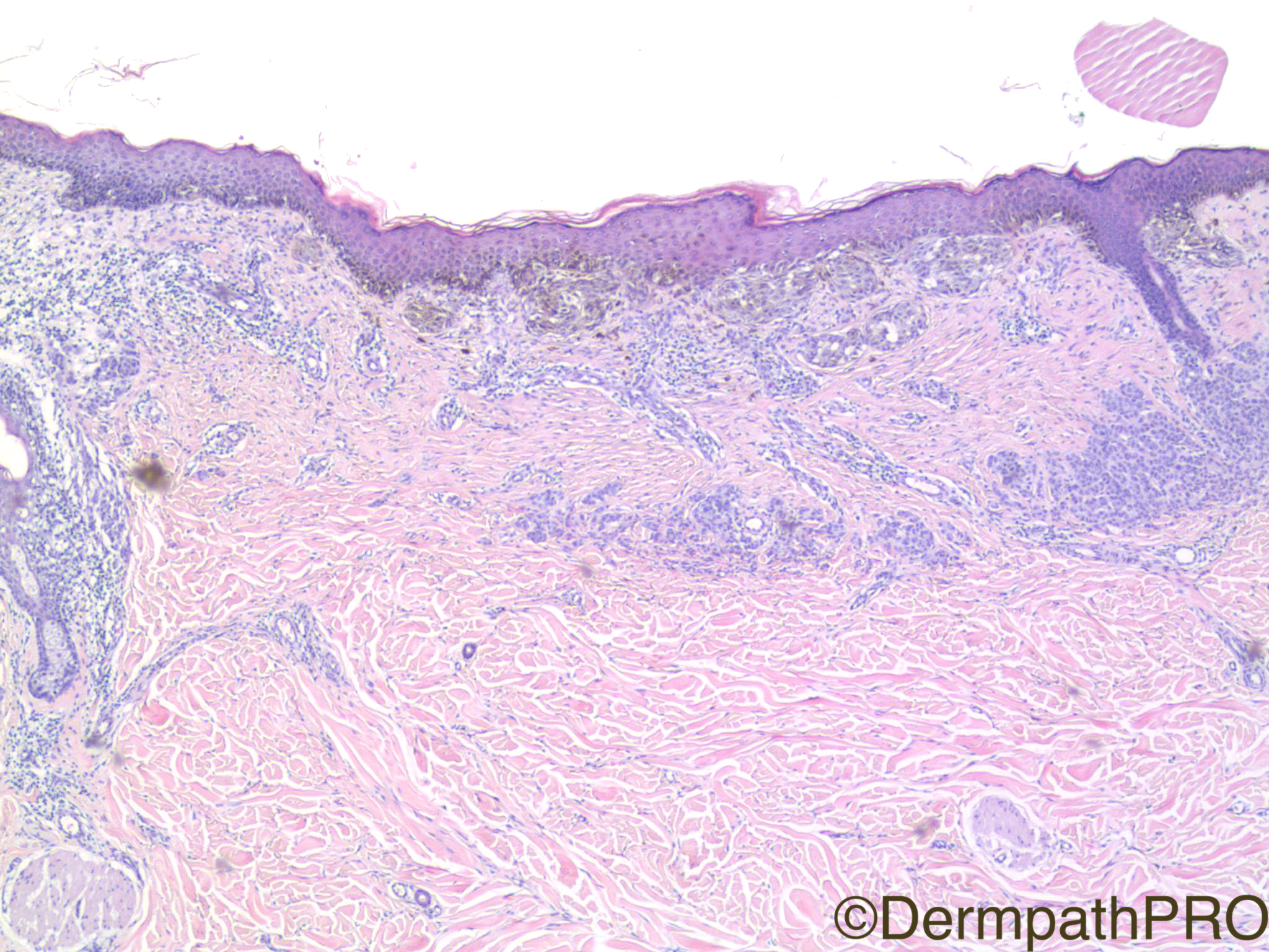
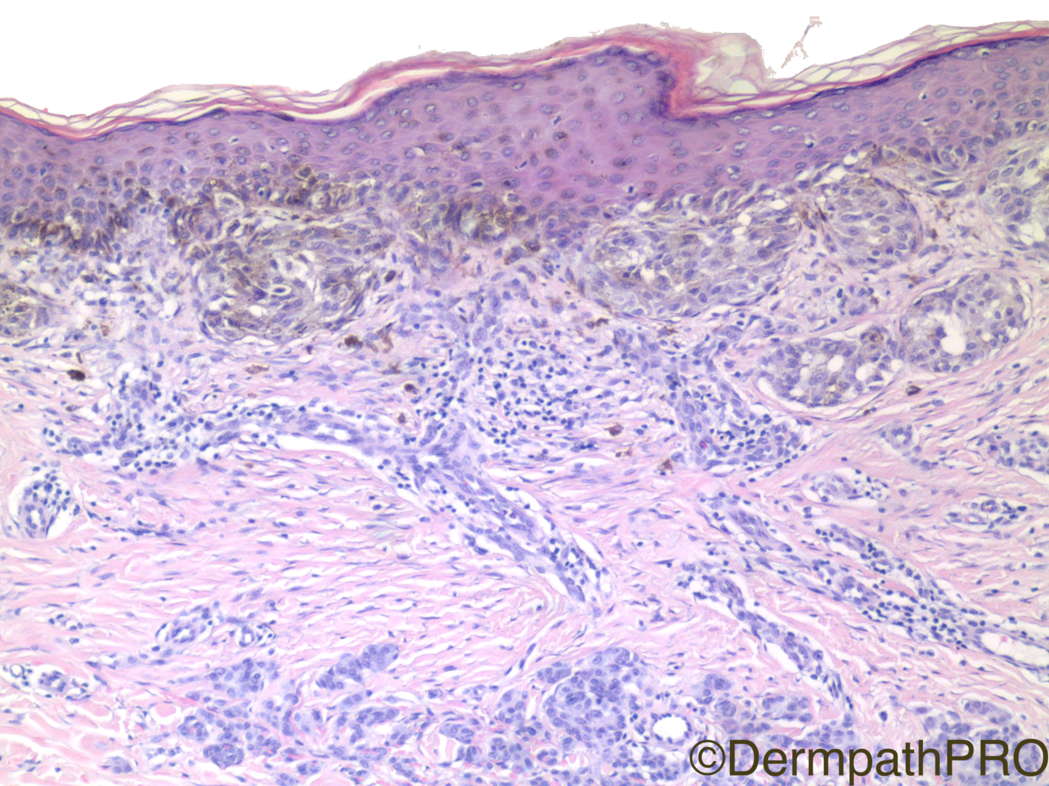
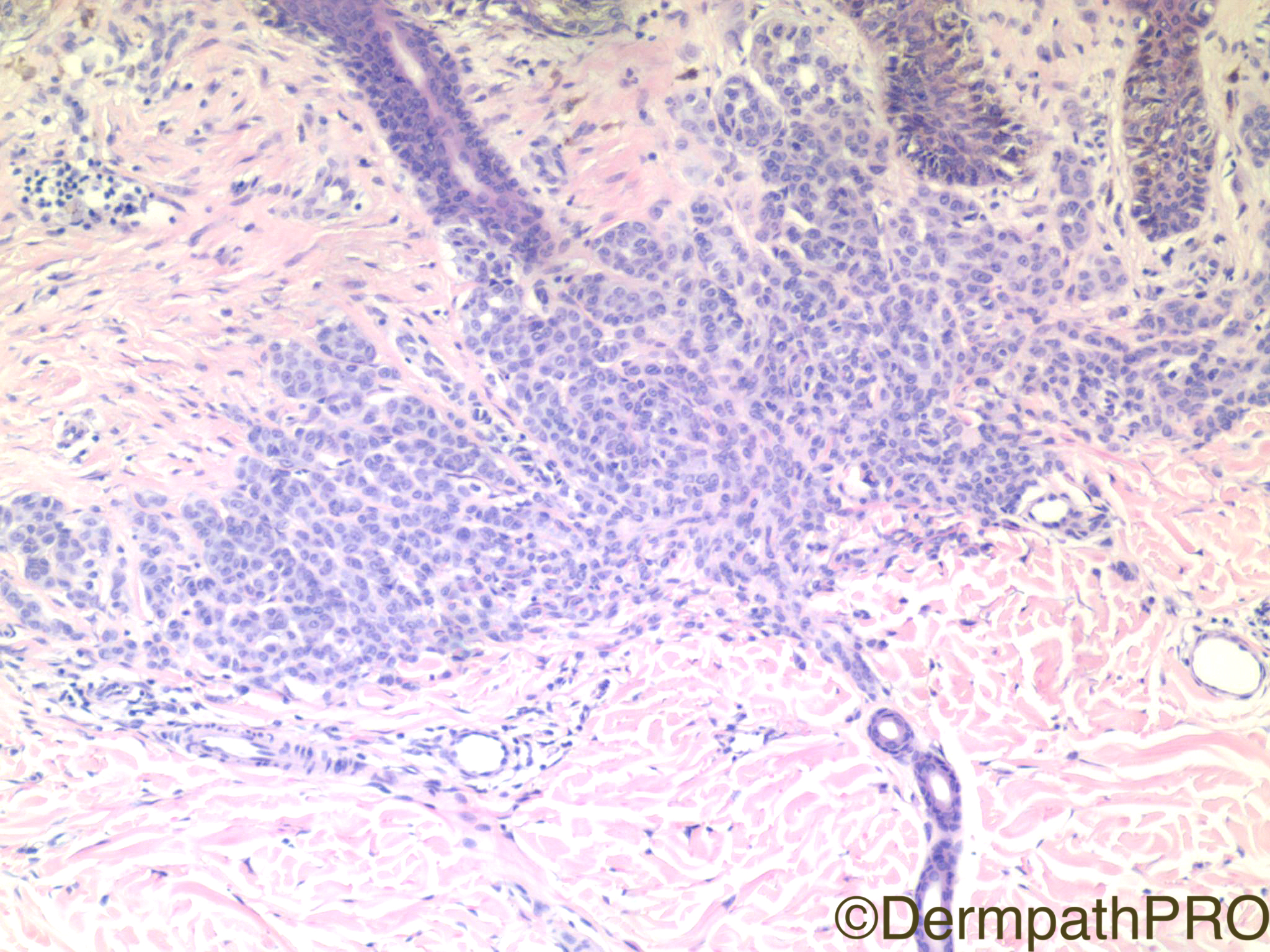
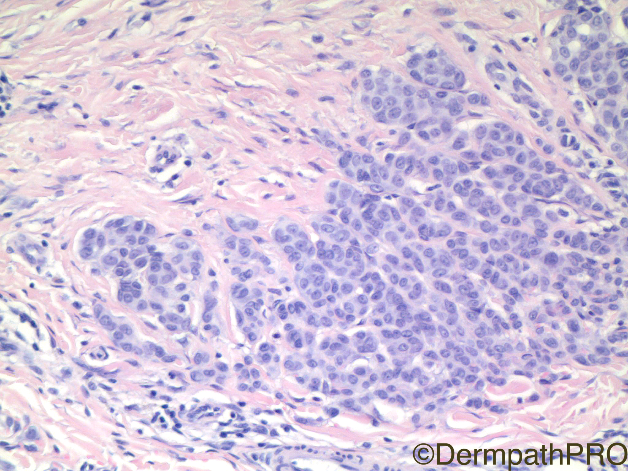
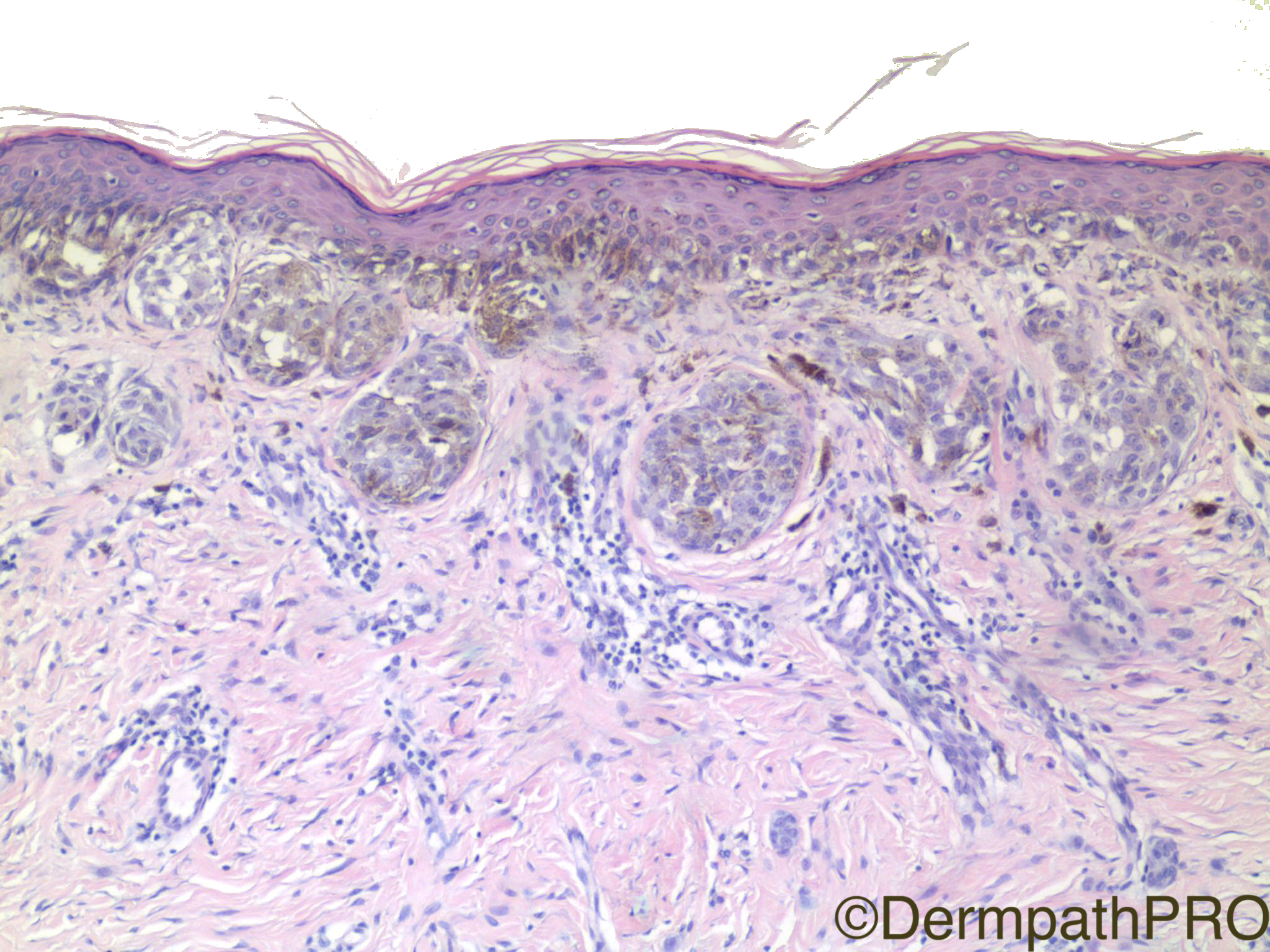
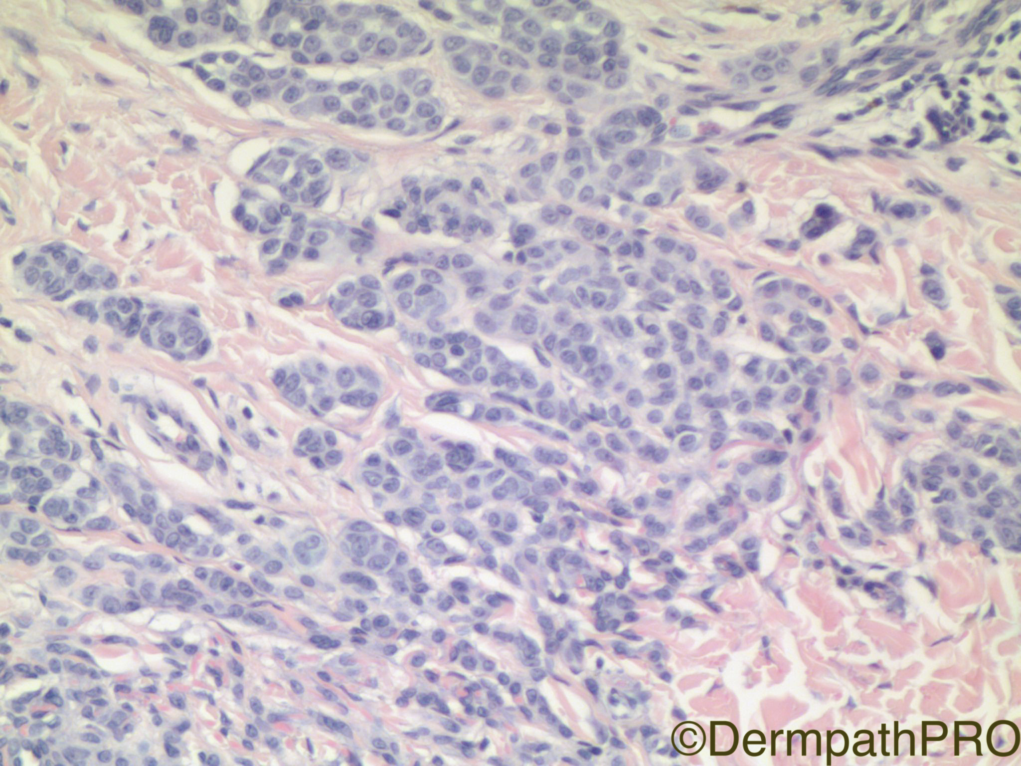
Clinical History: 50 year old woman with erythematous lesion, right rib cage.

Case Posted by Dr Uma Sundram


  Report Record

User Feedback


Raul Perret

Posted

There seems to be two independent melanocytic neoplasms. The junctional one is for me a melanoma in situ with irregular nests with prominent confluent growth and adnexal compromise. On the other hand, the dermal component looks more like a dermal melanocytic nevus as we do not see mitotic figures and there seems to be some maturation in pic 4 and 6. 

Share this comment


Link to comment
share_externally

sfwenson

Posted

Hmm, looks like superficial dermal scar to me.  My guess is compound congenital nevus with recurrent/traumatized nevus phenomenon.  Can't really tell if the junctional component goes far past the dermal component at the edges in these photos though.

Share this comment


Link to comment
share_externally

vincenzo polizzi

Posted

Looks like a recurrent/traumatized phenomenon...agree with sfwenson.


Share this comment


Link to comment
share_externally

IgorSC

Posted

I think there is a zonal disposition in this lesion that represents a recurrent nevus. There is also an activation of the lentiginous/juncional melanocytes.

Share this comment


Link to comment
share_externally

Dr. Mona Abdel-Halim

Posted

Recurrent nevus (pseudomelanoma) to me more than melanoma? Would HMB45 help?

Share this comment


Link to comment
share_externally

Mariantonieta Tirado

Posted (edited)

I think it is all benign. Agree with possible recurrent nevus (pseudomelanoma). I don't see epidermal upward scatter. No dermal mitoses. I would like to see better how the epidermal component behaves laterally beyond the "scar". A melan A would help to see easier symmetry and demarcation  . 

Edited by Mariantonieta Tirado

Share this comment


Link to comment
share_externally

Robledo F. Rocha

Posted

Agree with recurrent/persistent nevus. There is an architecturally disordered junctional melanocytic proliferation restricted to the underlying band area of dermal scar, with residual banal intradermal nevus at the bottom and on the sides.

I think reviewing the glass slide of the initial shaving biopsy is the best way to rule out melanoma. HMB-45 is useful when the initial biopsy is not available, with the caveat that recurrent/persistent nevus can lack obvious immunohistochemical maturation with this immunomarker, a pitfall that could be misconstrued as evidence of melanoma.

Share this comment


Link to comment
share_externally

Raul Perret

Posted

Yeah probably you guys are right, have to be very cautious with this kind of dx without CPC though

Share this comment


Link to comment
share_externally

Nitin Khirwadkar

Posted

Recurrent naevus/pseudomelanoma overlying a dermal scar. A banal intradermal naevus is present beneath the scar.

Share this comment


Link to comment
share_externally

Fernando Cabo

Posted

I'd like to see the borders. nested>>single. Only a few melanocytes above DEJ. Some pulverocytes.  traumatized nevus > melanoma on nevus 

Share this comment


Link to comment
share_externally

Uma Sundram

Posted

Recurrent nevus.  The lesion was laterally transected but we favored nevus over melanoma for all the reasons suggested. The lesion was re-excised.

Share this comment


Link to comment
share_externally


Join the conversation

You can post now and register later. If you have an account, sign in now to post with your account.

Guest
Add a comment...

×   Pasted as rich text.   Paste as plain text instead

  Only 75 emoji are allowed.

×   Your link has been automatically embedded.   Display as a link instead

×   Your previous content has been restored.   Clear editor

×   You cannot paste images directly. Upload or insert images from URL.

Loading...

×
×
  • Create New...