Jump to content
In this section we have spot diagnoses posted on a daily basis since June 2010, now over 4000! You can review the archived cases and read the suggested diagnoses by users and the final comment by the contributors.
Case are uploaded each week day by 10 am UK time with the correct diagnosis will generally be posted at 8 pm UK time. Why not view the most recent spot diagnosis and proffer a diagnosis?

Case Number : Case 1824 - 25 May - Dr Arti Bakshi Posted By: Guest

Please read the clinical history and view the images by clicking on them before you proffer your diagnosis.
Submitted Date :
   (0 reviews)

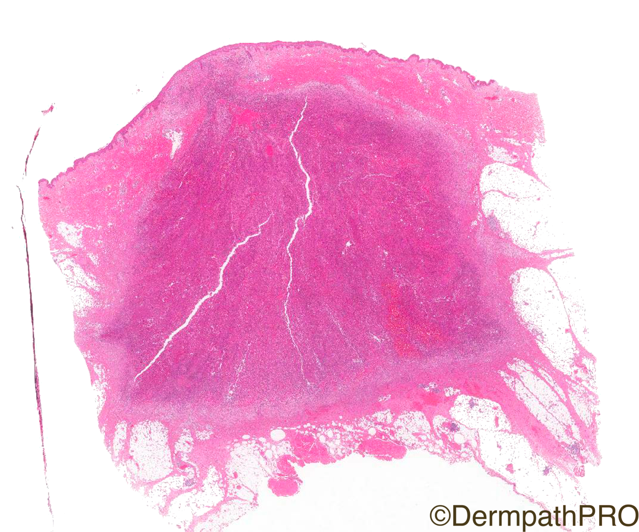
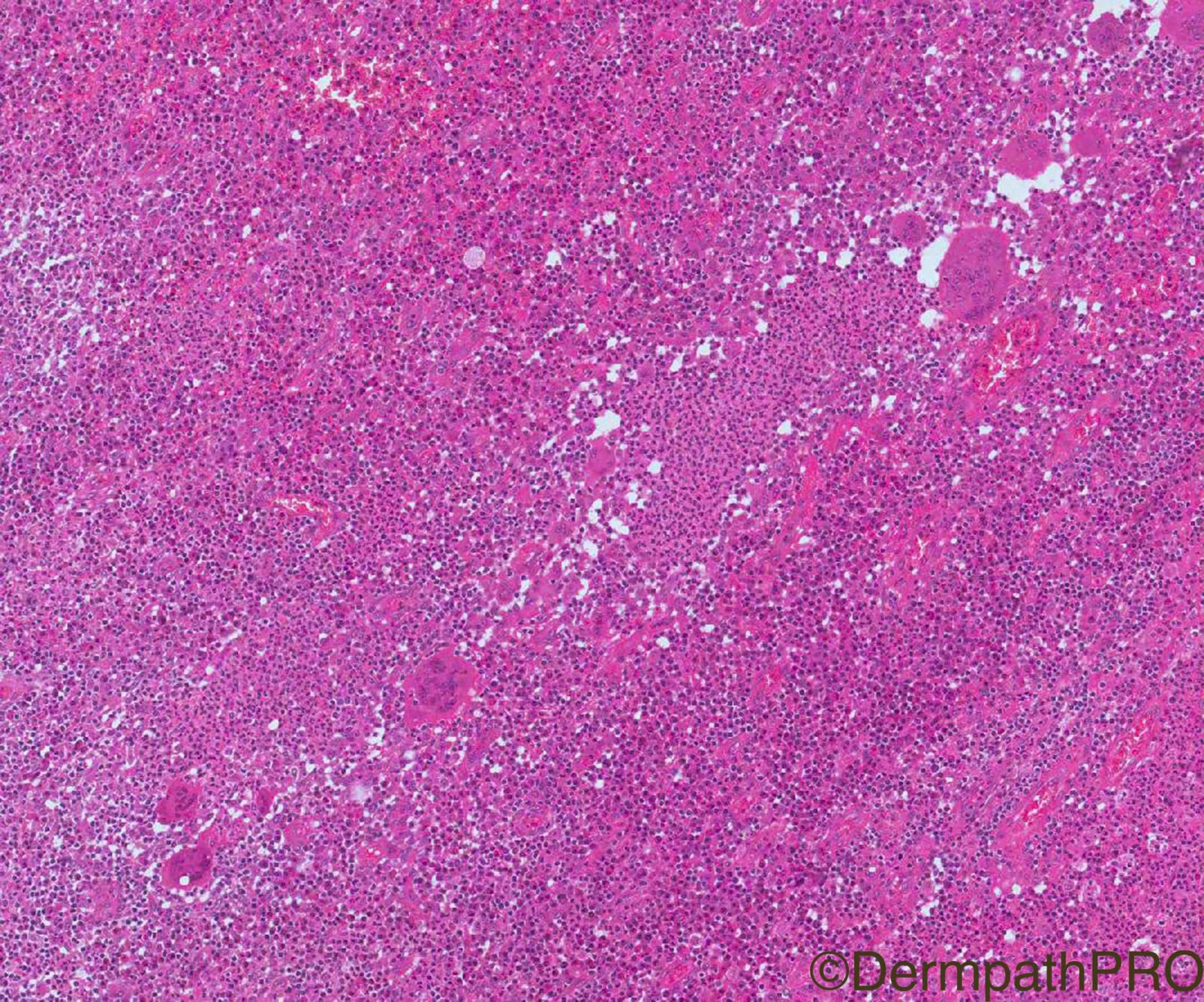
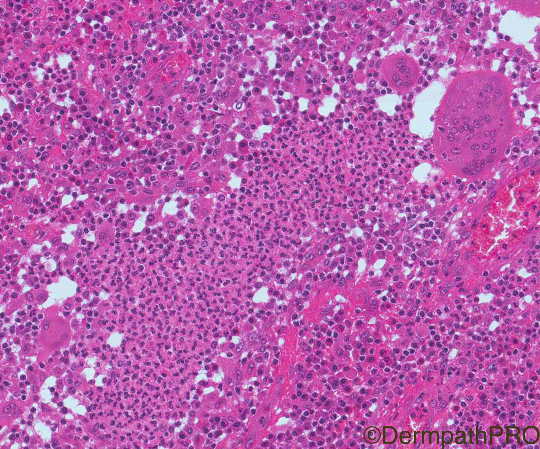
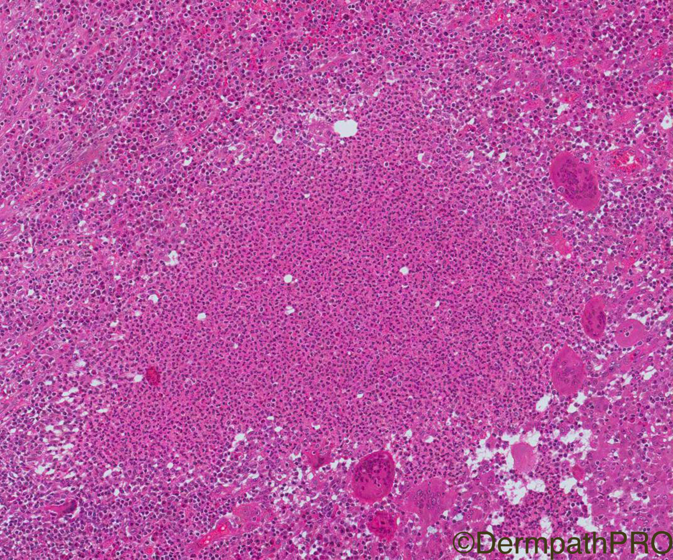
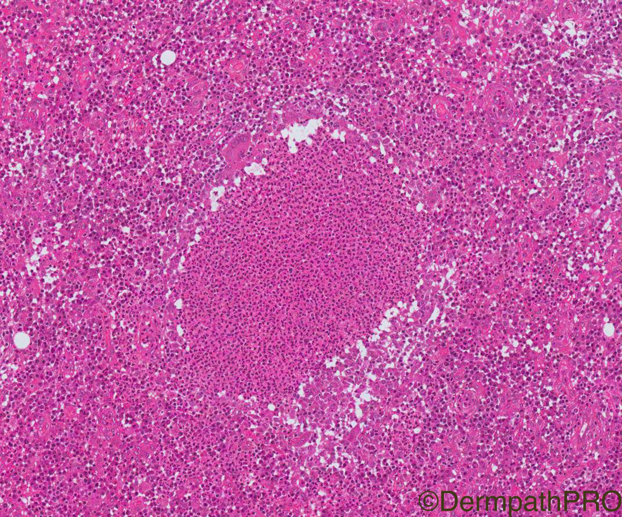
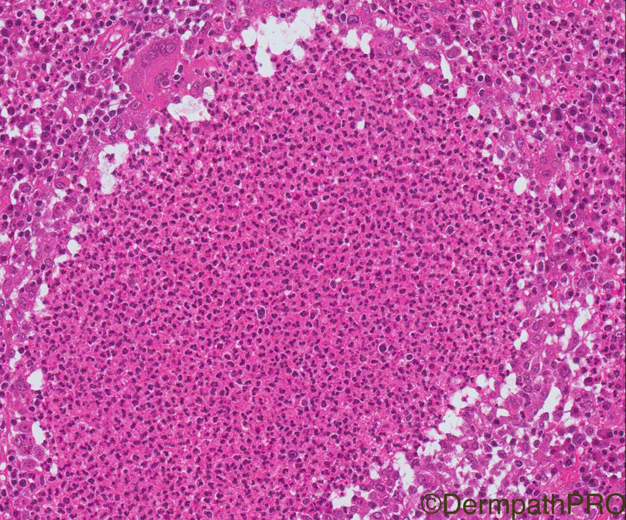
Clinical History: 38/F, erythematous plaques studded with pustules, shoulder and arm. Patient is on immunosuppressive therapy for rheumatoid arthritis.

Case Posted by Dr Arti Bakshi


  Report Record

User Feedback


Admin_Dermpath

Posted

Dr Arti Bakshi tells me that you have a treat of a Daily Spot Diagnosis Case today, enjoy.

Geoff Cross - DermpathPRO Projects

Share this comment


Link to comment
share_externally

Dr. Mona Abdel-Halim

Posted

Superficial vegetative (granulomatous) pyoderma gangrenosum.

Share this comment


Link to comment
share_externally

Dr. Richard Carr

Posted

I remember well a case of ?rheumatoid neutrophilic dermatosis in which I found acid fast bacilli!  Needs usual bug stains and cultures but certainly a granulomatous variant of pyoderma gangrenosum is a good suggestion. Rather impressive giant cells I thought.

Share this comment


Link to comment
share_externally

Anil Patki

Posted

Its a suppurative granuloma. Deep fungal infection to be ruled out in an immunocompromised patient.


Share this comment


Link to comment
share_externally

Saman Fatah

Posted

This looks like a suppurative granulomatous dermatitis in an immunocompromised host, infective causes is a priority to exclude through appropriate triple tissue cultures (mycobacterial, fungal and bacterial). Non-Tuberculous Mycobacterium (NTM) infections especially Mycobacterium Chelonae is frequently isolated in these patients (in the U.K. in my experience) particularly those on oral Prednisolone as part of their treatment regime. Once infective aetiology excluded, neutrophilic dermatosis is fairly reasonable to consider next including PG. 

Was this lady was on Prednisolone as part of therapy regime out of intertest? certainly it can be seen with any immunosuppressives. 

Thanks

Share this comment


Link to comment
share_externally

Robledo F. Rocha

Posted

Suppurative diffuse dermatitis with multinucleate foreign body giant cells. Agree with pyoderma gangrenosum, associated with rheumatoid arthritis.

Share this comment


Link to comment
share_externally

Fernando Cabo

Posted

It should be rule out infectious suppurative granulomatosa infection. Muy be superficial granulomatous pyoderma .

Share this comment


Link to comment
share_externally

IgorSC

Posted

Agree with special stains to search for microorganisms as well as culture. If both negative, it could be superficial vegetative pyoderma gangrenosum

Share this comment


Link to comment
share_externally

Arti Bakshi

Posted

Yes, this is a superficial granulomatous pyoderma gangrenosusm. So well done to all!

An infective eitiology, ofcourse, needs to be excluded in all such cases.The patient had multiple biopsies, all reported as granulomatous r/o infection. Special stains were repeatedly negative,tissue was sent for culture and PCR and these too were negative.The patient was on Adalimumab for RA.  Once a diagnosis of granulomatous PG was made in our CPC meeting, some more immunosuppressive agents were added and this led to rapid healing of the lesions. 

Although called superficial, lesions of superficial granulomatous PG can be fairly deep seated (as in this case). The zonal pattern of inflammation is typical and mutinucleate giants (plus plasma cells) are also characteristic. This variant typically follows a more indolent course than classic PG.

Thanks for all your comments!

Share this comment


Link to comment
share_externally

Arti Bakshi

Posted

Nice to see some new people putting in comments! Welcome to all!

Share this comment


Link to comment
share_externally

Saman Fatah

Posted

Thanks for the feedback on this interesting case with a good learning point and the welcome. Hope occasional comments from Clinicians is accepted in this platform among Dermpath colleagues and we learn from each other.

Share this comment


Link to comment
share_externally

Arti Bakshi

Posted

Ofcourse Saman! This website is for all with an interest in learning dermpath. There are many dermatologists who have contributed over the years and we have  learnt so much  from their  clinical perspective....it is this close link between dermatology and pathology which makes dermpath so unique (and undoubtedly the best subspeciality of histopathology!)

Share this comment


Link to comment
share_externally


Join the conversation

You can post now and register later. If you have an account, sign in now to post with your account.

Guest
Add a comment...

×   Pasted as rich text.   Paste as plain text instead

  Only 75 emoji are allowed.

×   Your link has been automatically embedded.   Display as a link instead

×   Your previous content has been restored.   Clear editor

×   You cannot paste images directly. Upload or insert images from URL.

Loading...

×
×
  • Create New...