Jump to content
In this section we have spot diagnoses posted on a daily basis since June 2010, now over 4000! You can review the archived cases and read the suggested diagnoses by users and the final comment by the contributors.
Case are uploaded each week day by 10 am UK time with the correct diagnosis will generally be posted at 8 pm UK time. Why not view the most recent spot diagnosis and proffer a diagnosis?

Case Number : Case 1826 - 29 May - Dr Limin Yu Posted By: Guest

Please read the clinical history and view the images by clicking on them before you proffer your diagnosis.
Submitted Date :
   (0 reviews)

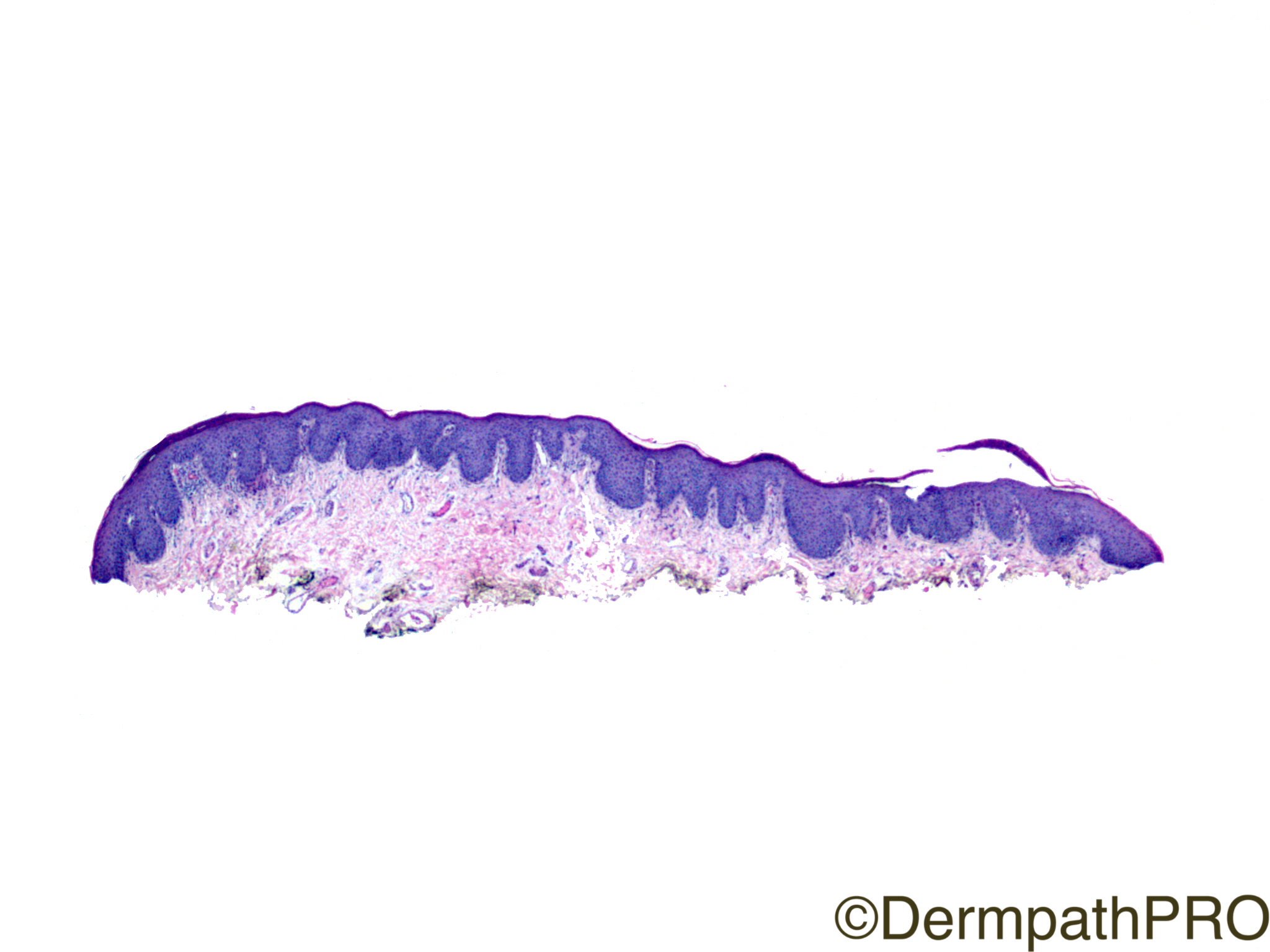
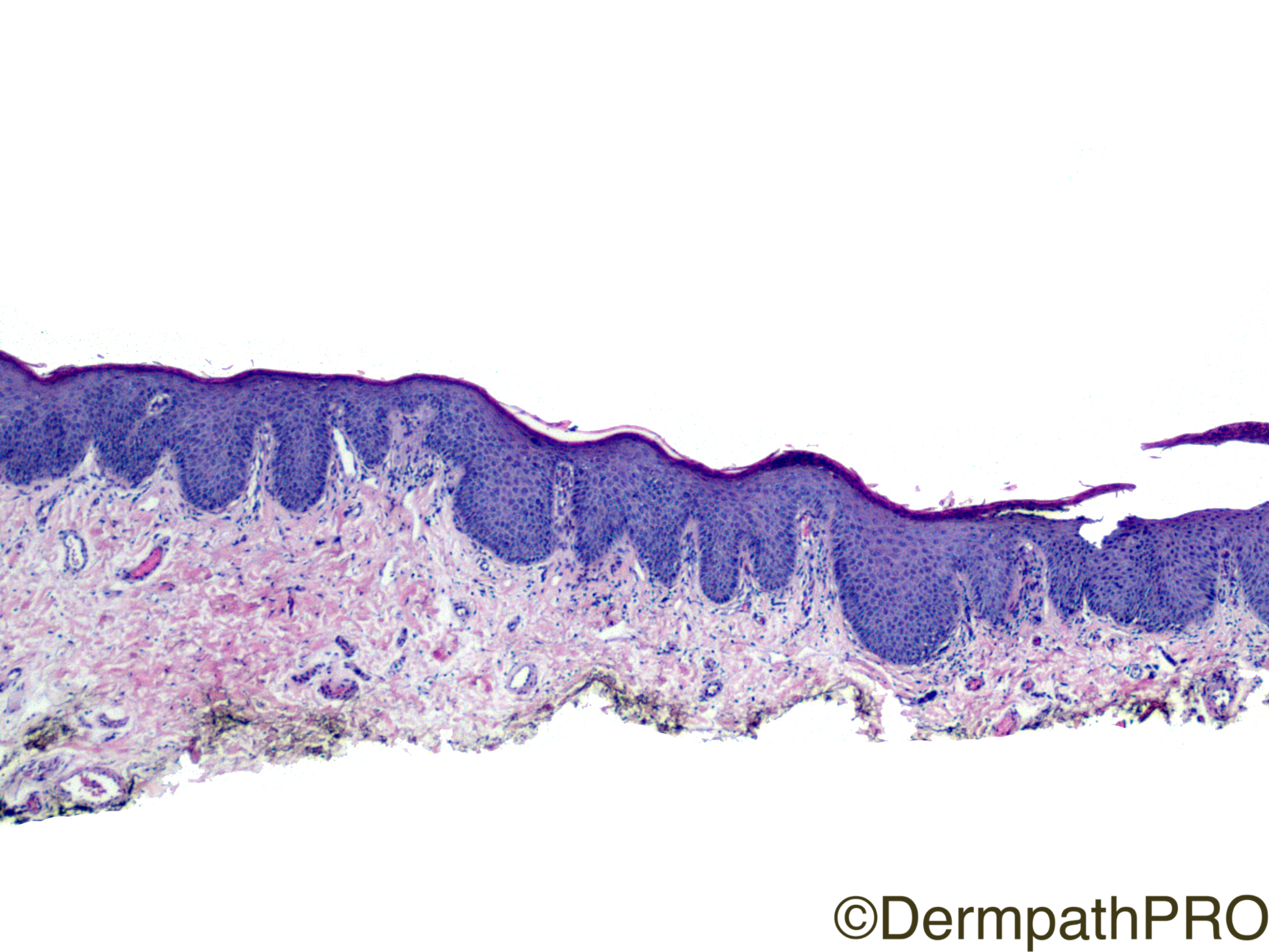
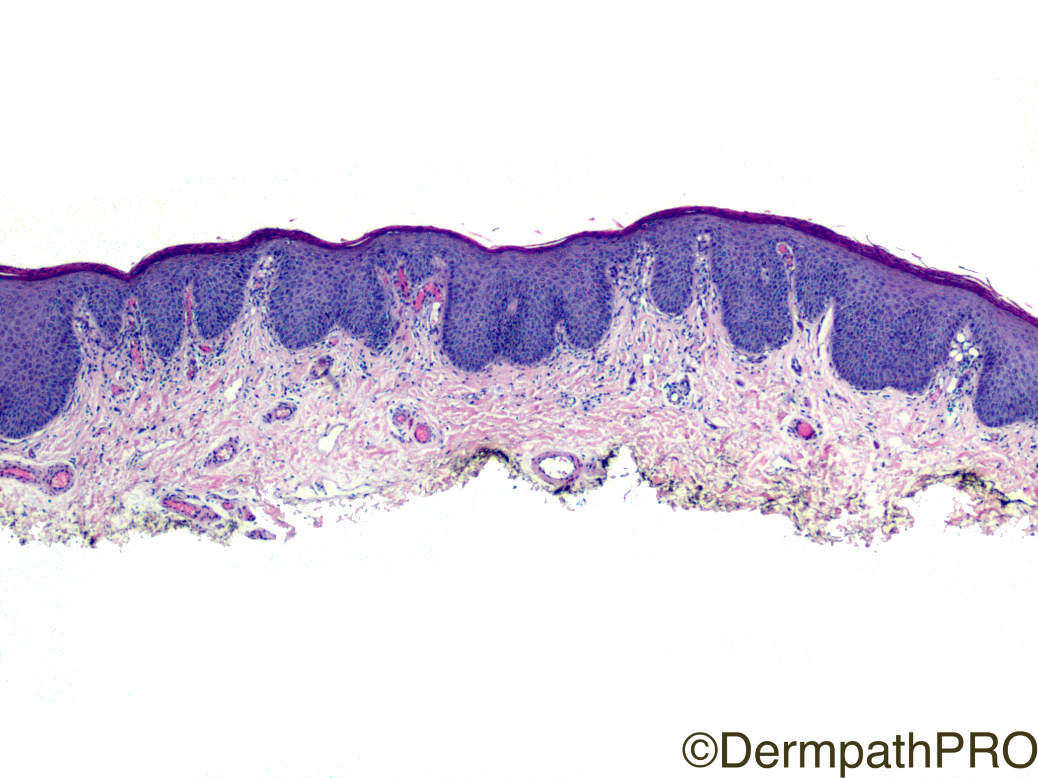
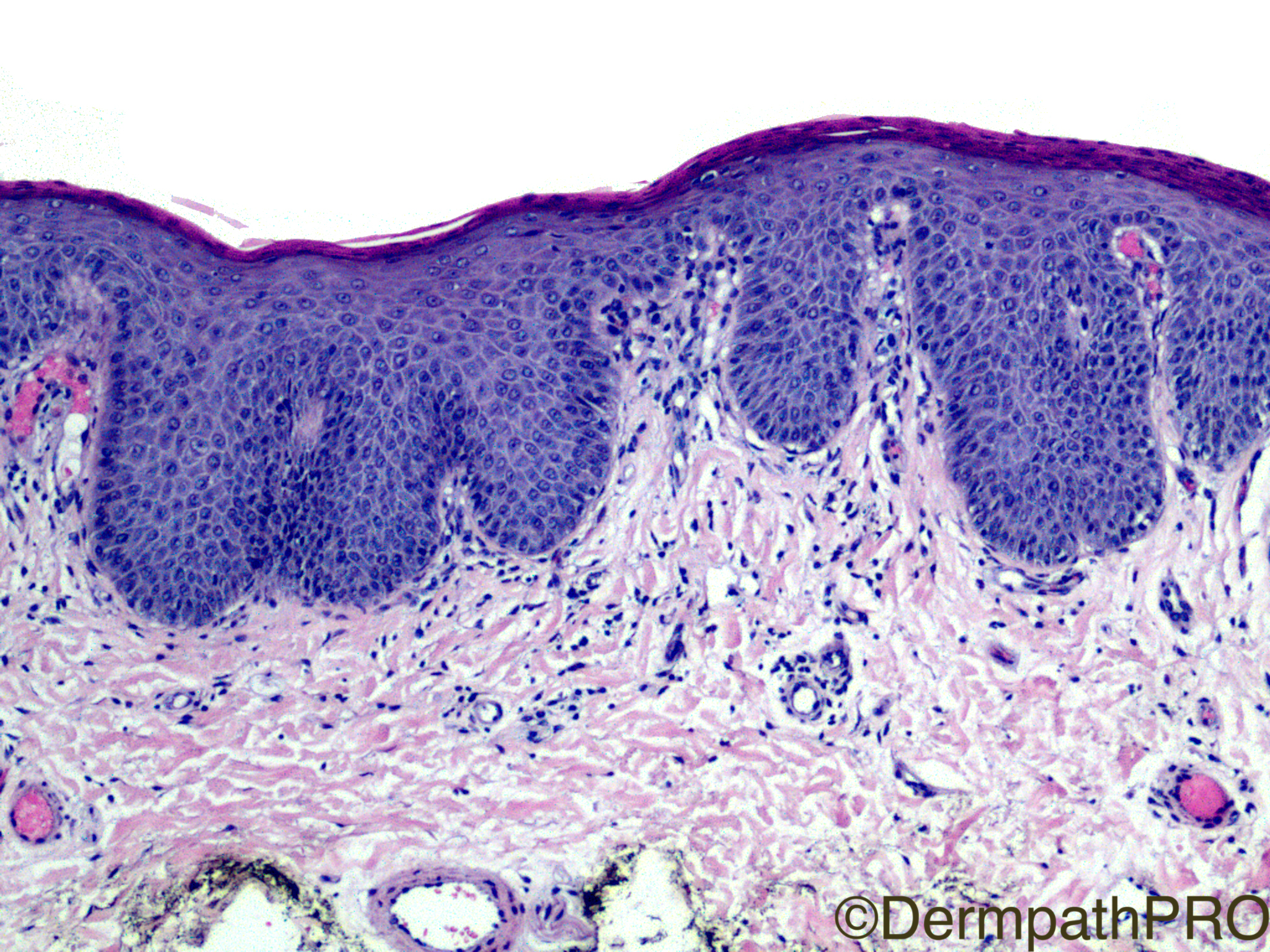
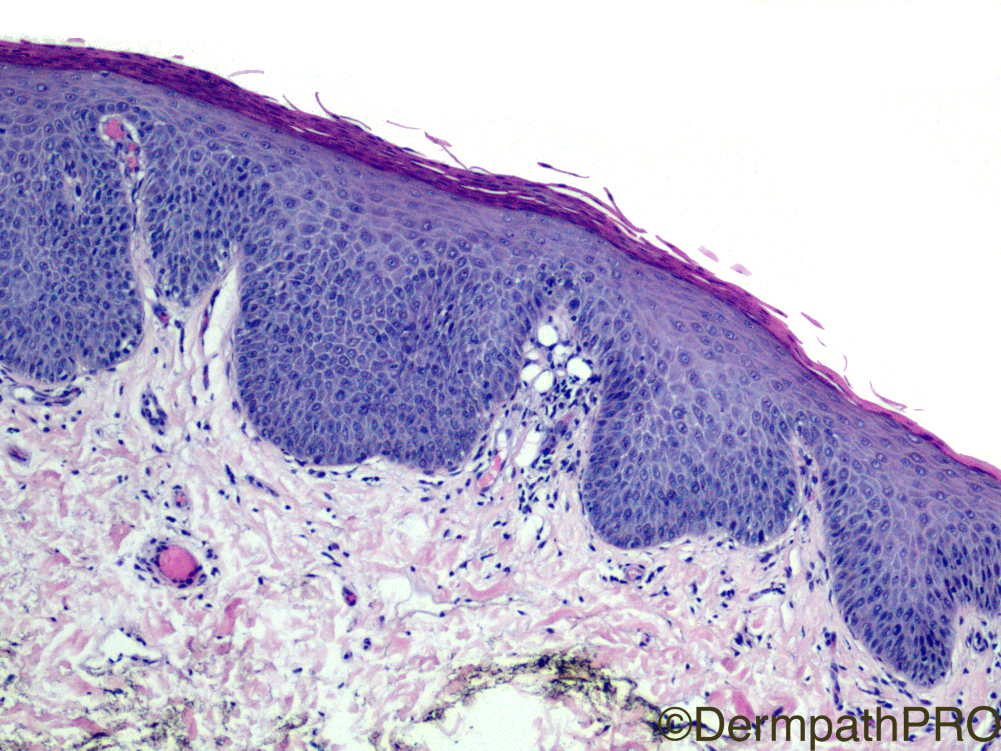
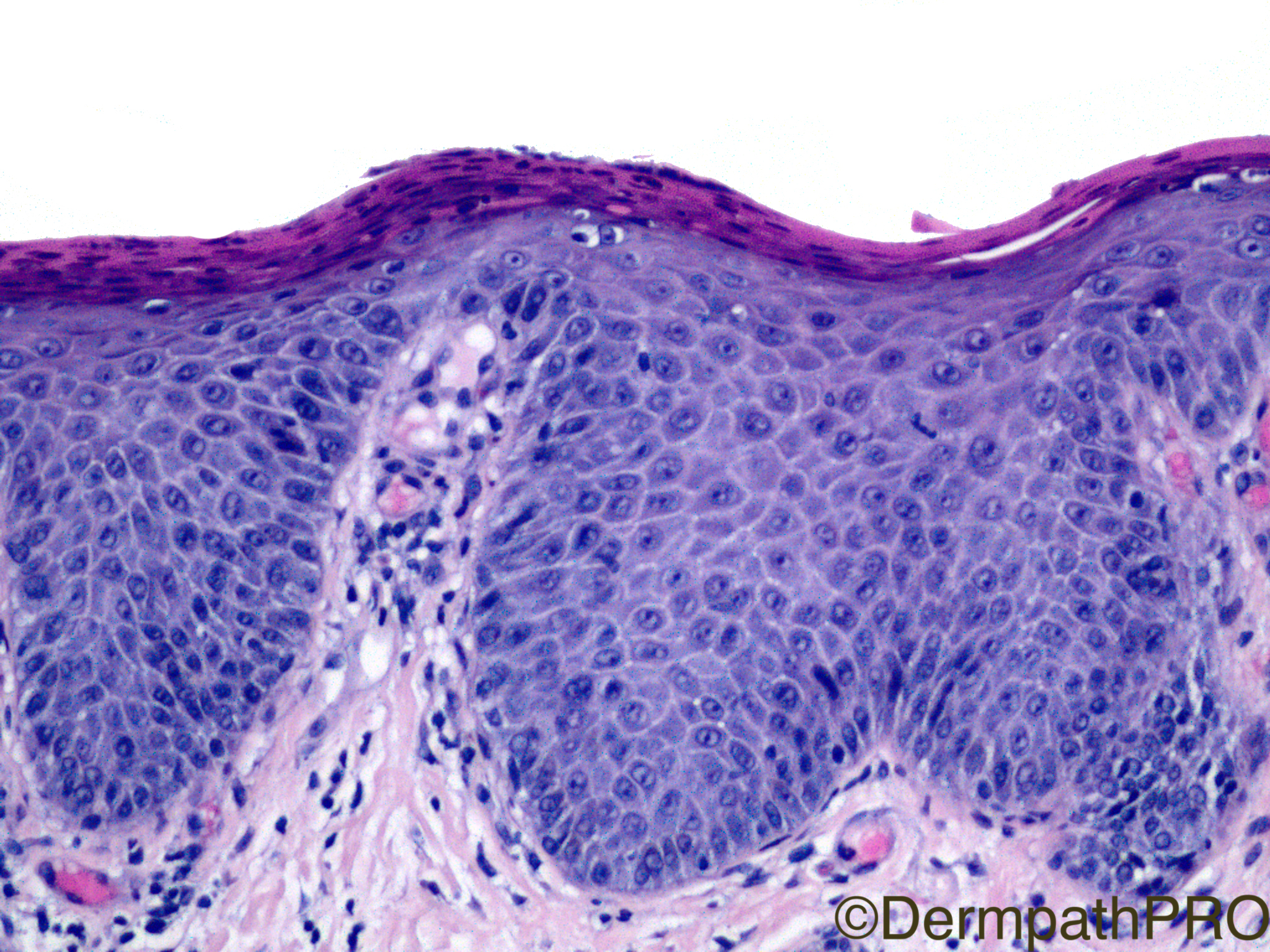
Clinical History: 27 year-old, penis.

Case Posted by Dr Limin Yu


  Report Record

User Feedback


Admin_Dermpath

Posted

Get your week off to a great start with a lovely case from Dr Limin Yu.

Geoff Cross - DermpathPRO Projects

Share this comment


Link to comment
share_externally

vincenzo polizzi

Posted

Mitotic figures in basal layers and above them, capillary loop in papillary dermis tip, thinning of sovrapapillary epidermis, loss of granular layer, and jumping parakeratotic scales (fig 1) are consistent with "genital psoriasis". 

Share this comment


Link to comment
share_externally

Dr. Mona Abdel-Halim

Posted

I was thinking of bowenoid papulosis ?

Share this comment


Link to comment
share_externally

urmilapandey

Posted (edited)

psoarisiform changes but distinct keratinocyte atypia and mitotic activity-not full thickness-would favour bowenoid papulosis


Edited by urmilapandey
typo error

Share this comment


Link to comment
share_externally

Robledo F. Rocha

Posted

A flat epidermal thickening due to crowded proliferation of keratinocytes that show nuclear hyperchromatism, arrest of maturation, and suprabasal mitoses. These microscopic features in combination with the clinical history of a genital lesion in a young adult makes me think of bowenoid papulosis.

Share this comment


Link to comment
share_externally

Fernando Cabo

Posted

Minimal inflamation, parakeratosis. Erytrasma? Woods's light?

Share this comment


Link to comment
share_externally

Raul Perret

Posted

Weedon, among others remarks in his talks that psoriasis is a disease with a high mitotic index. This elevated proliferation is one of the main causes of parakeratosis. Agree with Vincenzo, for me this is psoriasis

Share this comment


Link to comment
share_externally

IgorSC

Posted

I agree with Psoriasis as well. The dilated capillary vessels, regular acanthosis and hypogranulosis are well represented. DDx with bowenoid papulosis (bowen), but this is not the case. On last picture we can also see some neutrophils within the cornea.

Share this comment


Link to comment
share_externally

Neil Catterall

Posted

Unusual site but granular parakeratosis has been reported on penis

Share this comment


Link to comment
share_externally

Dr. Richard Carr

Posted

Agree psoriasis or candida - indistinguishable histologically.

Share this comment


Link to comment
share_externally

Limin Yu

Posted

I apologize for not providing my diagnosis in a timely manner. This is a genital psoriasis. There are increased mitotic figures, which can sometimes be seen in psoriasis. Thanks! 

Share this comment


Link to comment
share_externally


Join the conversation

You can post now and register later. If you have an account, sign in now to post with your account.

Guest
Add a comment...

×   Pasted as rich text.   Paste as plain text instead

  Only 75 emoji are allowed.

×   Your link has been automatically embedded.   Display as a link instead

×   Your previous content has been restored.   Clear editor

×   You cannot paste images directly. Upload or insert images from URL.

Loading...

×
×
  • Create New...