Jump to content
In this section we have spot diagnoses posted on a daily basis since June 2010, now over 4000! You can review the archived cases and read the suggested diagnoses by users and the final comment by the contributors.
Case are uploaded each week day by 10 am UK time with the correct diagnosis will generally be posted at 8 pm UK time. Why not view the most recent spot diagnosis and proffer a diagnosis?

Case Number : Case 1943 - 09 Nov Posted By: Iskander H. Chaudhry

Please read the clinical history and view the images by clicking on them before you proffer your diagnosis.
Submitted Date :
   (0 reviews)

55 Yr old Female

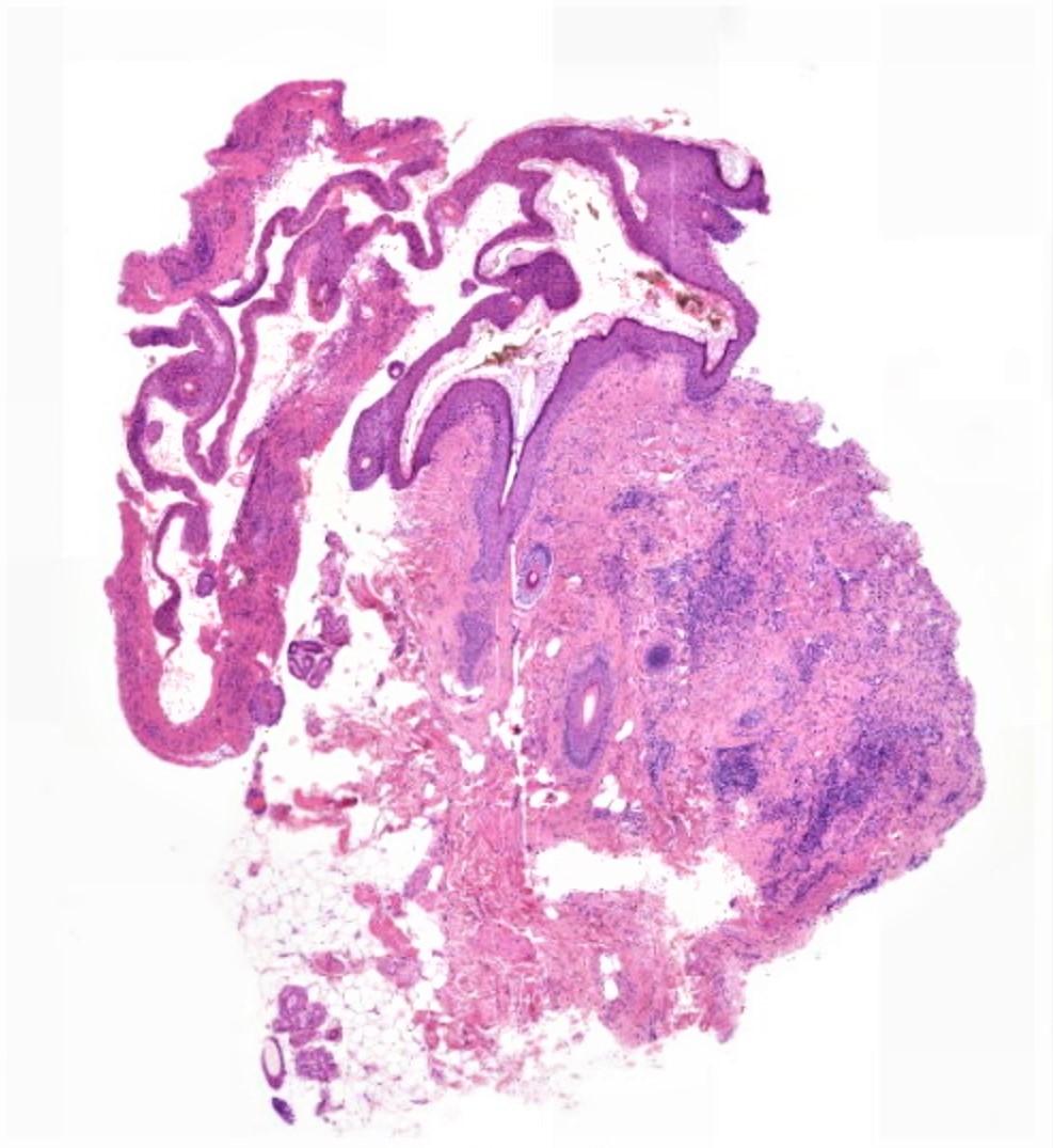
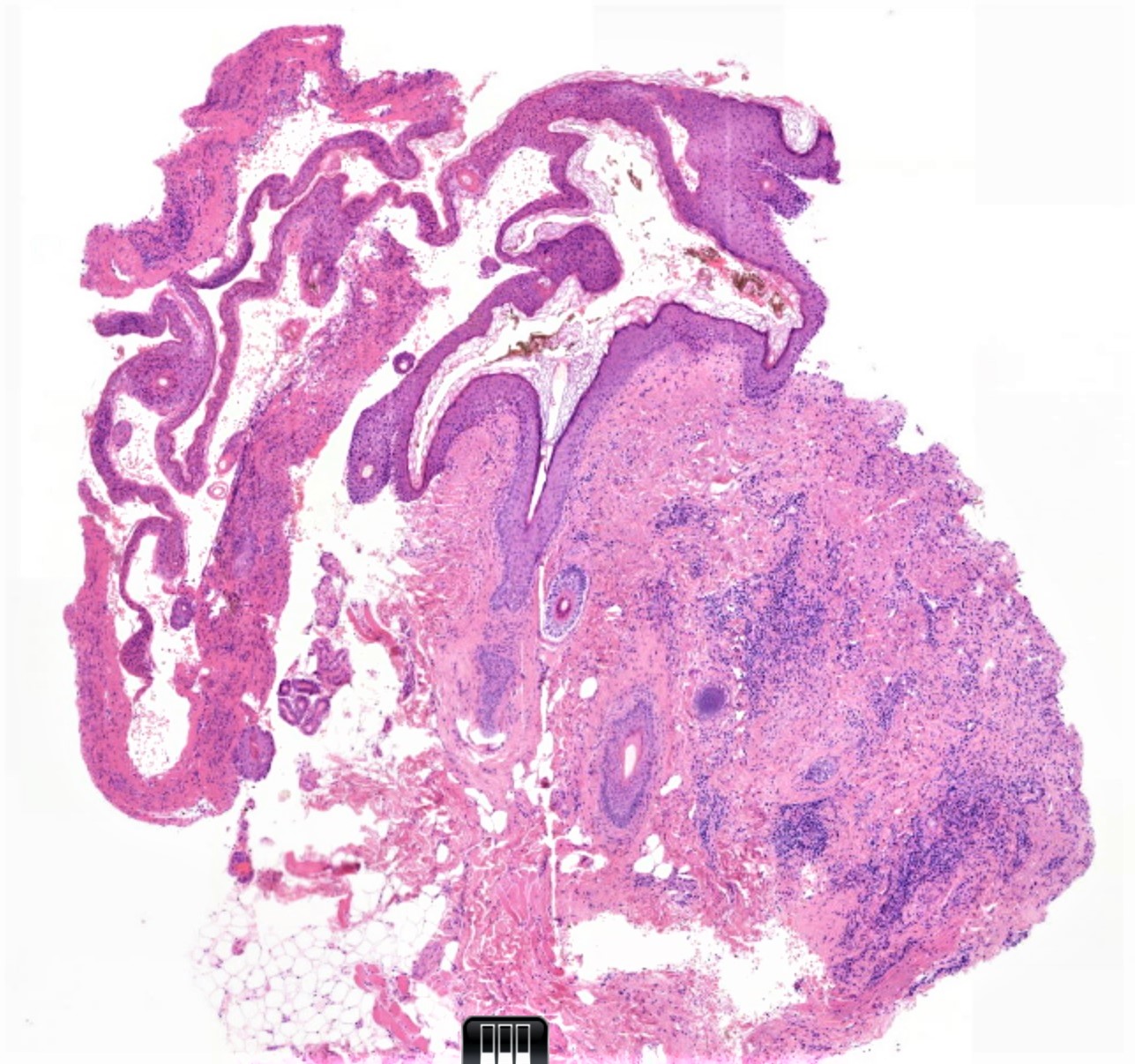
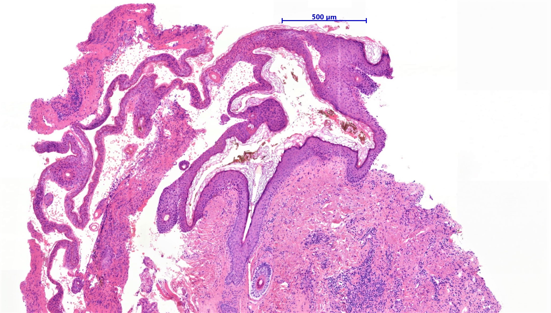
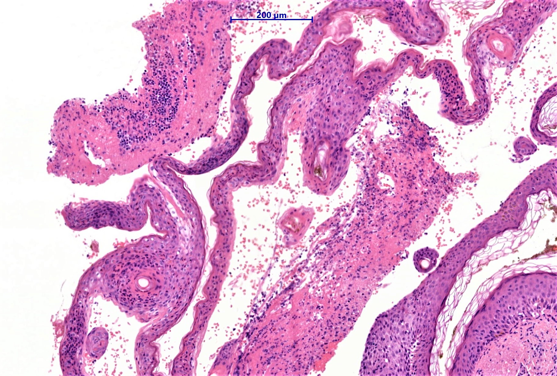
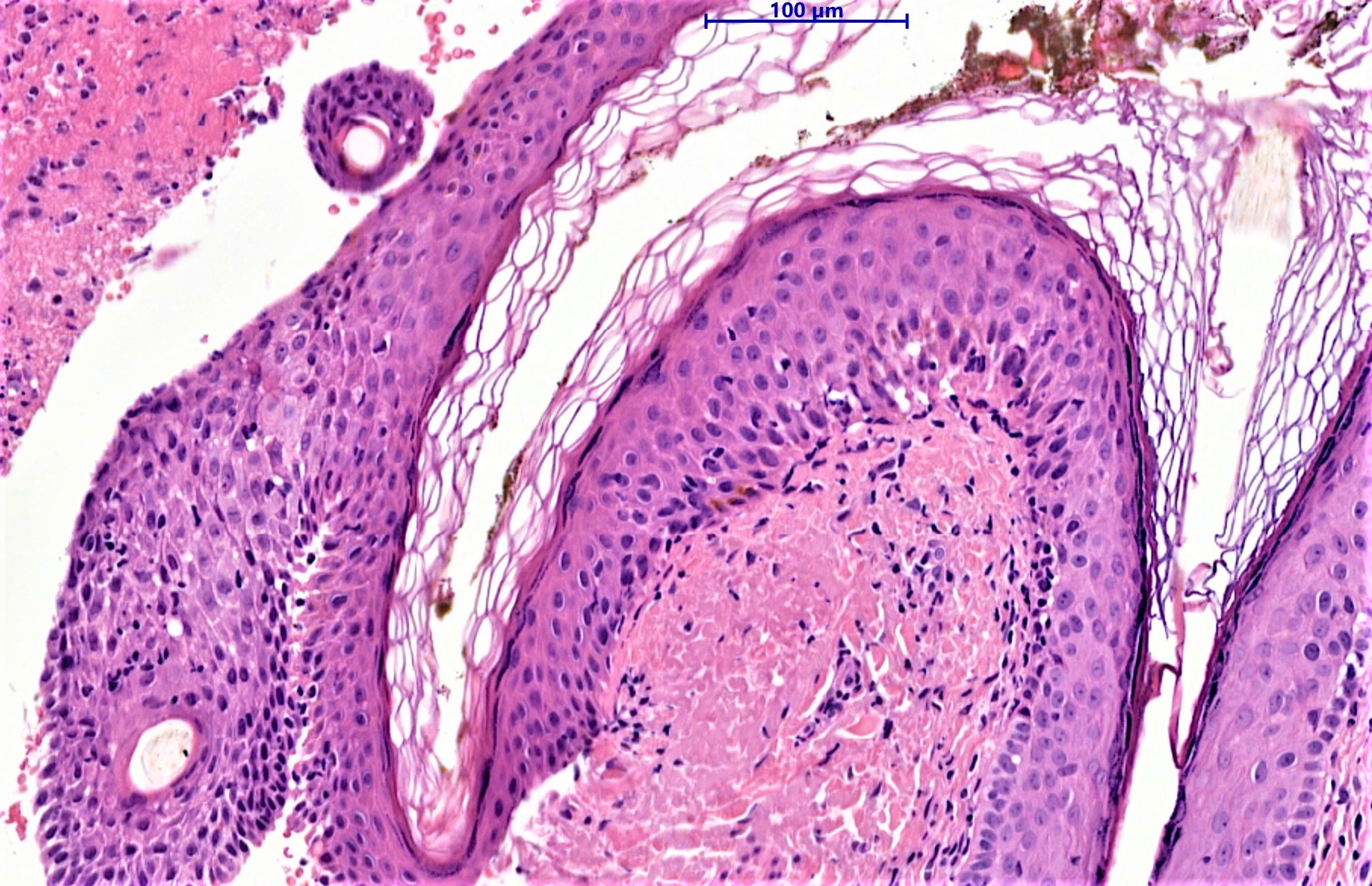
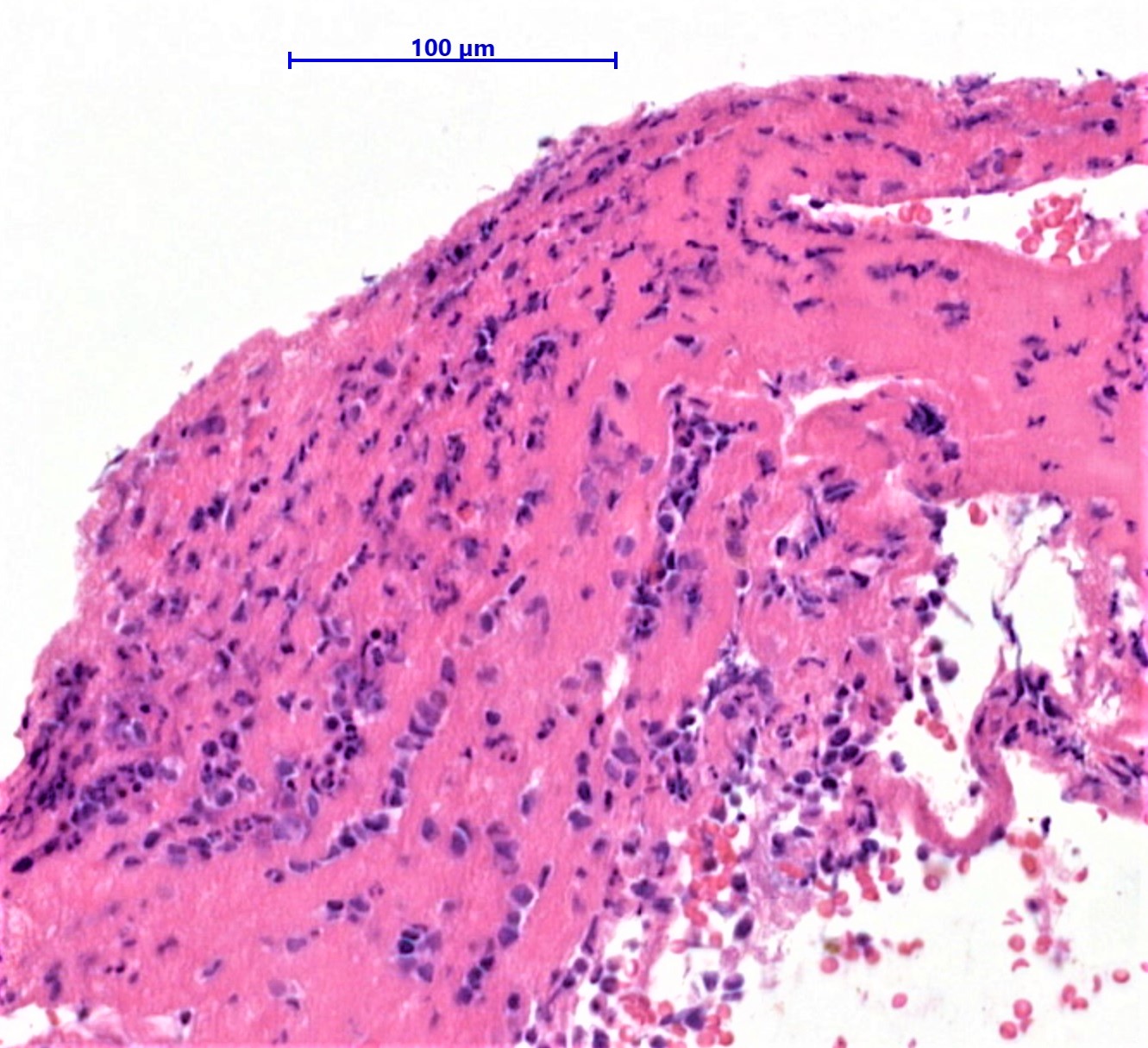
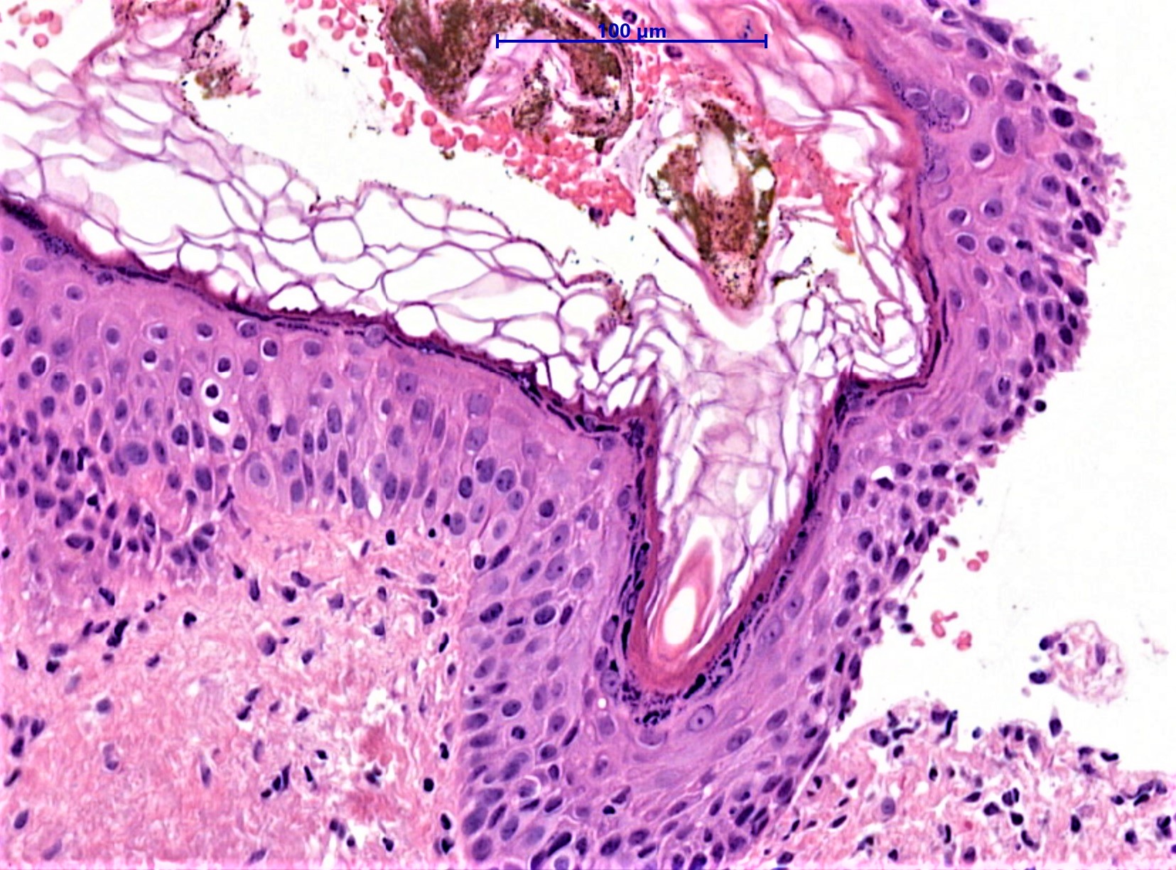
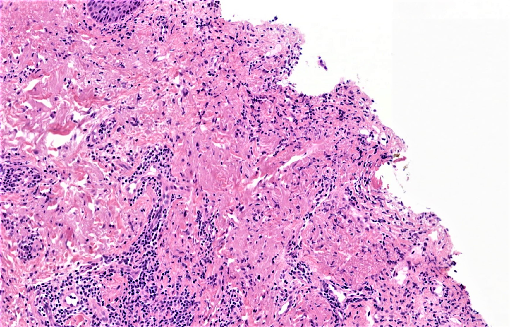
Incisional biopsy left upper lip.

  Report Record

User Feedback


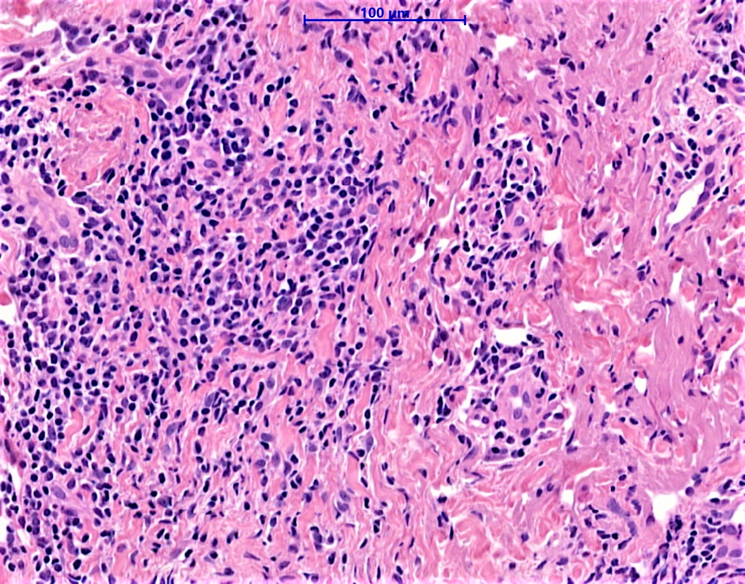
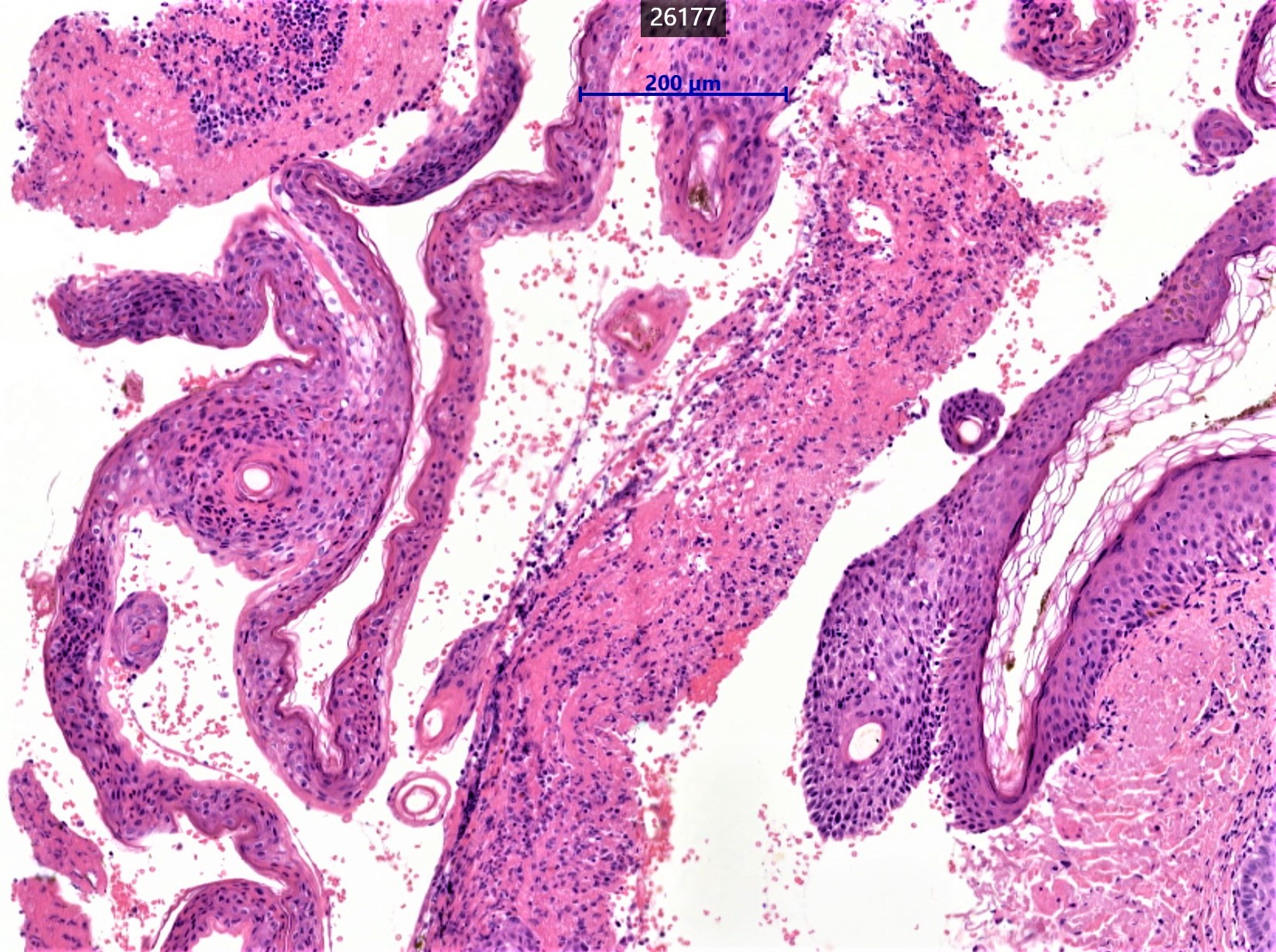
Cheilitis granulomatosa. There is dermal edema with mononuclear cell infiltrate. Obvious epithelioid cell granulomas may not be seen in all biopsies

Share this comment


Link to comment
share_externally

vincenzo polizzi

Posted

I was thinking instead of a mucous membrane pemphigoid/cicatricial pemphigoid...

Share this comment


Link to comment
share_externally

It is upper cutaneous lip. A lot depends on the clinical. Single lesion or rash, appearance, distribution, medical and medication history etc. If rash, it looks like subepidermal clefting with mixed dermal infiltrate including lymphocytes, plasma cells, neutrophils, and occasional eosinophils. Medication reactions such as bullous drug eruption and bullous fixed drug are more likely. Age is slightly younger for bullous pemphigoid, but BP is in the differential. Eos negate bullous SLE. Phototoxic, paraneoplastic, bullous bite reaction are all in the differential, so are other 100 things I can't think of or don't know about!

Share this comment


Link to comment
share_externally

Uma Sundram

Posted

I agree with Vparekh. I feel like there is something else going on here that I don't know about. My differential would be large as well.

Share this comment


Link to comment
share_externally

Iskander H. Chaudhry

Posted

Many thanks for the comments. The Direct immunofluorescence (DIF) of peri-lesional mucosa showed linear deposition of IgG, C3, and IgA along the basement membrane zone in keeping with Mucous membrane pemphigoid (MMP). In light of the IMF result the histology does fit although on its own it is difficult to discern from this biopsy! 

Share this comment


Link to comment
share_externally


Join the conversation

You can post now and register later. If you have an account, sign in now to post with your account.

Guest
Add a comment...

×   Pasted as rich text.   Paste as plain text instead

  Only 75 emoji are allowed.

×   Your link has been automatically embedded.   Display as a link instead

×   Your previous content has been restored.   Clear editor

×   You cannot paste images directly. Upload or insert images from URL.

Loading...

×
×
  • Create New...