Jump to content
In this section we have spot diagnoses posted on a daily basis since June 2010, now over 4000! You can review the archived cases and read the suggested diagnoses by users and the final comment by the contributors.
Case are uploaded each week day by 10 am UK time with the correct diagnosis will generally be posted at 8 pm UK time. Why not view the most recent spot diagnosis and proffer a diagnosis?

Case Number : Case 1946 - 14 Nov Posted By: Uma Sundram

Please read the clinical history and view the images by clicking on them before you proffer your diagnosis.
Submitted Date :
   (0 reviews)

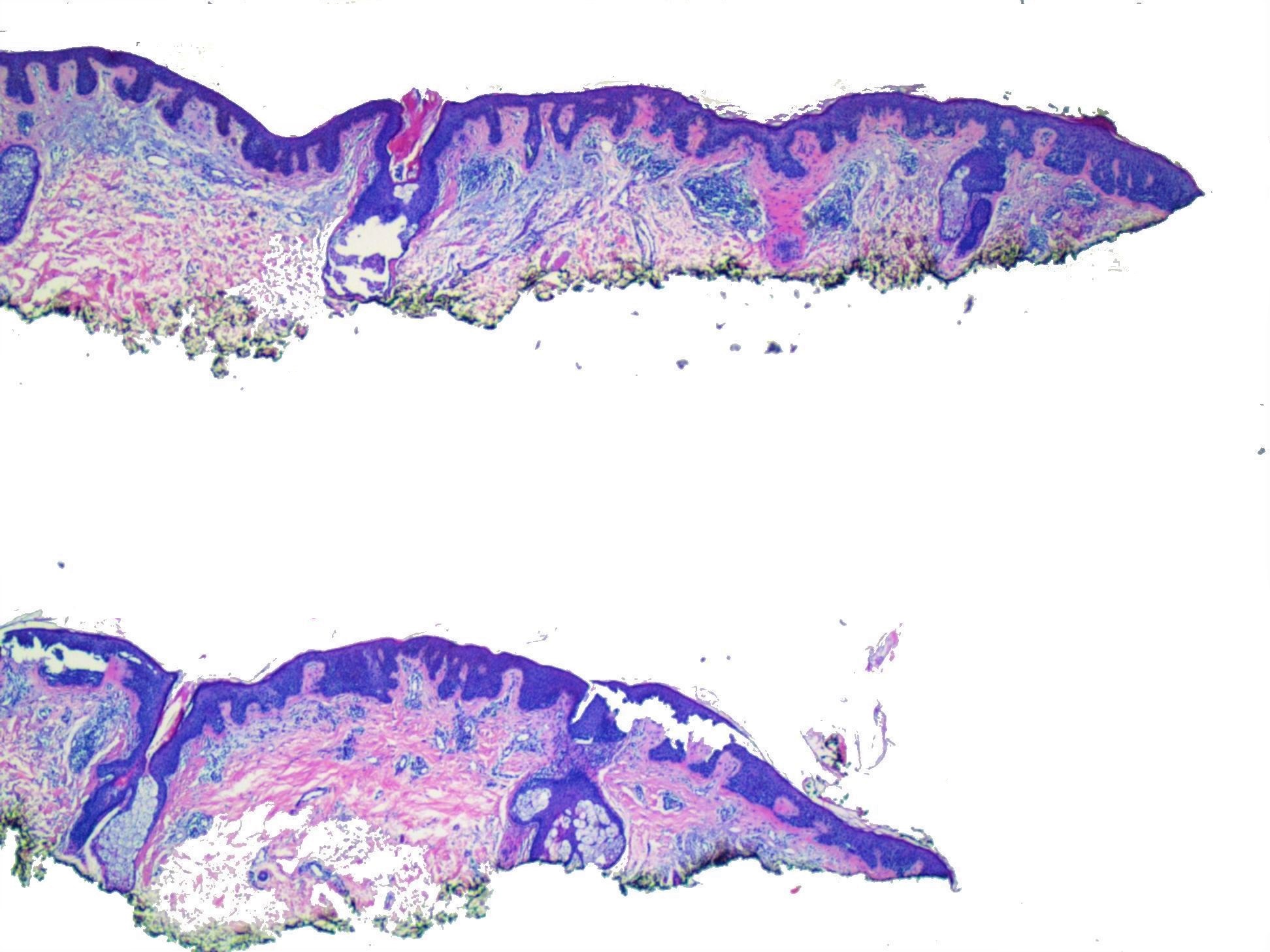
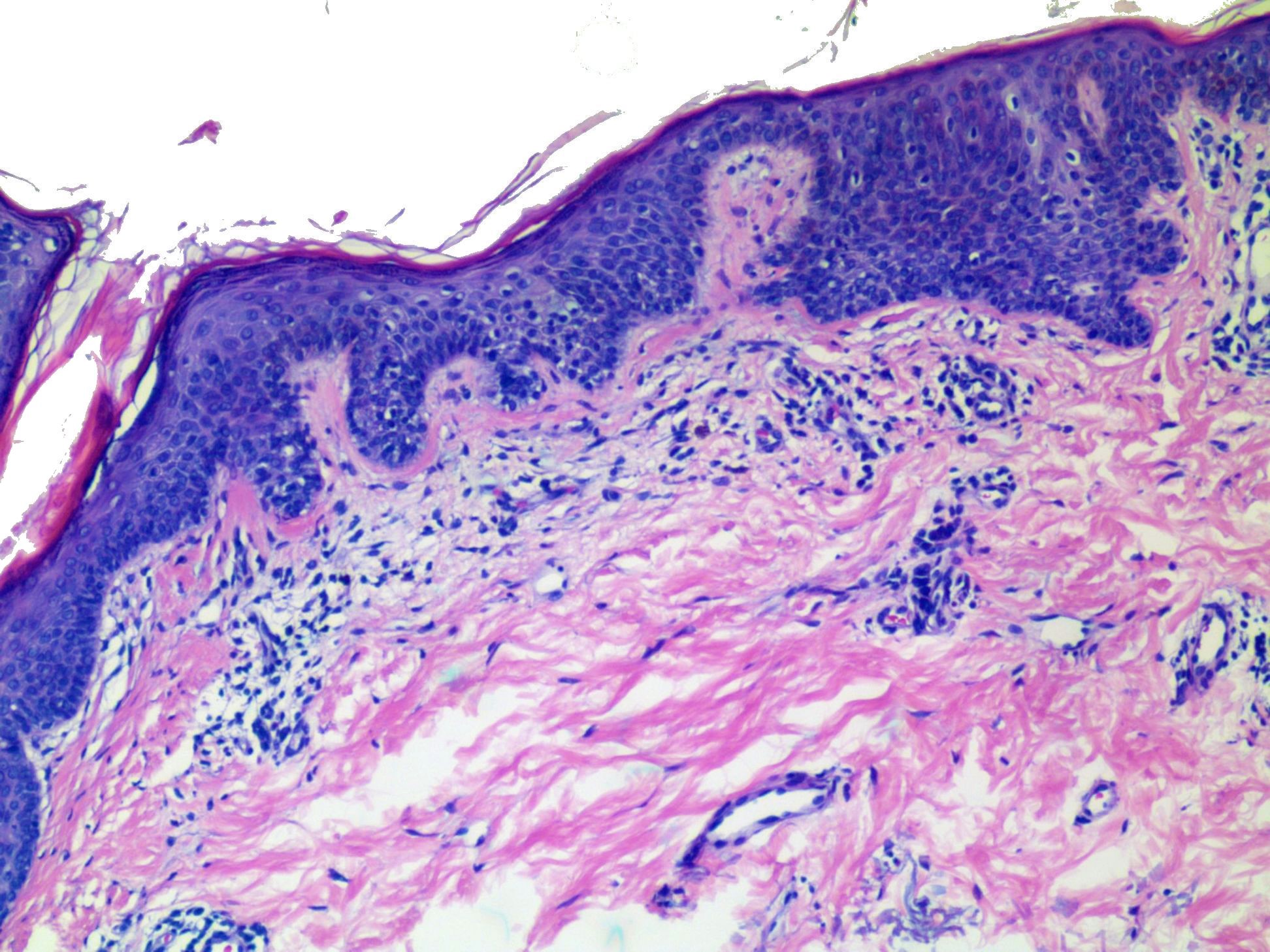
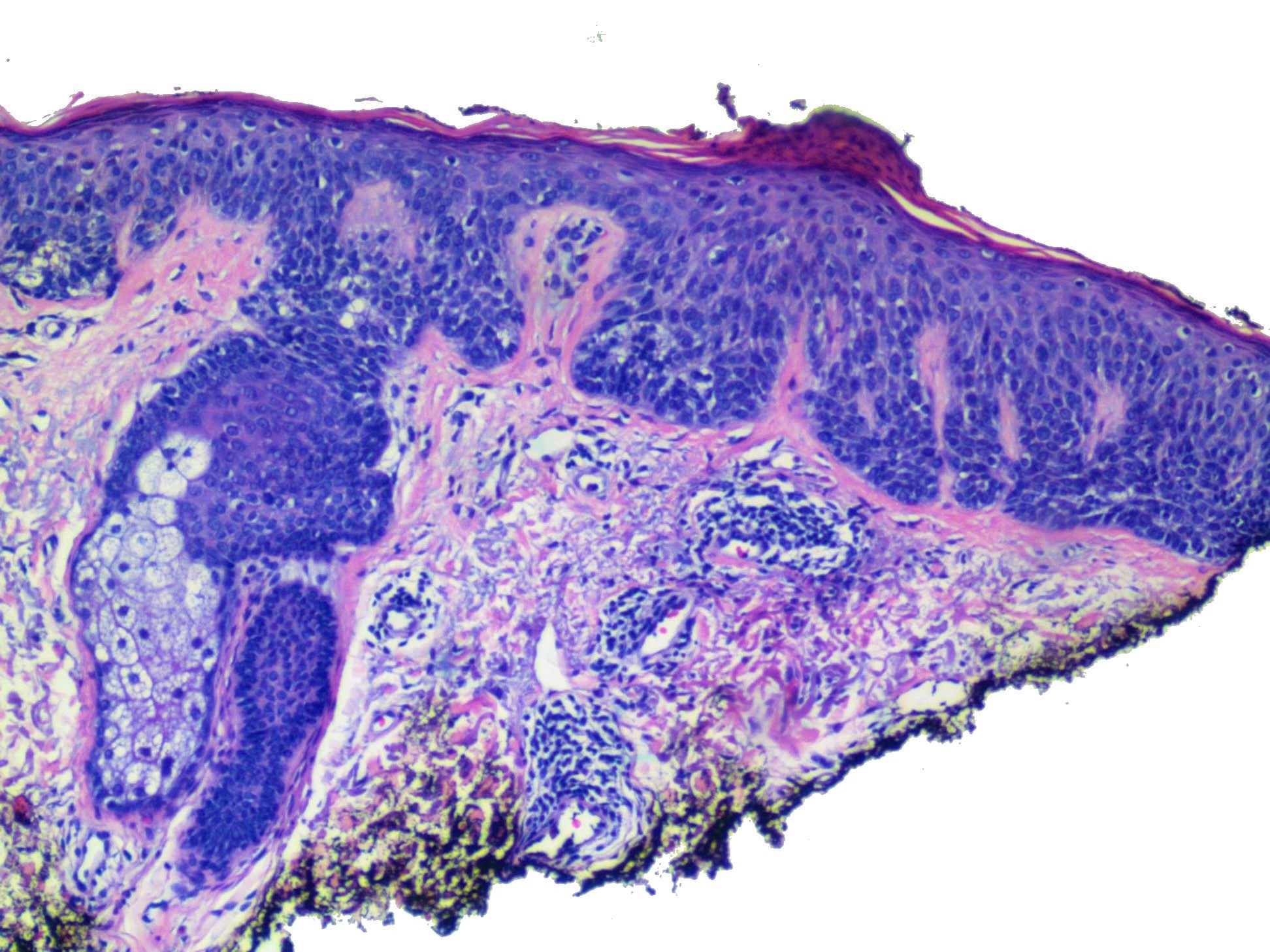
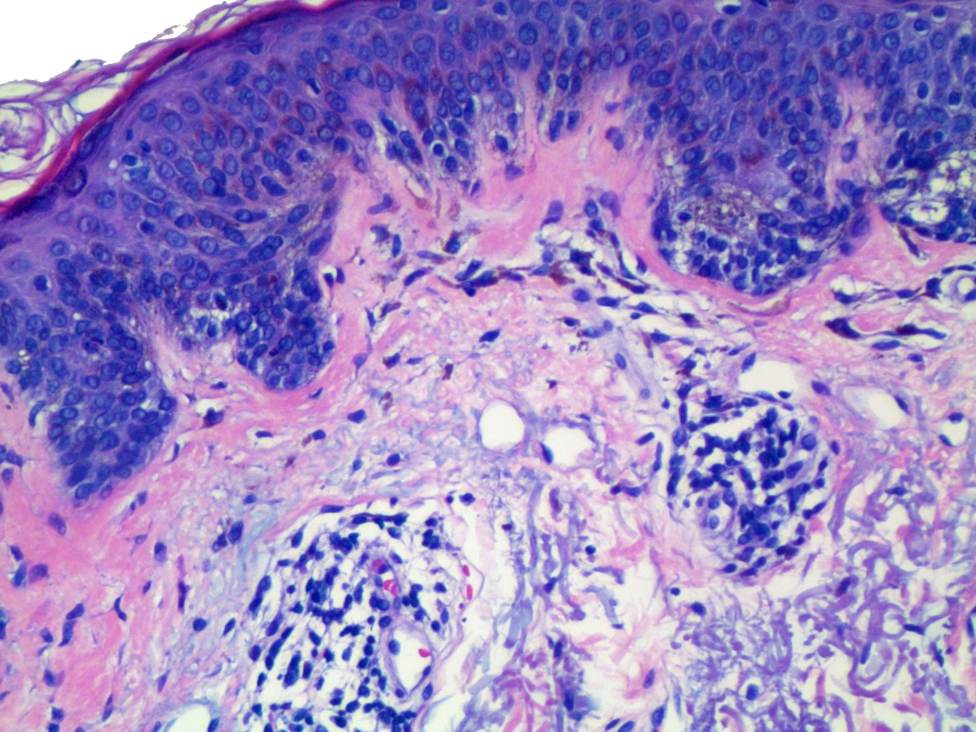
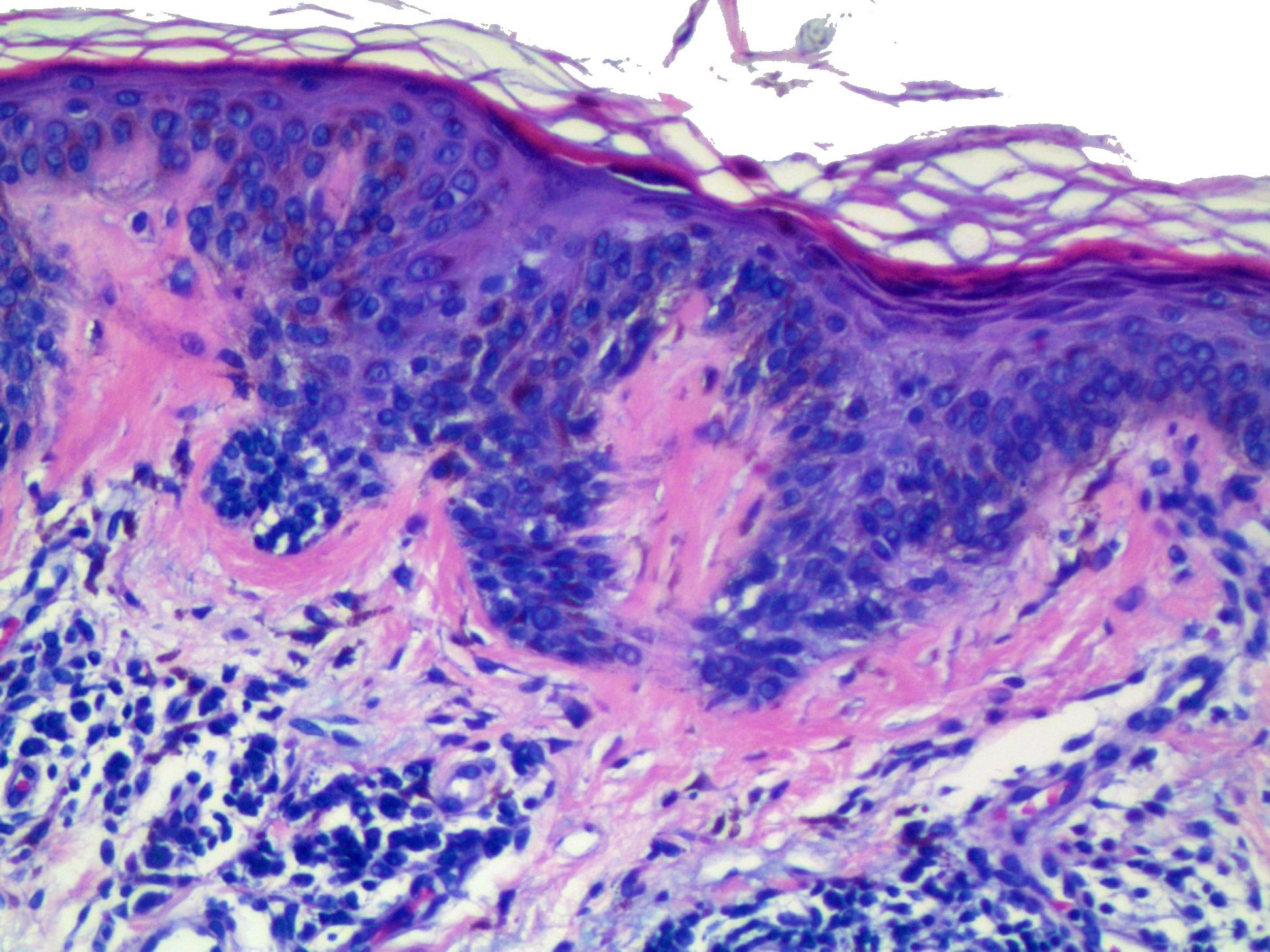
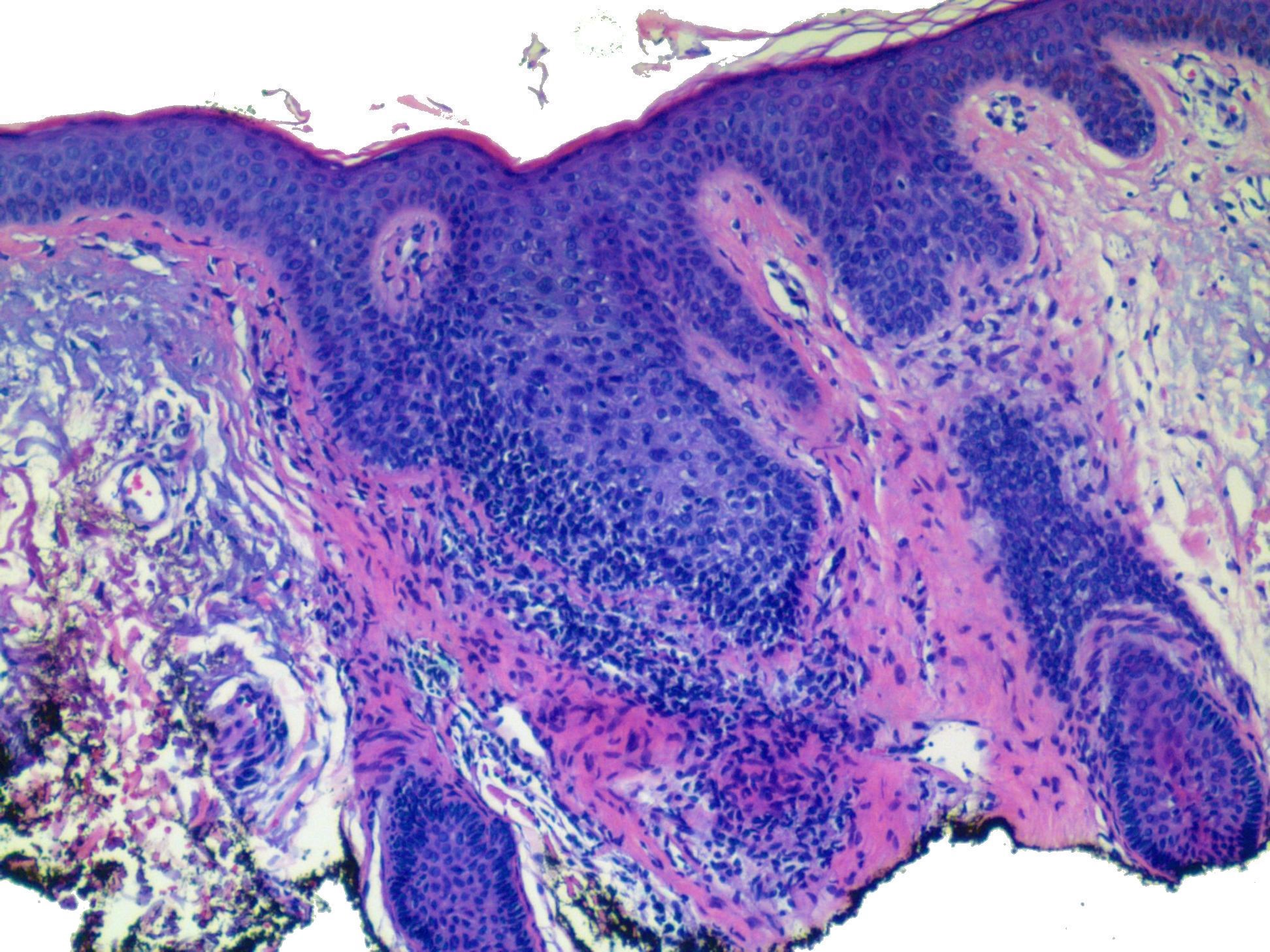
55 year old man with pigmented lesion on the left arm. What are your treatment recommendations?


  Report Record

User Feedback


urmilapandey

Posted (edited)

junctional melanocytic lesion with hyperplstic rete ridges, striking eosinophilic/lamellar fibroplasia, some lentiginous and nests of melanocytes with perhaps mild arhcitectural disarray not much of cytological atypia: ?dysplastic nevus with mild atypia. does extend to margins but given the mild atypia, not sure if a re-excision is warranted/prudent

Edited by urmilapandey

Share this comment


Link to comment
share_externally

Dr. Richard Carr

Posted

I note the IHC has now been posted for Case 1944 10th November - Enjoy!

Share this comment


Link to comment
share_externally

Dr. Richard Carr

Posted

I have started to take quite a draconian line on partial biopsies of thin melanocytic proliferations outwith specific clinicopathological settings. Not least when clinical information is [woefully] inadequate. I would not give a definitive diagnosis but merely confirm the lesion is melanocytic (as opposed to keratinocytic etc), comment on the degree of cytological atypia (looks mild here) and advise the clinical colleague to treat based on their clinical diagnosis including dermoscopy, now that they are aware there is a superficial melanocytic proliferation etc. This new information may at least allow the clinical colleague to make a probably more accurate diagnosis than our [limited] attempts at classification based on a few cells in the junction without clinical or dermatoscopic information. I would of course welcome a friendly chat about how to get the best out of their colleagues in histopathology and of course our limitations in such cases/specimens. Any clinically concerning (and therefore worthy of biopsy) lesion, especially in is sun-damaged skin, would be better excised completely for full examination with a clear margin - when practicable of course. We do of course see larger lesions and those in cosmetically sensitive sites (face, acral) where the biopsy can confirm a reasonably confident diagnosis (acral lentiginous and lentigo maligna) but these are frequently highly challenging and should be based on good clinicopathological correlations preferably (in UK at least) with multi-disciplinary team meeting/discussion. In practice I might have requested a melanA and a thin H&E to assess the proliferation first of course but it seems from the images to be at most only mildly atypical which includes a range of possibilities from benign to malignant [a spectrum of atypical junctional lentiginous melanocytic proliferations up to and including in situ melanoma].

Share this comment


Link to comment
share_externally

urmilapandey

Posted

23 minutes ago, Dr. Richard Carr said:

I have started to take quite a draconian line on partial biopsies of thin melanocytic proliferations outwith specific clinicopathological settings. Not least when clinical information is [woefully] inadequate. I would not give a definitive diagnosis but merely confirm the lesion is melanocytic (as opposed to keratinocytic etc), comment on the degree of cytological atypia (looks mild here) and advise the clinical colleague to treat based on their clinical diagnosis including dermoscopy, now that they are aware there is a superficial melanocytic proliferation etc. This new information may at least allow the clinical colleague to make a probably more accurate diagnosis than our [limited] attempts at classification based on a few cells in the junction without clinical or dermatoscopic information. I would of course welcome a friendly chat about how to get the best out of their colleagues in histopathology and of course our limitations in such cases/specimens. Any clinically concerning (and therefore worthy of biopsy) lesion, especially in is sun-damaged skin, would be better excised completely for full examination with a clear margin - when practicable of course. We do of course see larger lesions and those in cosmetically sensitive sites (face, acral) where the biopsy can confirm a reasonably confident diagnosis (acral lentiginous and lentigo maligna) but these are frequently highly challenging and should be based on good clinicopathological correlations preferably (in UK at least) with multi-disciplinary team meeting/discussion. In practice I might have requested a melanA and a thin H&E to assess the proliferation first of course but it seems from the images to be at most only mildly atypical which includes a range of possibilities from benign to malignant [a spectrum of atypical junctional lentiginous melanocytic proliferations up to and including in situ melanoma].

Thanks for sharing this Richard. As a generalist, I struggle with superficial biopsies of melanocytic lesions and often wonder if I am being too cautious or at times, too confident in giving an opinion. It is very assuring to see this approach from an expert and will follow suit.

Share this comment


Link to comment
share_externally

Iskander H. Chaudhry

Posted

Mildly dsyplastic lentiginous naevus 

Share this comment


Link to comment
share_externally

Uma Sundram

Posted

Great discussion and precisely the reason I post these cases and ask for people's thoughts. These cases highlight the significant differences that can be seen from one interpretation to another (justifiably), and the point raised by urmila about struggles over too conservative versus not conservative enough in terms of approaches to management. The struggles are identical within the US dermatopathology practitioner population. Significant variability exists between states and I am always curious about international approaches. So, this is not a right or wrong answer approach, but a chance to discuss best practices (as Richard has done so nicely).  The lesion, it turns out, was over 1 cm in size and clinically concerning for melanoma in situ; we recommended a re excision with 0.5 cm margins. Melan A highlighted a lentiginous population that traversed the entire biopsy and we were unable to assess for circumscription. Recognizing that evolving MMIS can have mild atypia in the middle of the lesion was key to the appropriate approach here.

Share this comment


Link to comment
share_externally

Dr. Richard Carr

Posted

Thanks Uma - a nice challenge of an every day problem we all face.  Seems we also agree pretty much on every point! Let's keep up the special relationship!

Share this comment


Link to comment
share_externally


Join the conversation

You can post now and register later. If you have an account, sign in now to post with your account.

Guest
Add a comment...

×   Pasted as rich text.   Paste as plain text instead

  Only 75 emoji are allowed.

×   Your link has been automatically embedded.   Display as a link instead

×   Your previous content has been restored.   Clear editor

×   You cannot paste images directly. Upload or insert images from URL.

Loading...

×
×
  • Create New...