Jump to content
In this section we have spot diagnoses posted on a daily basis since June 2010, now over 4000! You can review the archived cases and read the suggested diagnoses by users and the final comment by the contributors.
Case are uploaded each week day by 10 am UK time with the correct diagnosis will generally be posted at 8 pm UK time. Why not view the most recent spot diagnosis and proffer a diagnosis?

Case Number : Case 1947 - 15 Nov Posted By: Arti Bakshi

Please read the clinical history and view the images by clicking on them before you proffer your diagnosis.
Submitted Date :
   (0 reviews)

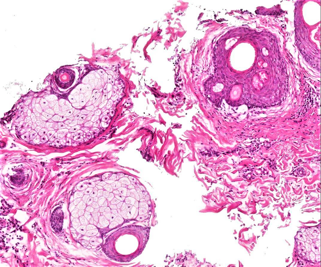
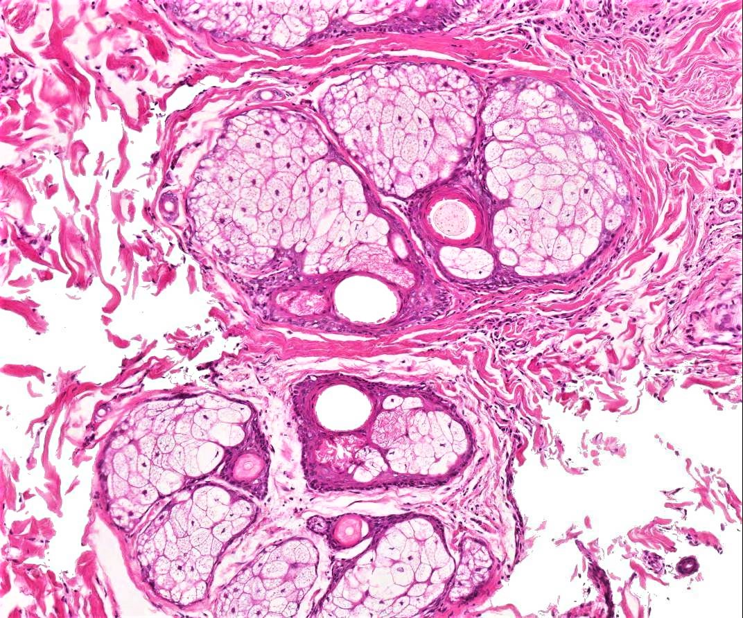
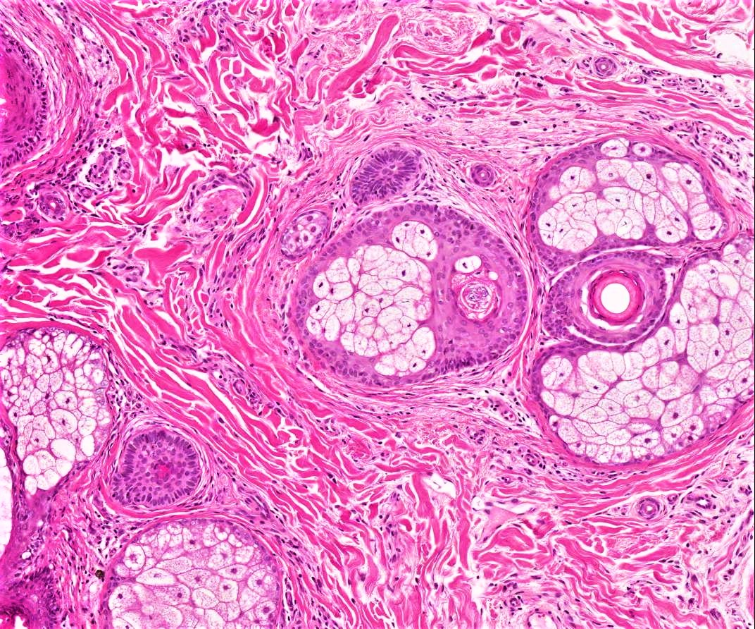
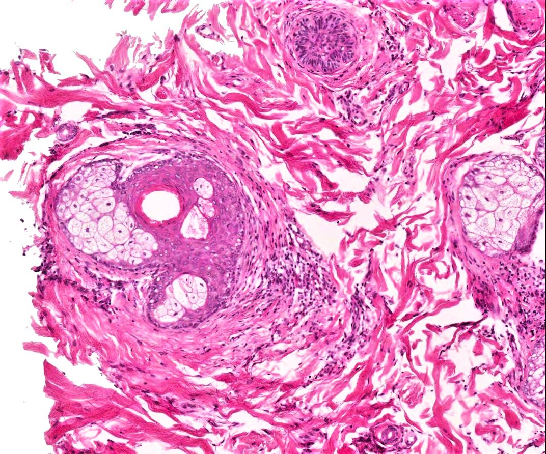
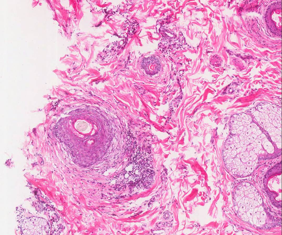
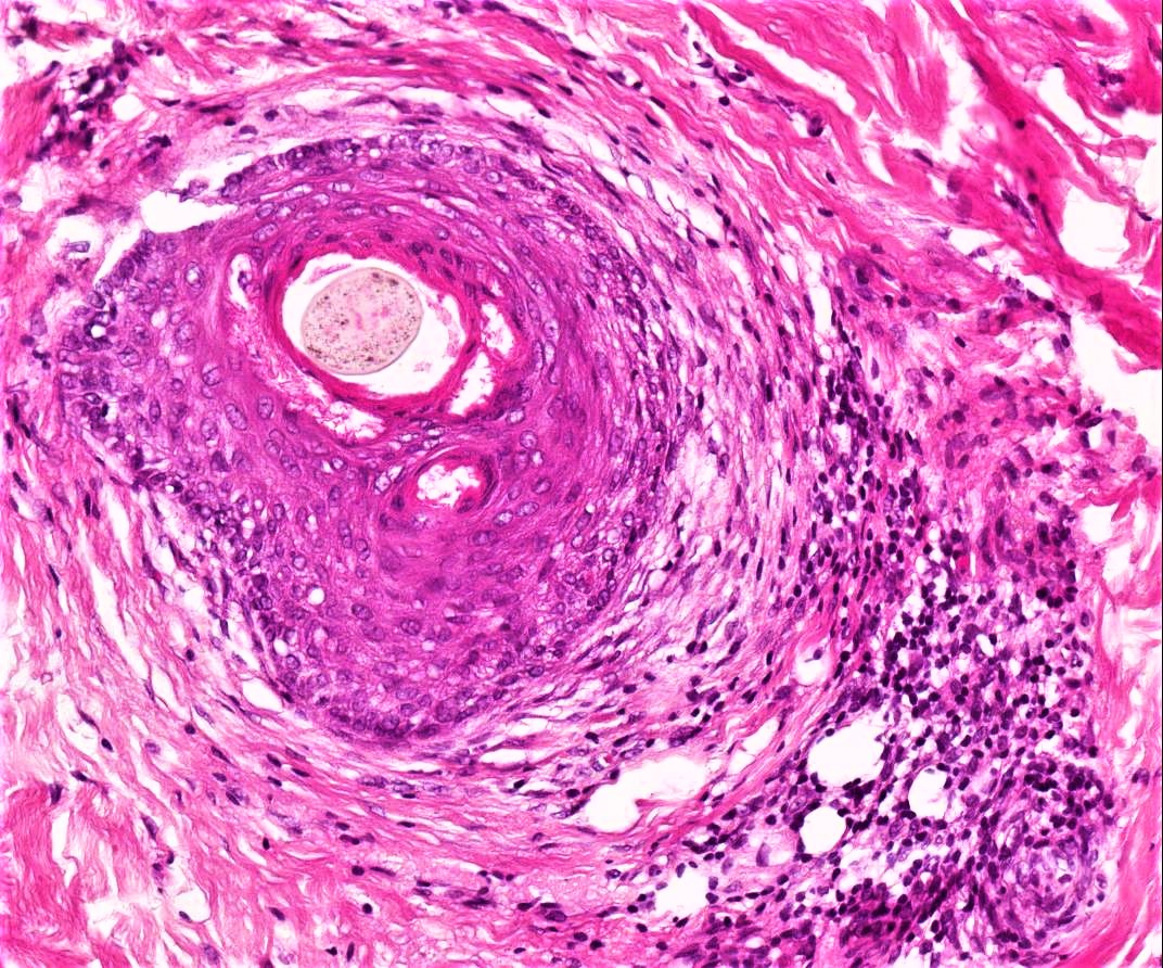
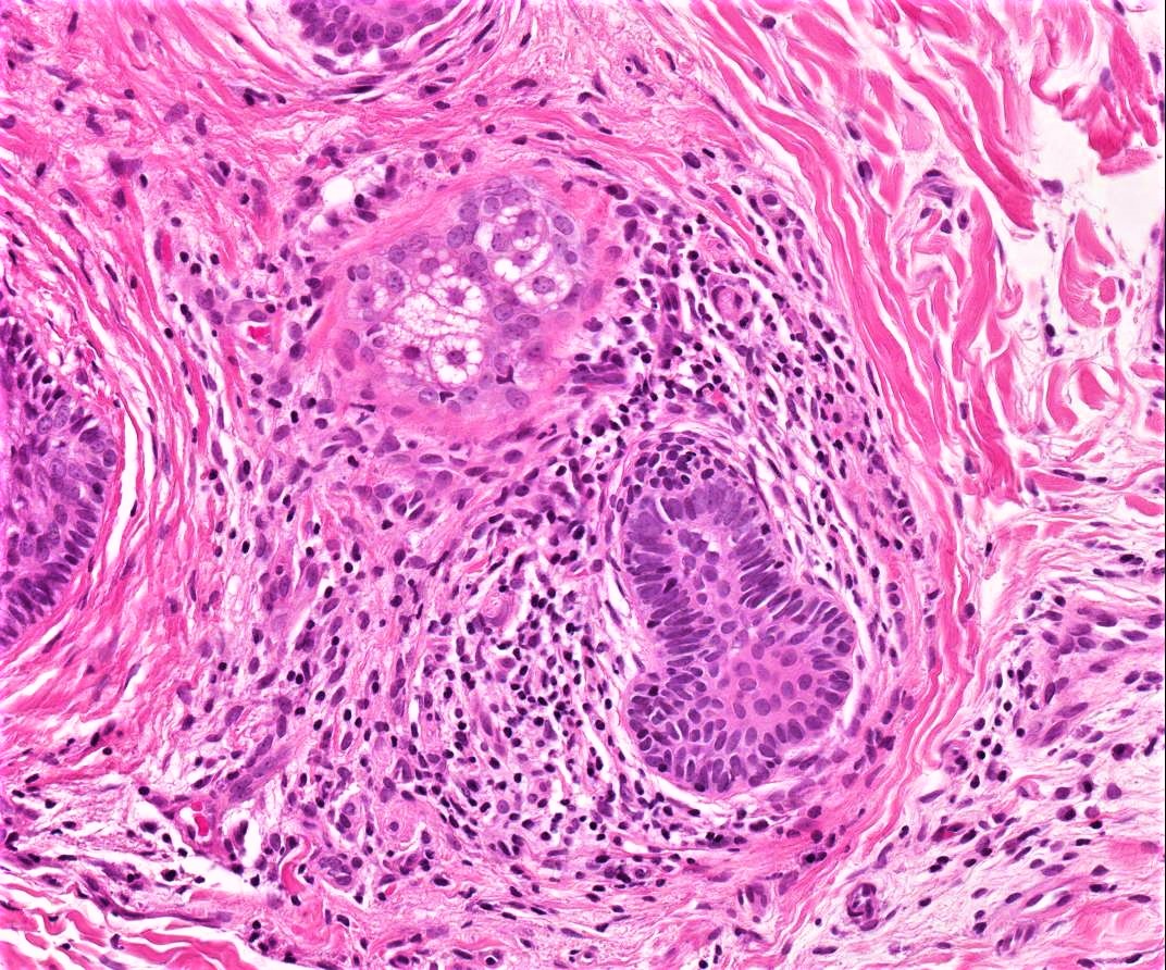
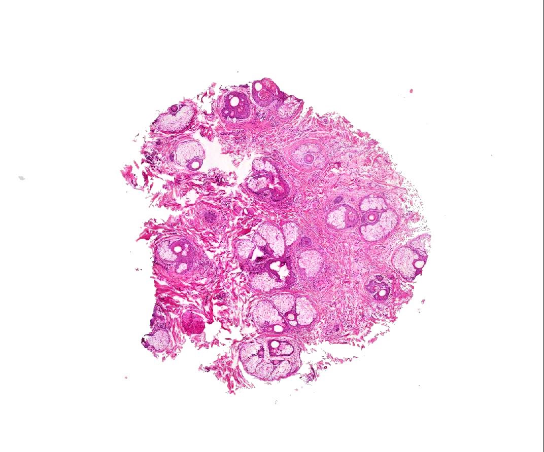
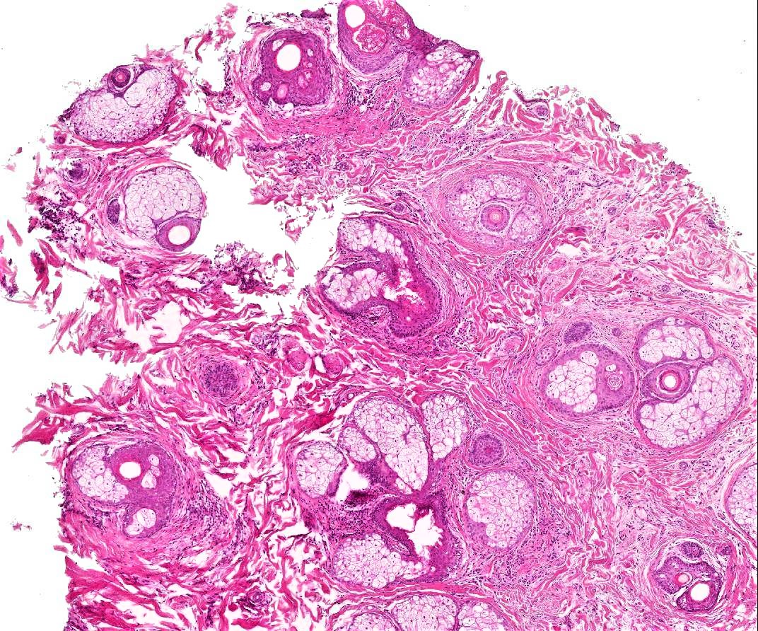
29/F hair loss to top of scalp, scaling and itchiness. ? lichen planopilaris ?female pattern hair loss ?diffuse alopecia areata

  • Like 1
  Report Record

User Feedback


Its a lymphocytic scarring alopecia. In picture 8, eccentric thinning of outer root sheath epithelium and concentric perifollicular lamellar fibroplasia like onion skin are evident.Since scaling and itching are not the features associated with alopecia areata and female pattern baldness, clinico-pathological correlation suggests central centrifugal cicatricial alopecia.

Share this comment


Link to comment
share_externally

vincenzo polizzi

Posted (edited)

I have no diagnosis but only questions: are there any miniaturized follicles(typical of Androgenetic Alopecia).  Is perifollicular fibrosis a streamer or a cicatricial one? And why so many sebaceous glands in a scarring alopecia? 


Edited by vincenzo polizzi

Share this comment


Link to comment
share_externally

Sasi Attili

Posted

Miniaturised hair follicles + perifollicular lymphocytic infiltrate- combination of diffuse a. areata and And. Alopecia

Share this comment


Link to comment
share_externally

A vellus hair has a diameter which is equal to or smaller than the thickness of the inner root sheath. They will be visible only when the section passes through upper dermis as their roots are above the roots of normal hair..Sebaceous glands will be visible in the early stages of scarring alopecia

Share this comment


Link to comment
share_externally

Dr. Mona Abdel-Halim

Posted

I think this is diffuse AA. There is increased miniaturized hair follilces and telogen and lymphocytic infiltrate. Also I think there is compensatory hypertrophy of sebaceous lobules.

Share this comment


Link to comment
share_externally

Need a deeper level section to see hair bulbs in the subcutaneous adipose tissue to see whether there are lymphocytes around hair bulbs which would confirm AA. From the histologic pictures with concentric perifollicular fibrosis and perifollicular lymphocytic infiltrate, would favor lichen planopilaris or central centrifugal cicatricial alopecia.

Share this comment


Link to comment
share_externally

Dr. Richard Carr

Posted

Agree a confusing case. I think we do however have fairly good features of alopecia areata for the reasons outlined by colleagues (striking miniaturisation, some telogen & focal peri-bulbar lymphocytic infiltrate). I'm not so confident about the scarring side but would consider a dual pathology for reasons cited by colleagues.

Share this comment


Link to comment
share_externally

Arti Bakshi

Posted

Yes, this is a difficult case and clearly one that needed close CPC. The history given is what I had on the request card with the additional information that the hair loss was ongoing for 5 years. The perifollicular inflammation is confusing and makes one wonder about a LPP, but as someone rightly pointed the sebaceous glands are well preserved which would be odd for a scarring alopecia, particularly given that the hair loss was long standing. The main feature is prominent miniaturisation of hair follicles (terminal: vellus ration was 1:1) and some excess of telogen (a formal count revealed 20%). My main possibility on this histology was that of an androgenic alopecia (with perhaps an element of telogen effluvium). A diffuse alopecia areata was in my differential but I was not convinced of definite peribulbar inflammation nor were there any inflammatory cells in the stella. The last image was the only focus in the bx (examined at multiple levels), where one gets an impression of peribulbar infalmmation, but I suspect this is just a tangentially cut vellus follicle and the inflammation is not strictly peribulbar.

The case was discussed in our inflammatory MDT and a senior dermatologist reviewed the patient. He felt the pattern of hair loss was typical of androgenic alopecia. The 'scaling and itchiness' mentioned in the request card were not a significant finding to him. There was no inflammation or scarring clinically.

The main point, which I wanted to highlight in this case, is that some degree of peri-follicular inflammation (particularly at the infundibular level) can be seen as a non specific finding in androgenic alopecias. I see it fairly commonly and it is easy to fall into the trap of calling these cases early scarring alopecias. Beware of this finding and always discuss these cases with clinical colleagues, preferably with an interest in alopecias.. Remember patients may have been seen by clinicians less experienced in this area, who may provide misleading information!

Thanks for all your comments.

Share this comment


Link to comment
share_externally

vincenzo polizzi

Posted

15 hours ago, arti bakshi said:

Yes, this is a difficult case and clearly one that needed close CPC. The history given is what I had on the request card with the additional information that the hair loss was ongoing for 5 years. The perifollicular inflammation is confusing and makes one wonder about a LPP, but as someone rightly pointed the sebaceous glands are well preserved which would be odd for a scarring alopecia, particularly given that the hair loss was long standing. The main feature is prominent miniaturisation of hair follicles (terminal: vellus ration was 1:1) and some excess of telogen (a formal count revealed 20%). My main possibility on this histology was that of an androgenic alopecia (with perhaps an element of telogen effluvium). A diffuse alopecia areata was in my differential but I was not convinced of definite peribulbar inflammation nor were there any inflammatory cells in the stella. The last image was the only focus in the bx (examined at multiple levels), where one gets an impression of peribulbar infalmmation, but I suspect this is just a tangentially cut vellus follicle and the inflammation is not strictly peribulbar.

The case was discussed in our inflammatory MDT and a senior dermatologist reviewed the patient. He felt the pattern of hair loss was typical of androgenic alopecia. The 'scaling and itchiness' mentioned in the request card were not a significant finding to him. There was no inflammation or scarring clinically.

The main point, which I wanted to highlight in this case, is that some degree of peri-follicular inflammation (particularly at the infundibular level) can be seen as a non specific finding in androgenic alopecias. I see it fairly commonly and it is easy to fall into the trap of calling these cases early scarring alopecias. Beware of this finding and always discuss these cases with clinical colleagues, preferably with an interest in alopecias.. Remember patients may have been seen by clinicians less experienced in this area, who may provide misleading information!

Thanks for all your comments.

Thanks Arthi. I love this website because it’s a constant incentive to update. 

Share this comment


Link to comment
share_externally


Join the conversation

You can post now and register later. If you have an account, sign in now to post with your account.

Guest
Add a comment...

×   Pasted as rich text.   Paste as plain text instead

  Only 75 emoji are allowed.

×   Your link has been automatically embedded.   Display as a link instead

×   Your previous content has been restored.   Clear editor

×   You cannot paste images directly. Upload or insert images from URL.

Loading...

×
×
  • Create New...