Jump to content
In this section we have spot diagnoses posted on a daily basis since June 2010, now over 4000! You can review the archived cases and read the suggested diagnoses by users and the final comment by the contributors.
Case are uploaded each week day by 10 am UK time with the correct diagnosis will generally be posted at 8 pm UK time. Why not view the most recent spot diagnosis and proffer a diagnosis?

Case Number : Case 1948 - 16 Nov 2017 Posted By: Iskander H. Chaudhry

Please read the clinical history and view the images by clicking on them before you proffer your diagnosis.
Submitted Date :
   (0 reviews)

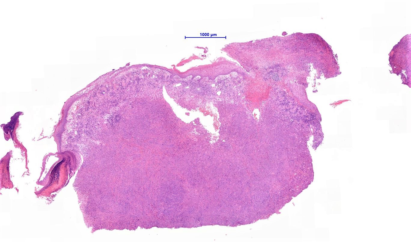
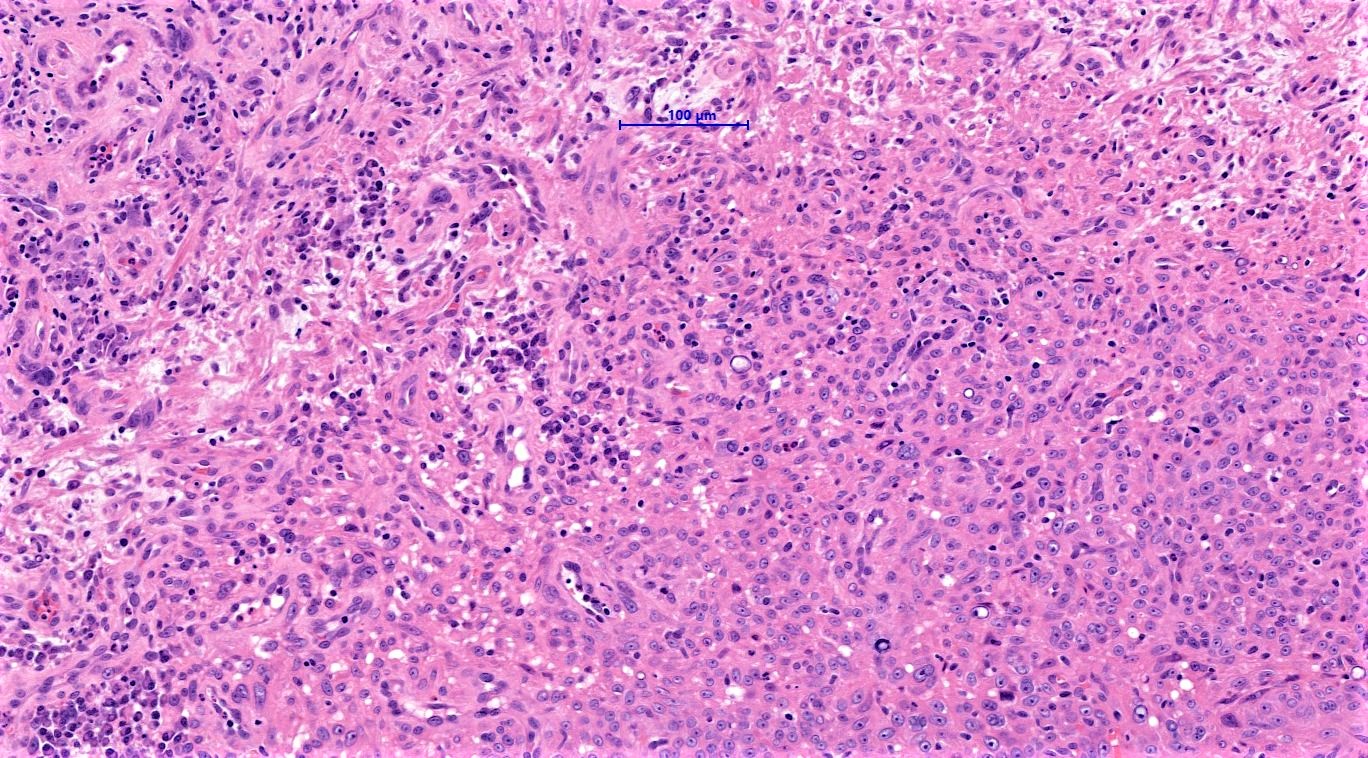
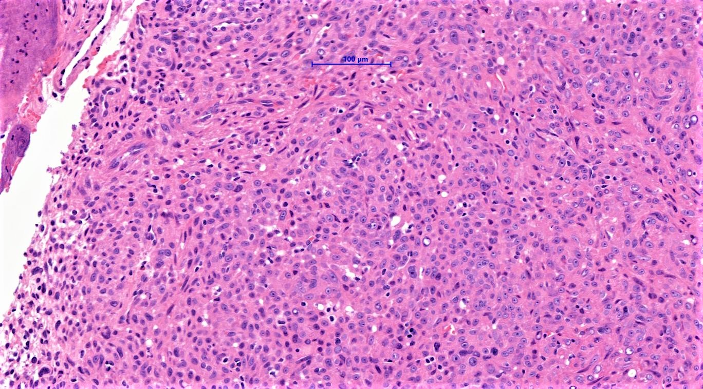
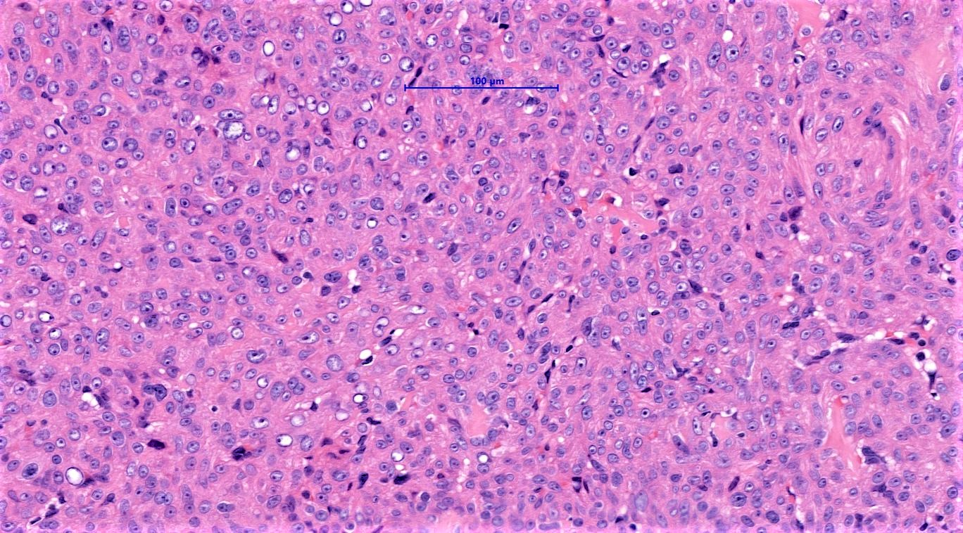
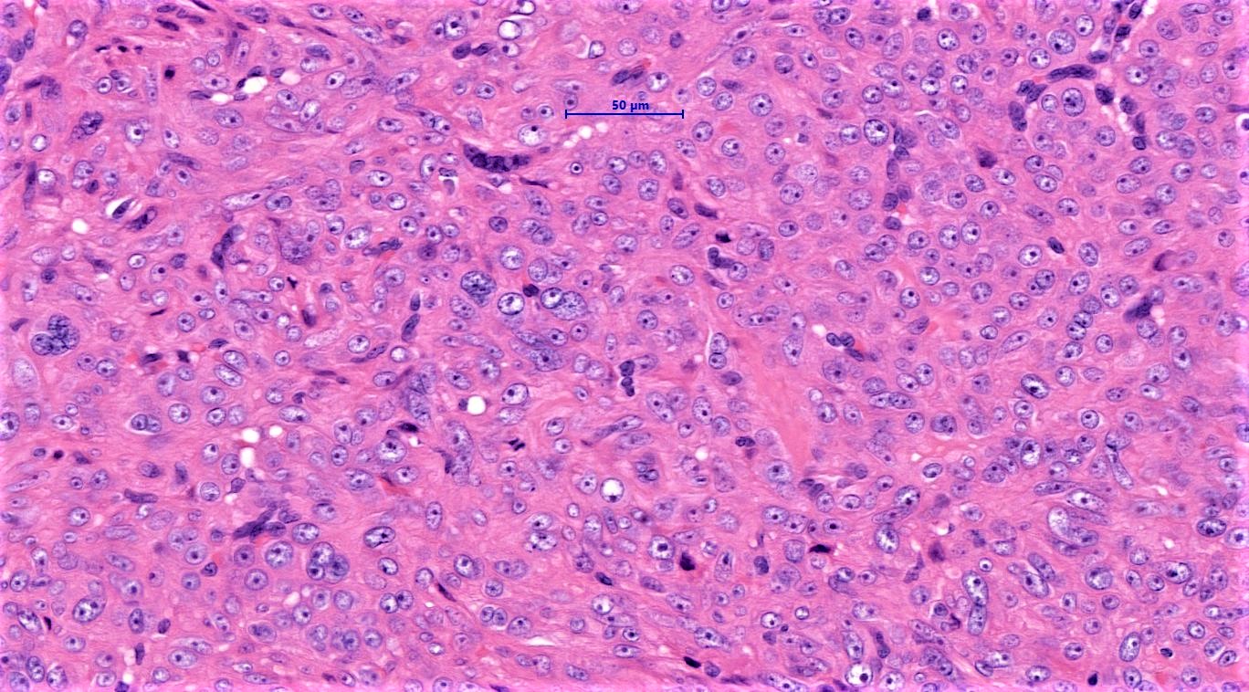
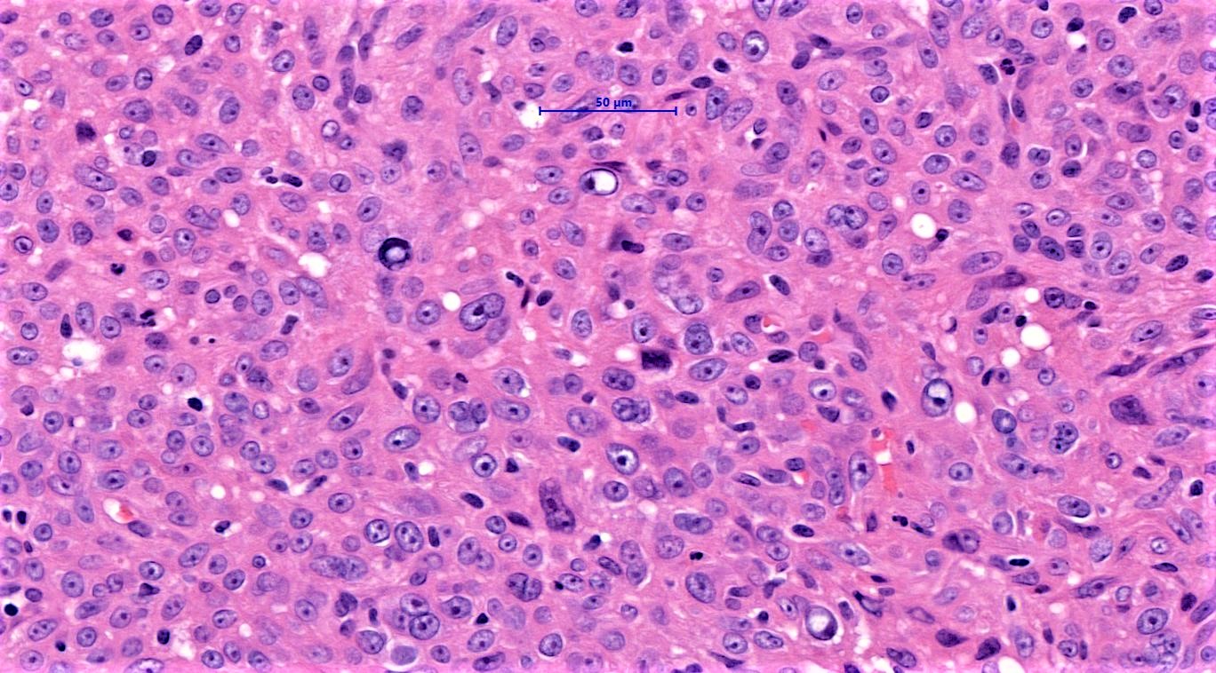
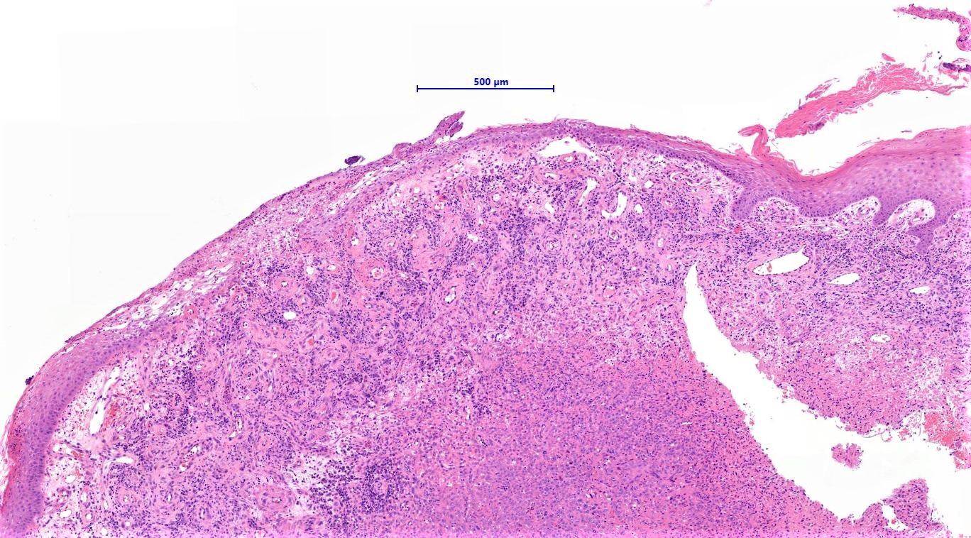
65 year old, female: C+C right third toe.
?P.G. ?Malignancy. 12 year history.
Edited by Admin_Dermpath


  Report Record

User Feedback


Dr. Mona Abdel-Halim

Posted

Thinking epithelioid hemangioendothelioma?

Share this comment


Link to comment
share_externally

vincenzo polizzi

Posted

Atypical glomus tumor is my first thought. Second one is epithelioid perineurioma.

Share this comment


Link to comment
share_externally

Dr. Richard Carr

Posted

Tricky. I wondered about epithelioid cell histiocytoma, PECOMA, epithelioid angiomatous nodule or epithelioid H.E. Like suggestion of glomus - I was wondering about a perivascular type tumour and it also crossed my mind. Needs IHC!

Share this comment


Link to comment
share_externally

Don't really appreciate cytoplasmic lumen or red blood cells commonly in vascular lesions. Epithelioid histiocytoma?

Share this comment


Link to comment
share_externally

Admin_Dermpath

Posted

Dr Chaudhry's response: 


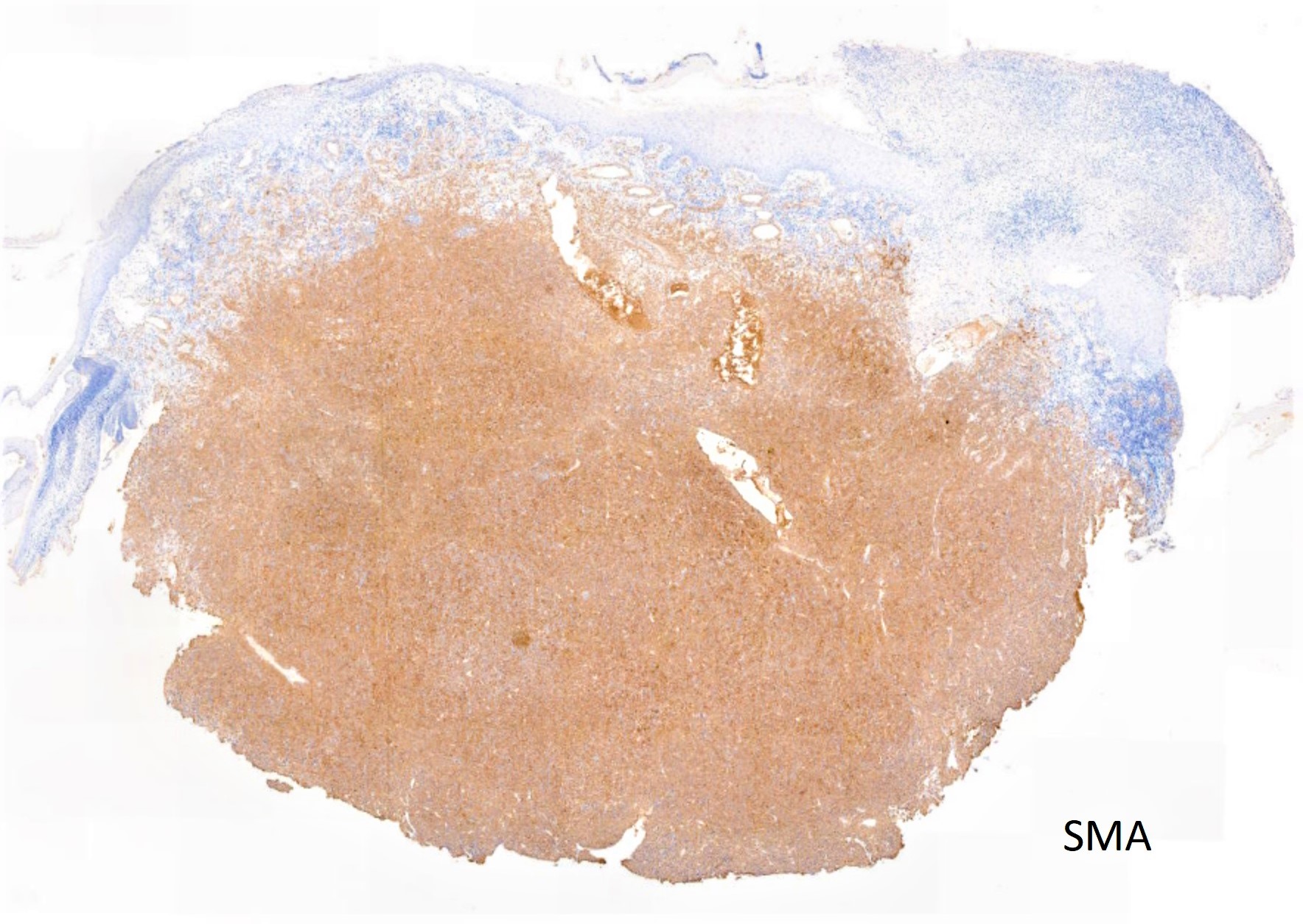
The tumour is negative for S100, p63, EMA, Desmin and AE1AE3. Before I disclose the final immuno results what is your preferred diagnosis? 

Share this comment


Link to comment
share_externally

Raul Perret

Posted

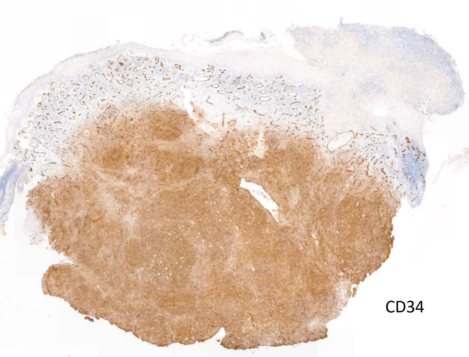
I was wondering about ALK-1 in this case too. Before answering I would have liked to see H-caldesmon, CD34, ERG, and ki67. For me this is not the expected morphology of Glomus tumor. Without any immunos my first hypothesis would have been epithelioid fibrous hystiocytoma.

Share this comment


Link to comment
share_externally

Arti Bakshi

Posted

Epithelioid fibrous histiocytoma vs epithelioid angiomatous nodule, favouring latter.

Share this comment


Link to comment
share_externally

Just wonder how much controversy such tumours must have created in the pre- immunostaining days! 

Share this comment


Link to comment
share_externally

vincenzo polizzi

Posted

In the preimmunoAge many entities as CEAN ECH PECOMA didn’t exist...:-))

Share this comment


Link to comment
share_externally

Even usual conditions created controversy. Remember"The Final Diagnosis" by Arthur Hailey?

Share this comment


Link to comment
share_externally

vincenzo polizzi

Posted

Yes off course...pathology is the Controversy Science...and the disputes have not been settled by new Ancillary technologies as 

Immuno and Molecular but, if possible, incremented.

Share this comment


Link to comment
share_externally

Nitin Khirwadkar

Posted

Epithelioid fibrous histiocytoma vs epithelioid CEAN vs glomus. ALK, SMA, ERG.

Share this comment


Link to comment
share_externally

Admin_Dermpath

Posted

Dr Chaudhry's comments:

Please see the additional immuno!  The tumour is negative for CD10, CD31, Scattered CD68 + cells and low proliferation index for Ki67. 

So the final diagnosis is ........

Share this comment


Link to comment
share_externally

Very difficult to make sense of the immunohistochemical staining pattern. Most glomus tumors are negative for CD34. But rarely glomus tumors can be positive for CD34 (CD34-positive glomus tumor). That's my best interpretation of this case. But I have never seen one before.

CD34-positive glomus tumor: clinicopathologic and immunohistochemical analysis of six cases with myxoid stromal changes.

Mentzel T, Hügel H, Kutzner H.

J Cutan Pathol. 2002 Aug;29(7):421-5


Share this comment


Link to comment
share_externally

vincenzo polizzi

Posted

Expression of CD34 in glomus tumors.

Hatori M, et al. Tohoku J Exp Med. 1997.

Share this comment


Link to comment
share_externally

Dr. Mona Abdel-Halim

Posted

CD34 positive Glomus sounds good idea!


Share this comment


Link to comment
share_externally

Admin_Dermpath

Posted

Dr Chaudhry : Final diagnosis

CD34-positive glomus tumour

Many thanks for the excellent discussion points 


Share this comment


Link to comment
share_externally


Join the conversation

You can post now and register later. If you have an account, sign in now to post with your account.

Guest
Add a comment...

×   Pasted as rich text.   Paste as plain text instead

  Only 75 emoji are allowed.

×   Your link has been automatically embedded.   Display as a link instead

×   Your previous content has been restored.   Clear editor

×   You cannot paste images directly. Upload or insert images from URL.

Loading...

×
×
  • Create New...