Jump to content
In this section we have spot diagnoses posted on a daily basis since June 2010, now over 4000! You can review the archived cases and read the suggested diagnoses by users and the final comment by the contributors.
Case are uploaded each week day by 10 am UK time with the correct diagnosis will generally be posted at 8 pm UK time. Why not view the most recent spot diagnosis and proffer a diagnosis?

Case Number : Case 1958 - 30 Nov 2017 Posted By: Arti Bakshi

Please read the clinical history and view the images by clicking on them before you proffer your diagnosis.
Submitted Date :
   (0 reviews)

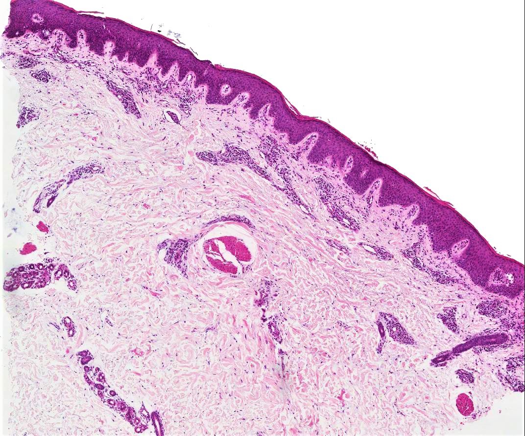
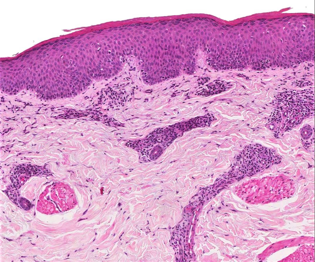
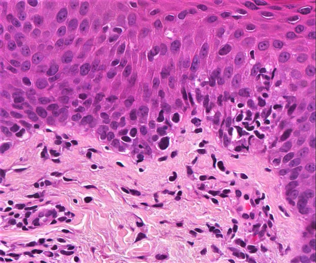
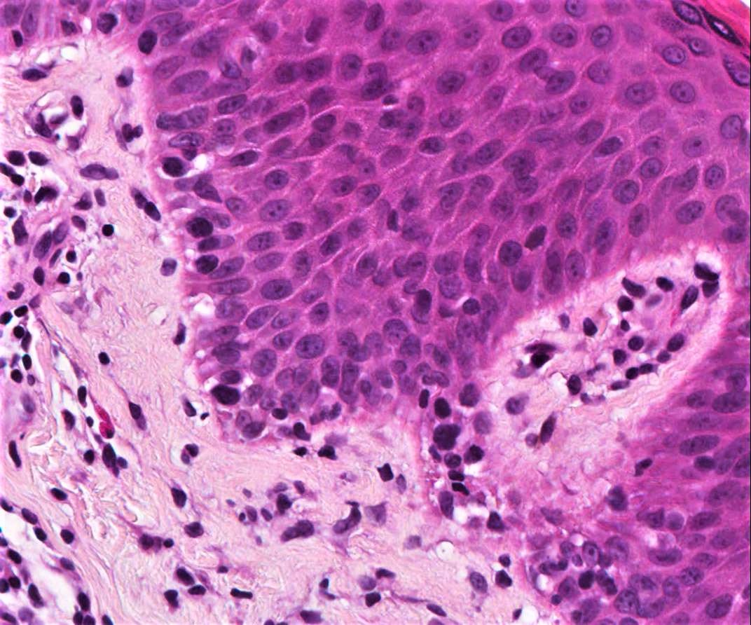
48/M, widespread papular rash

Edited by Admin_Dermpath


  Report Record

User Feedback


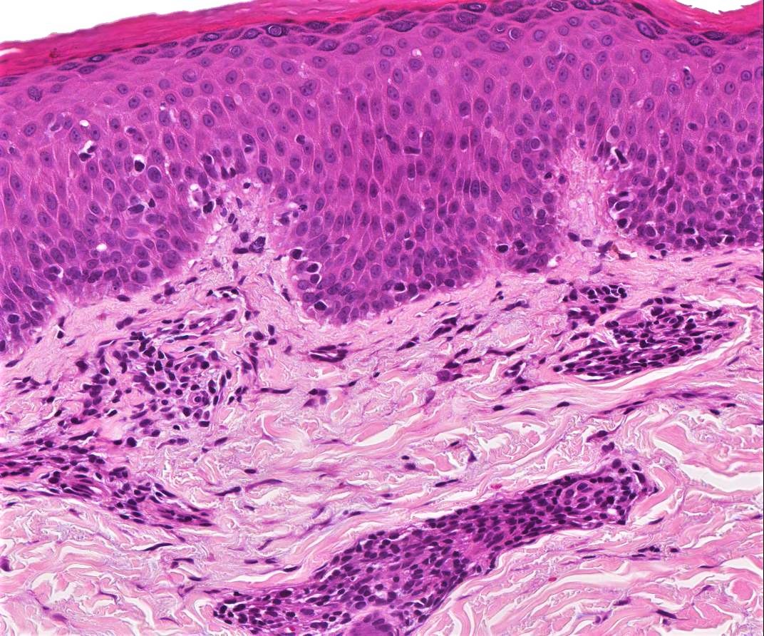
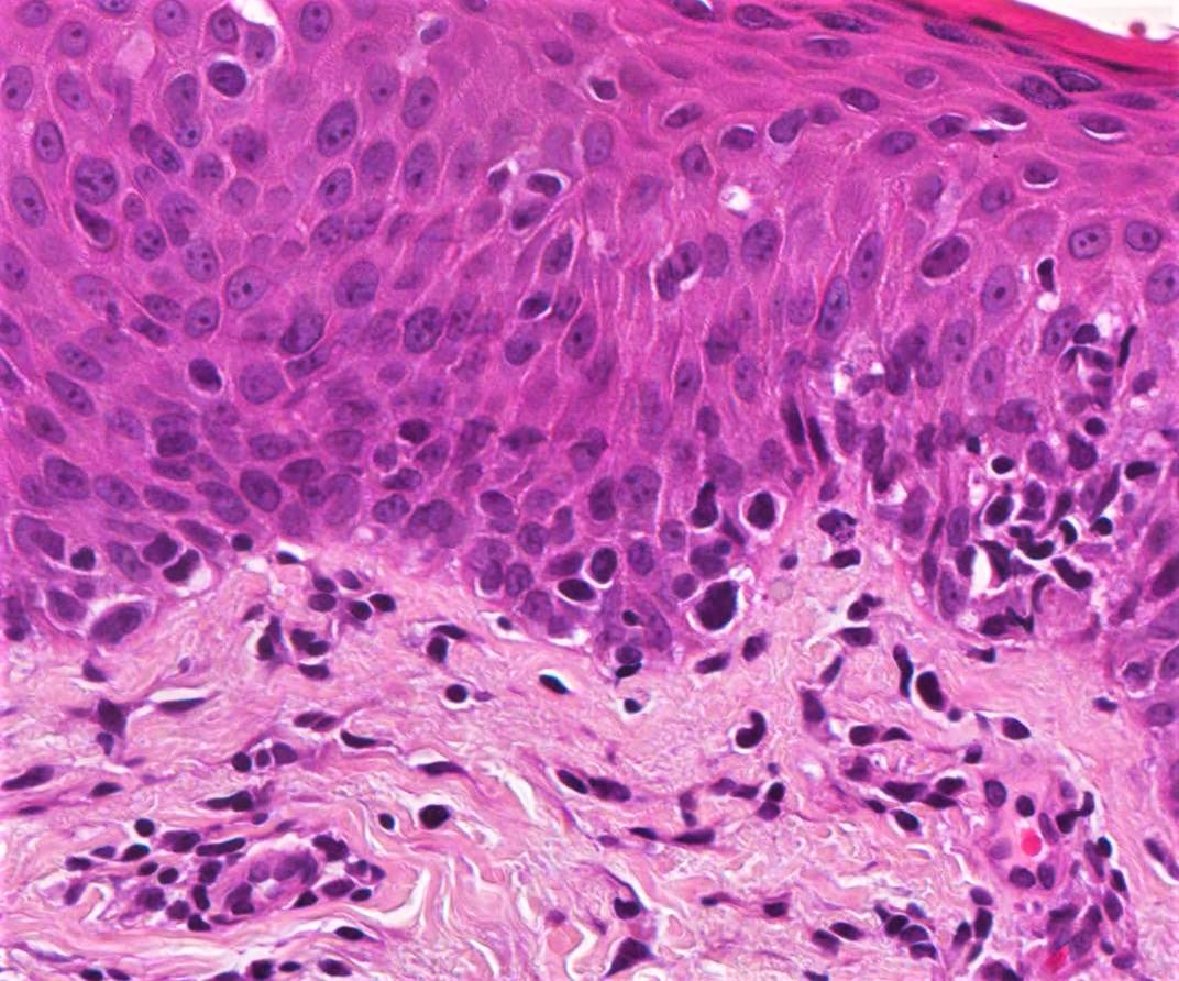
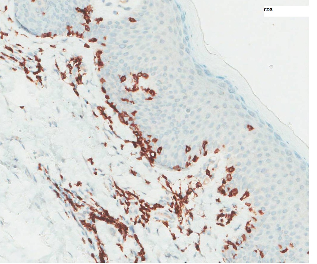
Epidermotropism with lymphocytes in the epidermis larger than those in the dermis,  Pautrier microabscess and history of widespread rash, all point towards MF going into Sezari syndrome

Share this comment


Link to comment
share_externally

vincenzo polizzi

Posted

Yes. At least 3 clues to MF/SS: 1) hyperkeratosis with some papillary dermal fibrosis point toward a long time lesion...2) intraepidermal lymphocytic nuclear sizes 3) exocytosis without spongiosis.

Share this comment


Link to comment
share_externally

Uma Sundram

Posted

MF/SS is certainly part of the differential, but one must also consider drug eruptions within the differential diagnosis; without clinical correlation, we would not be able to tell the difference.

Share this comment


Link to comment
share_externally

Saman Fatah

Posted

I think more information is needed before one suggest an appropriate diagnosis, the perivascular lymphocytic infiltrate is strikingly tight "coat-sleeve like".

Do these papules wax and wane, leave scars i.e Lymphomatoid papulosis course especially type B or fixed and not evolving?

Is he on any medication, as lymphomatoid drug eruption "pseudolymphoma" can be papular and look like MF on histology?

If above is negative, then one can consider MF but this will be a "papular" MF which a very rare sub-variant, one still have to examine the patient for any patches or plaques of the disease somewhere else, occasionally only observation and the time course will clarify.


Share this comment


Link to comment
share_externally

Dr. Mona Abdel-Halim

Posted

CPC is needed, Papular MF tops the list


Share this comment


Link to comment
share_externally

vincenzo polizzi

Posted

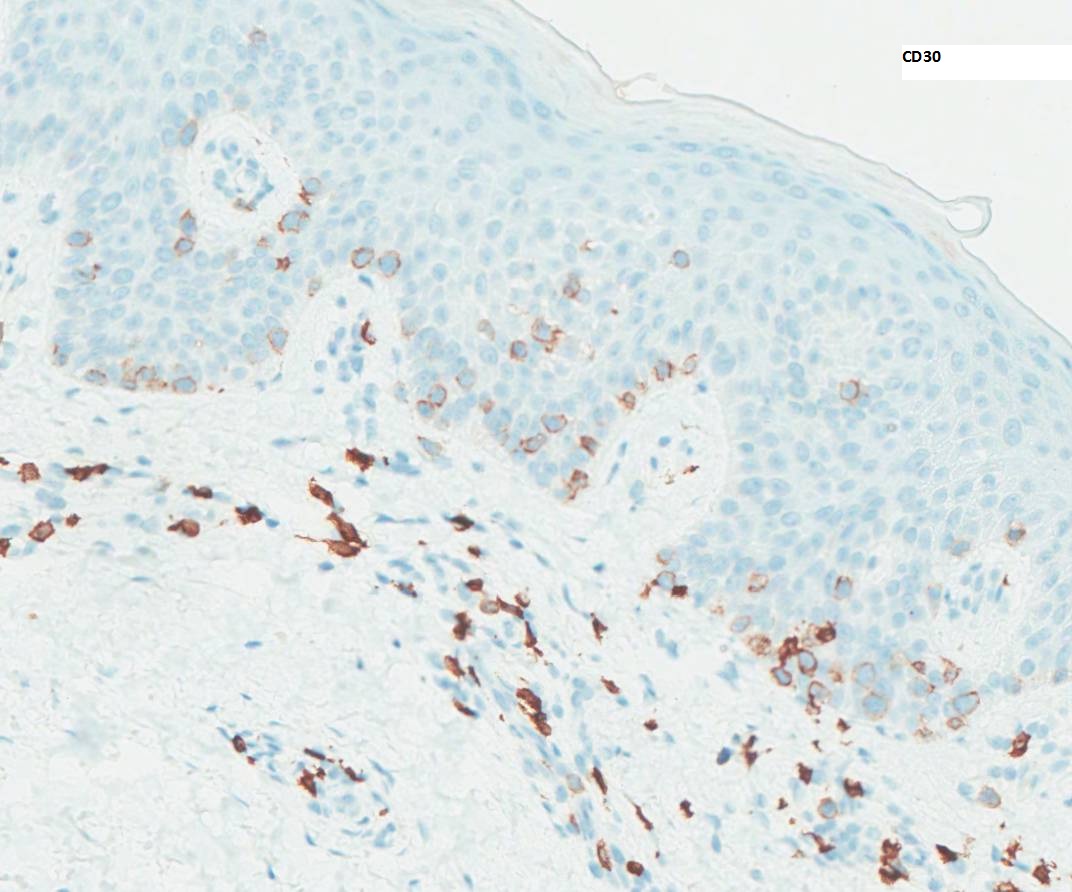
Intriguing case. CD30+ intraepidermal cells could be more in keeping with type B LyP... But I don’t see any pleomorphic dermal infiltrate ( also the monomorphic variant of LyP as Type B shows some pleomorphism ). The papular variant of MF should well be the transformation early stage of MF, so I favour PAPULAR MYCOSIS FUNGOIDES. 

Share this comment


Link to comment
share_externally

Arti Bakshi

Posted

Great discussion!

This is a case of LyP type B. The similarity to MF is striking and essentially, the 2 are indistinguishable histologically. Clinically, the lesions were papular and underwent spontaneous regression.There were no patches or plaques. 

This case shows CD30 positivity in the intraepidermal cells, but remember LyP type B can be entirely CD30 negative, making the diagnosis even more difficult. As always, CPC is key to the diagnosis.

Share this comment


Link to comment
share_externally

Dr. Richard Carr

Posted

Fantastic case Arti - I have not seen a case personally (if you know what I mean).

Share this comment


Link to comment
share_externally


Join the conversation

You can post now and register later. If you have an account, sign in now to post with your account.

Guest
Add a comment...

×   Pasted as rich text.   Paste as plain text instead

  Only 75 emoji are allowed.

×   Your link has been automatically embedded.   Display as a link instead

×   Your previous content has been restored.   Clear editor

×   You cannot paste images directly. Upload or insert images from URL.

Loading...

×
×
  • Create New...