Jump to content
In this section we have spot diagnoses posted on a daily basis since June 2010, now over 4000! You can review the archived cases and read the suggested diagnoses by users and the final comment by the contributors.
Case are uploaded each week day by 10 am UK time with the correct diagnosis will generally be posted at 8 pm UK time. Why not view the most recent spot diagnosis and proffer a diagnosis?

Case Number : Case 1910 - 25 Sept - Dr Limin Yu Posted By: Guest

Please read the clinical history and view the images by clicking on them before you proffer your diagnosis.
Submitted Date :
   (0 reviews)

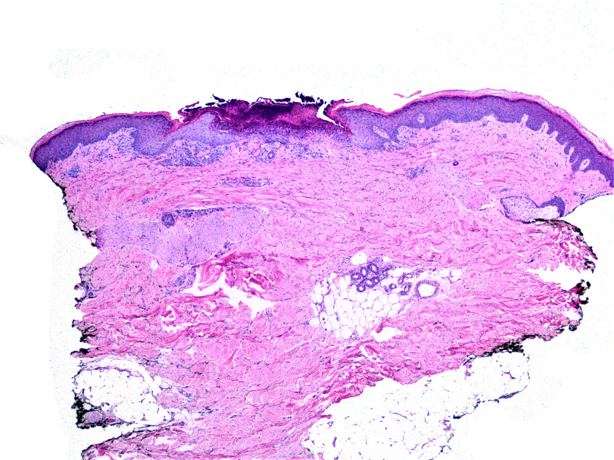
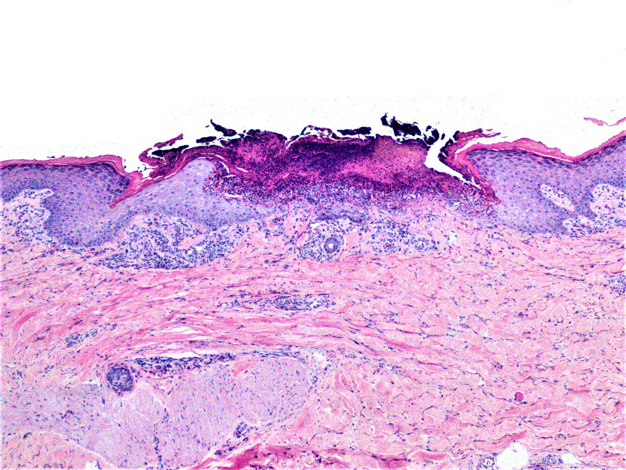
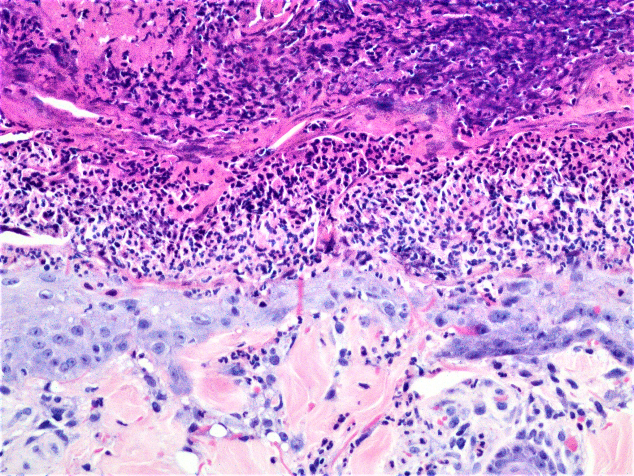
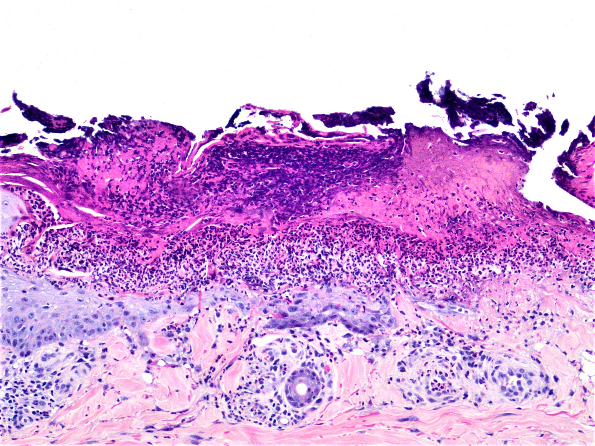
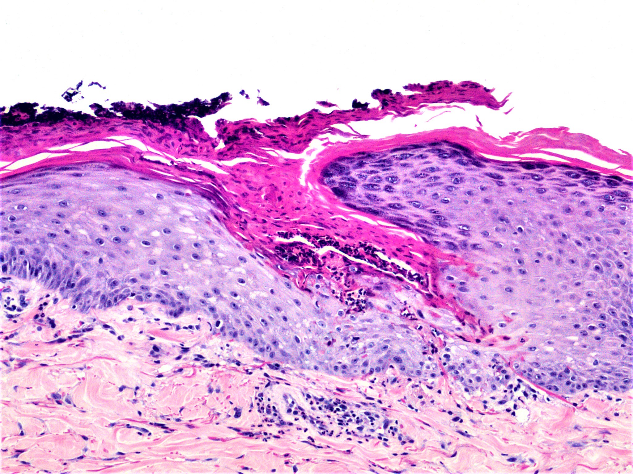
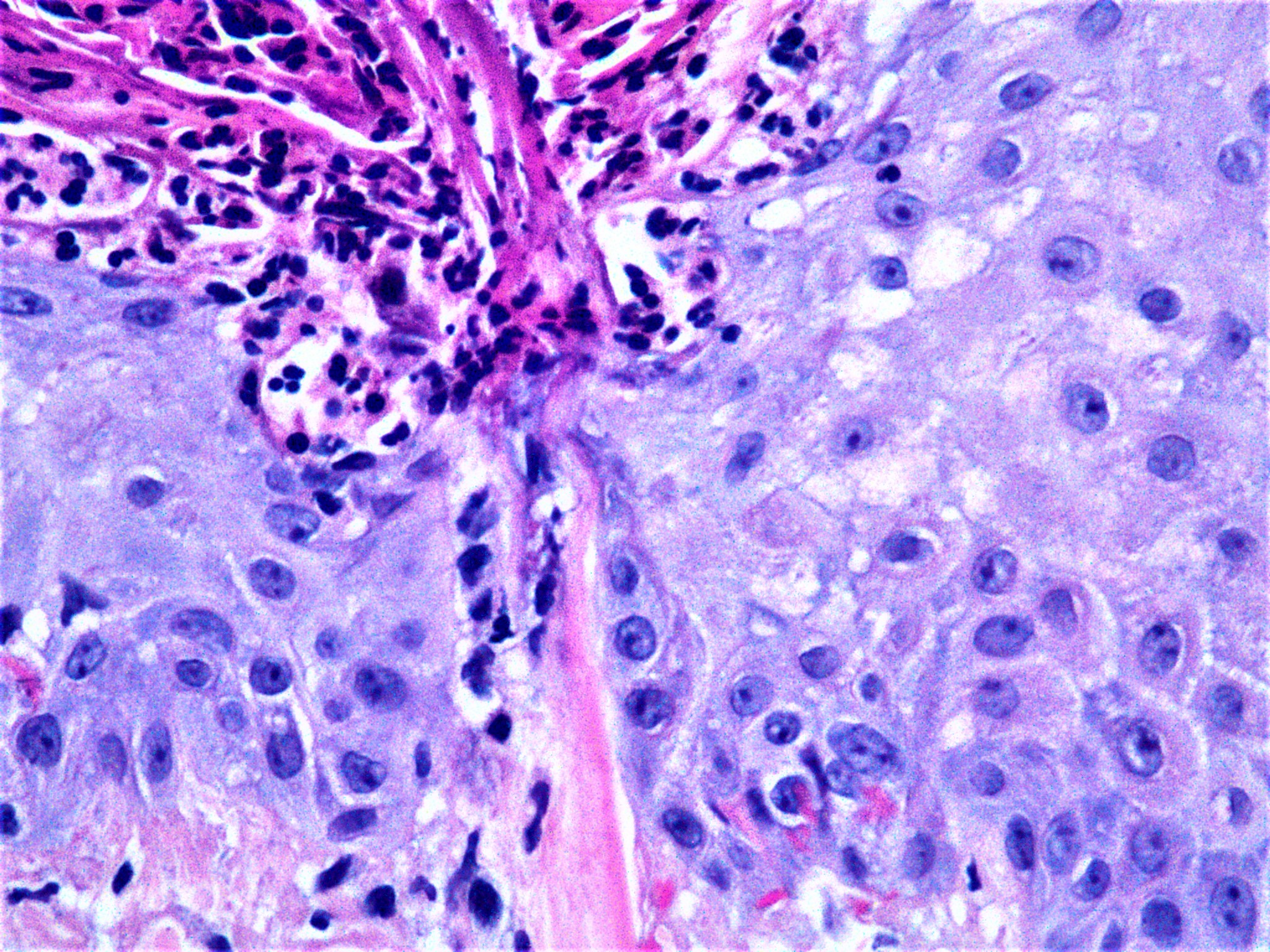
70 yo M, h/o diabetic. Lateral mid thigh biopsy


  • Like 1
  Report Record

User Feedback


vincenzo polizzi

Posted

Agree with Anil.    There are 4  perforating disorders, usually diabetes-associated: Kyrle's disease, perforating folliculitis, elastosis perforans serpiginosa, and reactive perforating collagenosis. I think this perforating material is collagen, so favor acquired reactive perforating collagenosis. But I'm not able to distinguish between Kirle's disease and ARPC. CPC is important of course.

 Very nice case.

Share this comment


Link to comment
share_externally

Raul Perret

Posted

The pictures are fantastic. Agree with the colleagues, acquired perforating dermatosis, probably acquired reactive perforating collagenosis for CPC.

Share this comment


Link to comment
share_externally

Saman Fatah

Posted

Very nice example of perforating dermatosis, likely RPC as transepidermal elimination of collagen can be seen in some of the photos.

Relatively straight to Spot diagnose but no so easy to treat.

Share this comment


Link to comment
share_externally

Nitin Khirwadkar

Posted

Agree, perforating collagenosis. Lovely pictures!

Share this comment


Link to comment
share_externally

Dr. Richard Carr

Posted

The last image shows vertically orientated light pink collagen (I think) in the centre and I'm pretty sure I can see some subtle more basophilic collagen in the crust too (polarising microscopy or trichrome will show it nicely - not required). The slender darker pink (hmm I'm struggling for the best colour for elastic fibres!) are elastic fibres. Collagen is a lighter pink than elastic (perhaps "bubble gum" pink). The elastic fibres are wavy in the dermis but often look vertical and straighter when perforating. The basophilic change to the collagen and neutrophils in the crust is highly typical of perforating collagenosis. EVG will show the elastic but again not sure it's required here.

Share this comment


Link to comment
share_externally

Thank everyone for insightful discussion. I agree with all of you. This is a perforating disease and acquired reactive perforating collagenosis is the diagnosis. 

I emailed trichrome stain images to Admin. He/she didn't post them, I guess, not to make it too straightforward.  The stain shows the blue colored collagen perforation.  Unfortunately I can't attach the image file in this reply due to the low limit of file size (less than 0.11 MB) in attachment. 


Thanks!


Share this comment


Link to comment
share_externally


Join the conversation

You can post now and register later. If you have an account, sign in now to post with your account.

Guest
Add a comment...

×   Pasted as rich text.   Paste as plain text instead

  Only 75 emoji are allowed.

×   Your link has been automatically embedded.   Display as a link instead

×   Your previous content has been restored.   Clear editor

×   You cannot paste images directly. Upload or insert images from URL.

Loading...

×
×
  • Create New...