Jump to content
In this section we have spot diagnoses posted on a daily basis since June 2010, now over 4000! You can review the archived cases and read the suggested diagnoses by users and the final comment by the contributors.
Case are uploaded each week day by 10 am UK time with the correct diagnosis will generally be posted at 8 pm UK time. Why not view the most recent spot diagnosis and proffer a diagnosis?

Case Number : Case 1912 - 27 Sept - Dr Richard Carr Posted By: Guest

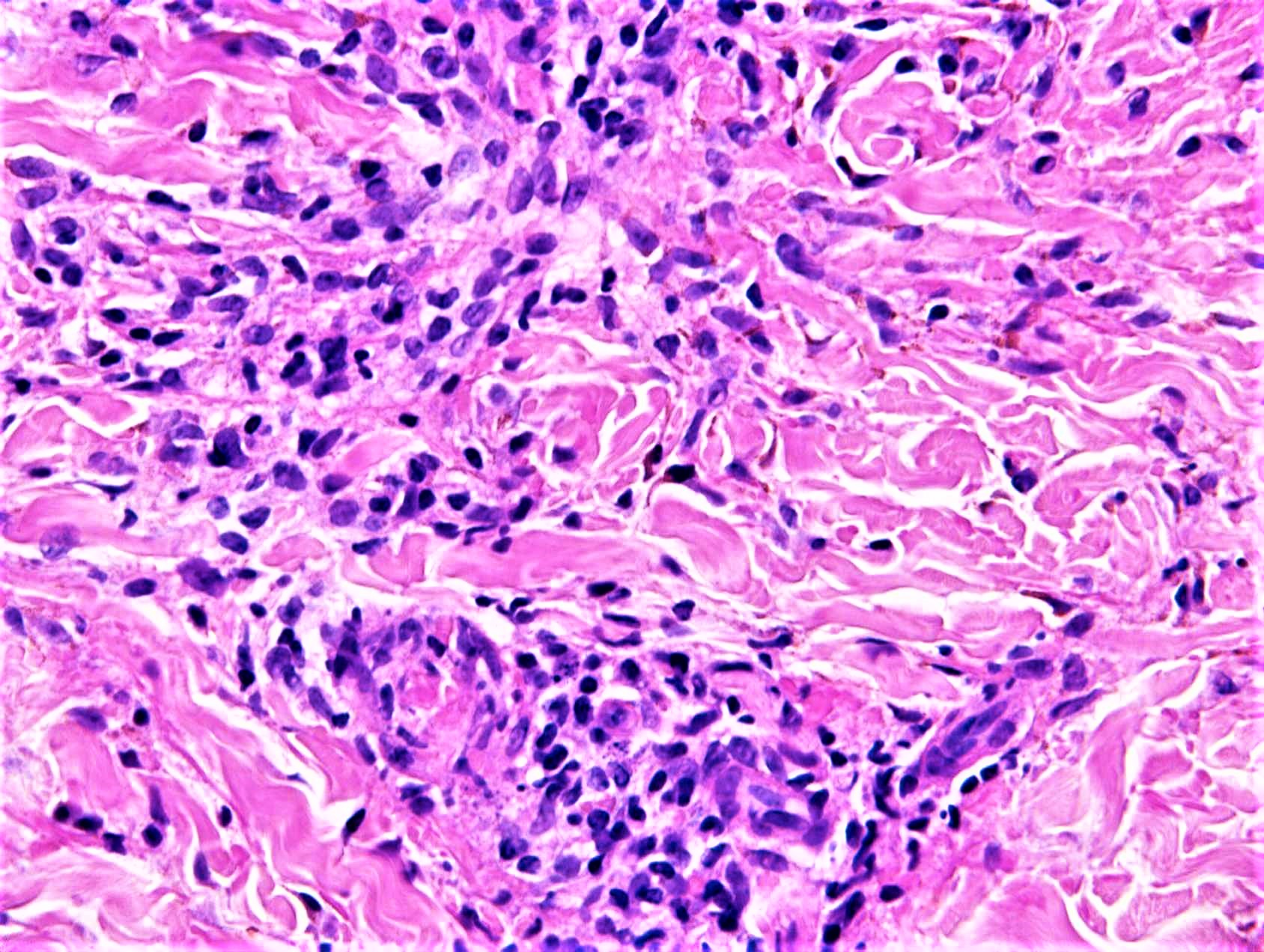
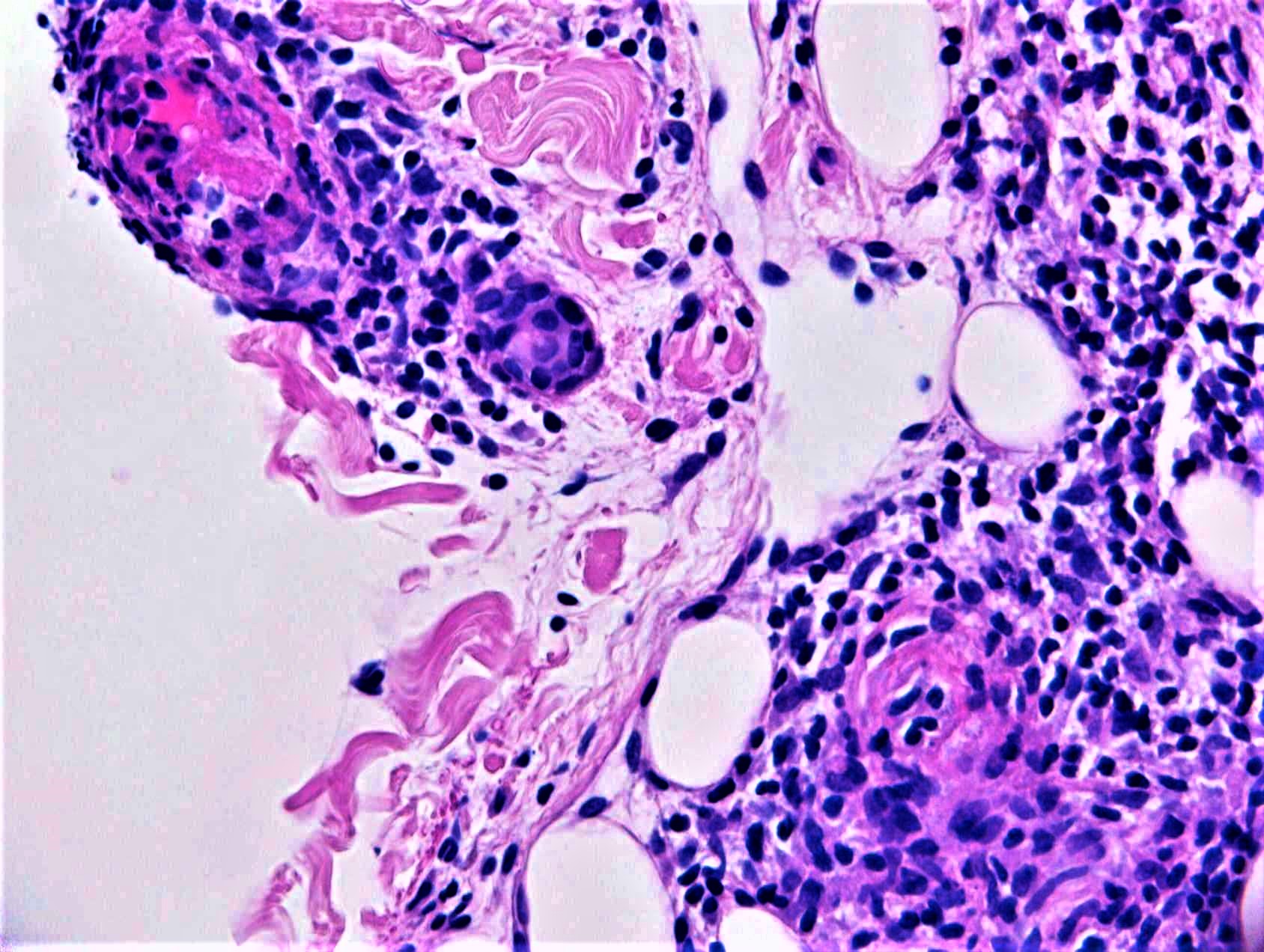
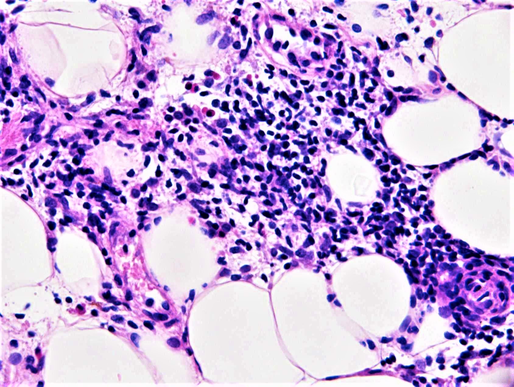
Please read the clinical history and view the images by clicking on them before you proffer your diagnosis.
Submitted Date :
   (0 reviews)

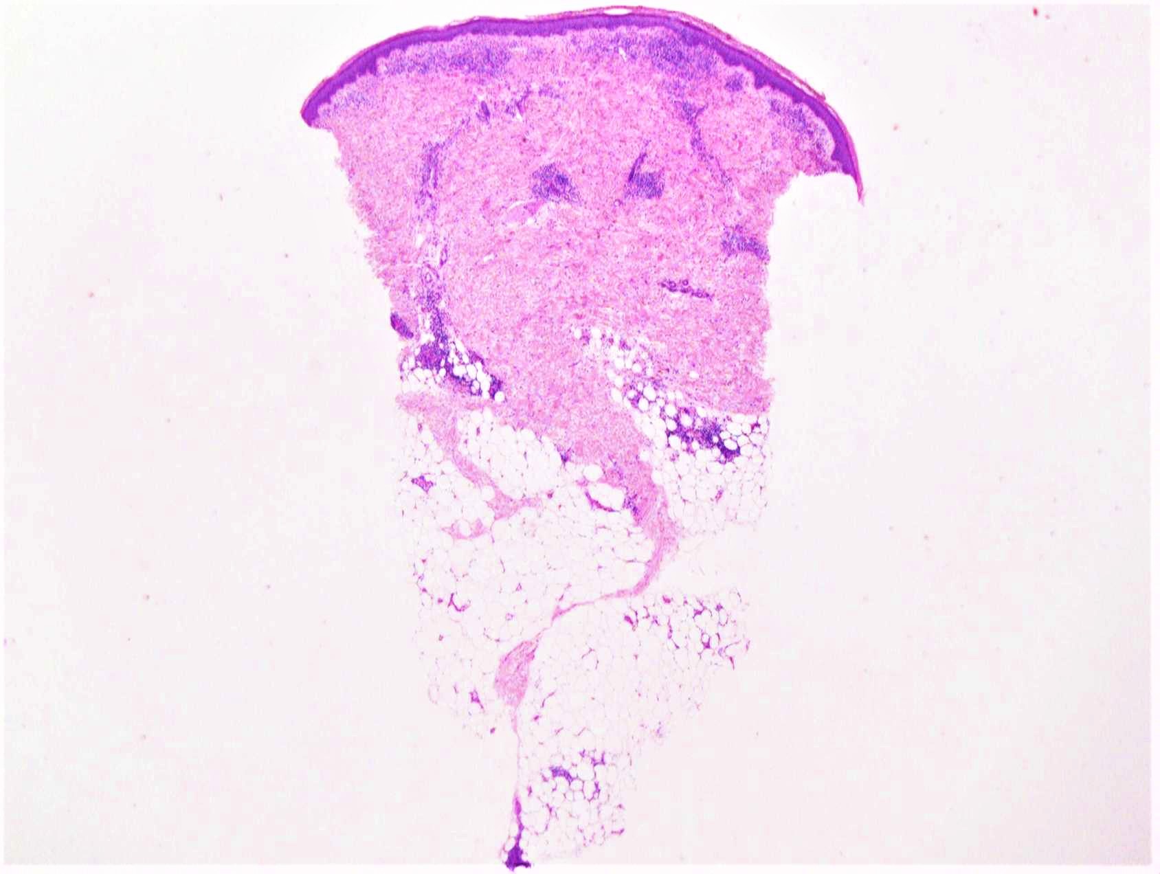
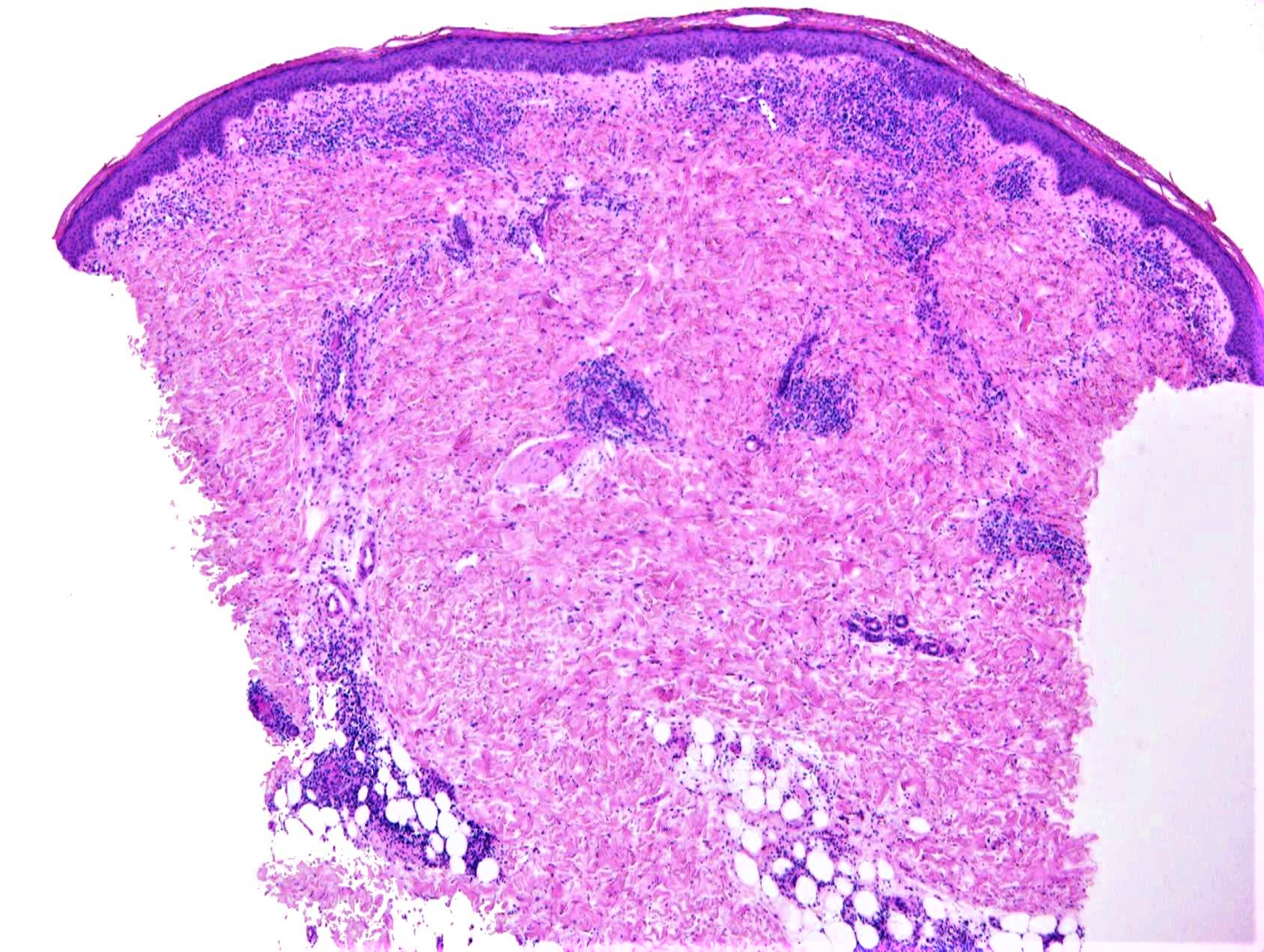
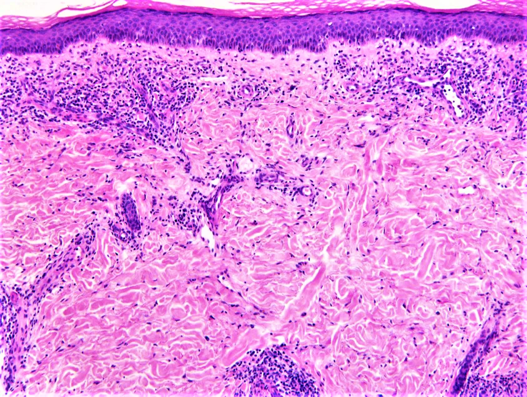
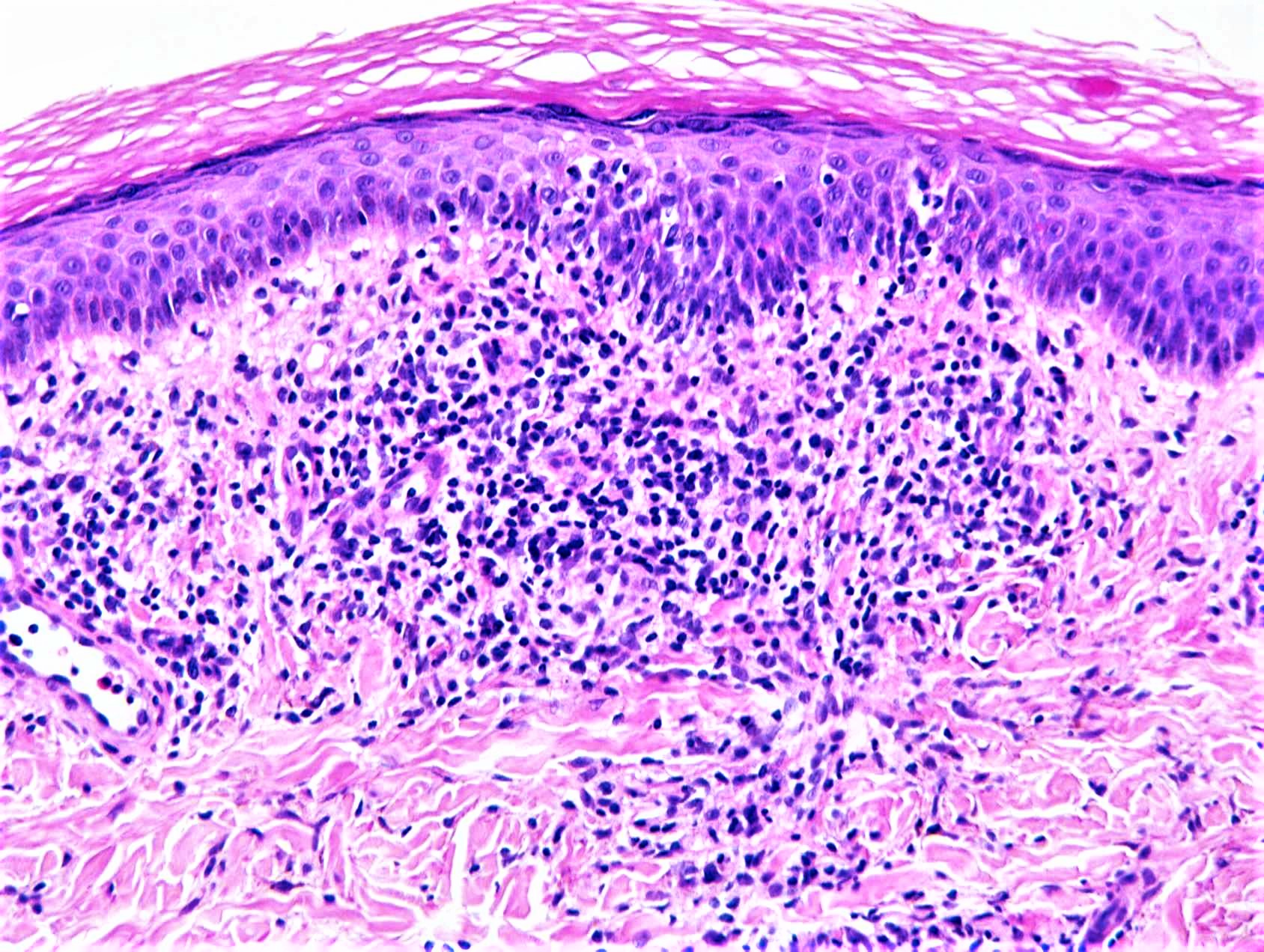
Clinical Details: M10. Erythematous macule on the calf for 12 to 18 months.


  Report Record

User Feedback


vincenzo polizzi

Posted

First thought : Lichen striatus.

May be this is a long standing lesion ( 18 month ) so the interfacie active component is relatively regressed.

Share this comment


Link to comment
share_externally

vincenzo polizzi

Posted

But now, on my computer, I want to change the LS spot. Realizing the deep dermal inflammation is vasculitis (lymphocytic vasculitis), not perieccrine infiltrate.

So this could be a chilblain lupus. Frank ymphocytic vasculitis is usually more evident in CL than in idiopatic chilblain.


Share this comment


Link to comment
share_externally

Raul Perret

Posted

Like Vincenzo I thought of  "lymphocytic vasculitis" but I was thinking it could be one of the PPDs like Schamberg's disease which is occasionally seen in children.

Share this comment


Link to comment
share_externally

vincenzo polizzi

Posted

Like your comment Raul. PPD is a good (and maybe the correct one) differential.

I was just thinking of chilblain lupus based on deep sited infiltrate and obvious vascular damage. But realizing that 12/18 months are more in keeping with PPD.

Share this comment


Link to comment
share_externally

Saman Fatah

Posted

Pigmented Purpuric Dermatoses, likely Lichen Aureus sub-variant given the clinical description and age of the patient but clearly needs correlation as this group will have overlapping clinical/histological features.

The density and the depth of the infiltrate is rather impressive. 

Share this comment


Link to comment
share_externally

Superficial and deep perivacular lymphocytic cuffing, a little spongiosis and panniculitis to add to the confusion!. To me it looks like a deep gyrate erythema- either erythema annulare centrifugum or erythema chronicum migrans if there is a history of tick bite.

Share this comment


Link to comment
share_externally

Admin_Dermpath

Posted

Many thanks everyone


The final diagnosis is:


Lichen aureus

Dr Richard Carr


Share this comment


Link to comment
share_externally


Join the conversation

You can post now and register later. If you have an account, sign in now to post with your account.

Guest
Add a comment...

×   Pasted as rich text.   Paste as plain text instead

  Only 75 emoji are allowed.

×   Your link has been automatically embedded.   Display as a link instead

×   Your previous content has been restored.   Clear editor

×   You cannot paste images directly. Upload or insert images from URL.

Loading...

×
×
  • Create New...