Jump to content
In this section we have spot diagnoses posted on a daily basis since June 2010, now over 4000! You can review the archived cases and read the suggested diagnoses by users and the final comment by the contributors.
Case are uploaded each week day by 10 am UK time with the correct diagnosis will generally be posted at 8 pm UK time. Why not view the most recent spot diagnosis and proffer a diagnosis?

Case Number : Case 1914 - 29 Sept - Dr Richard Carr Posted By: Guest

Please read the clinical history and view the images by clicking on them before you proffer your diagnosis.
Submitted Date :
   (0 reviews)

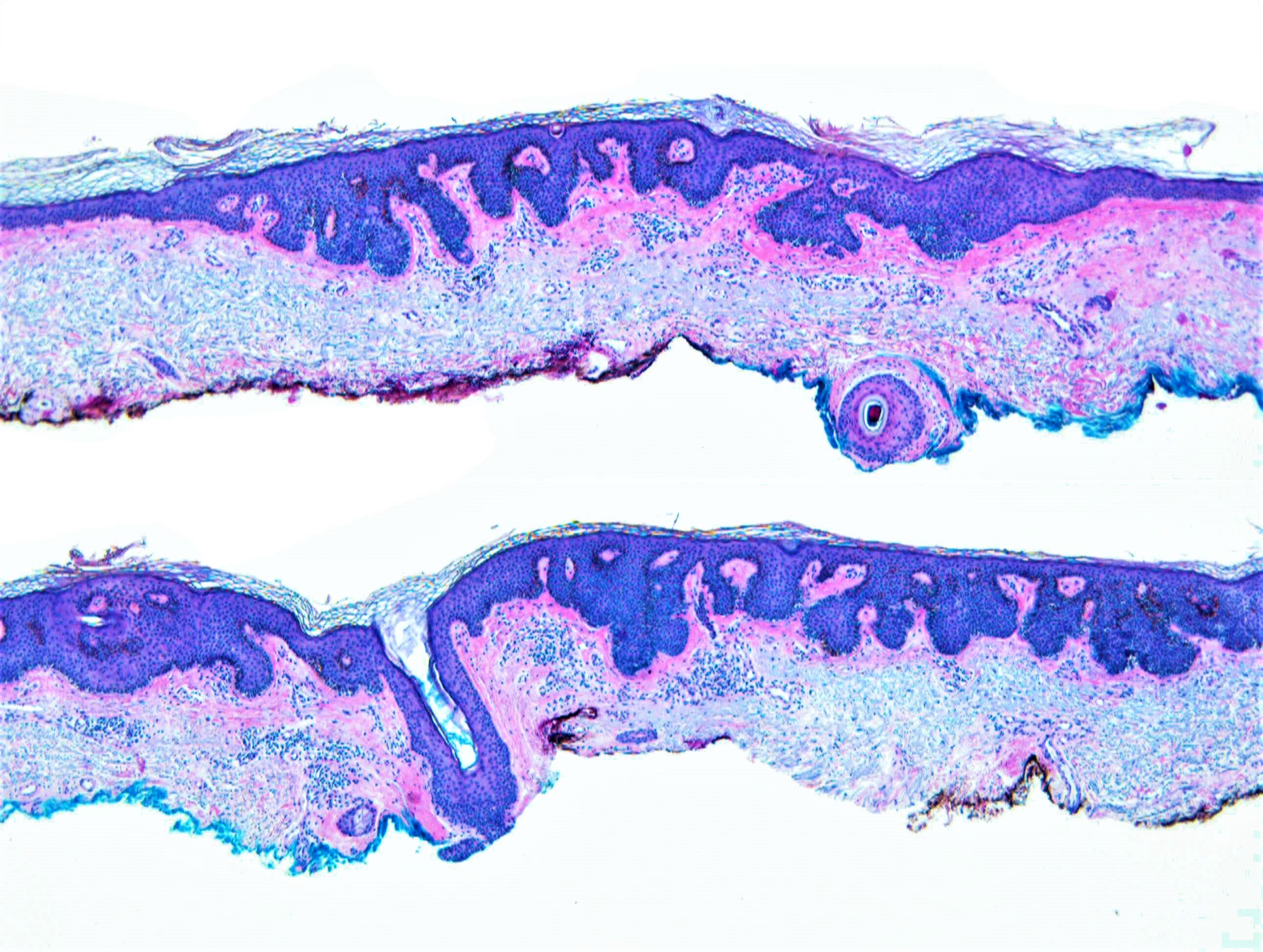
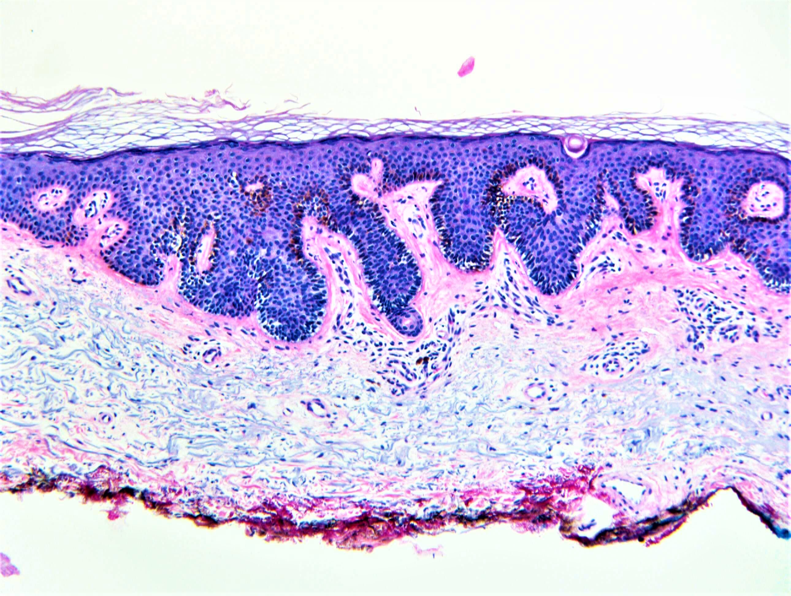
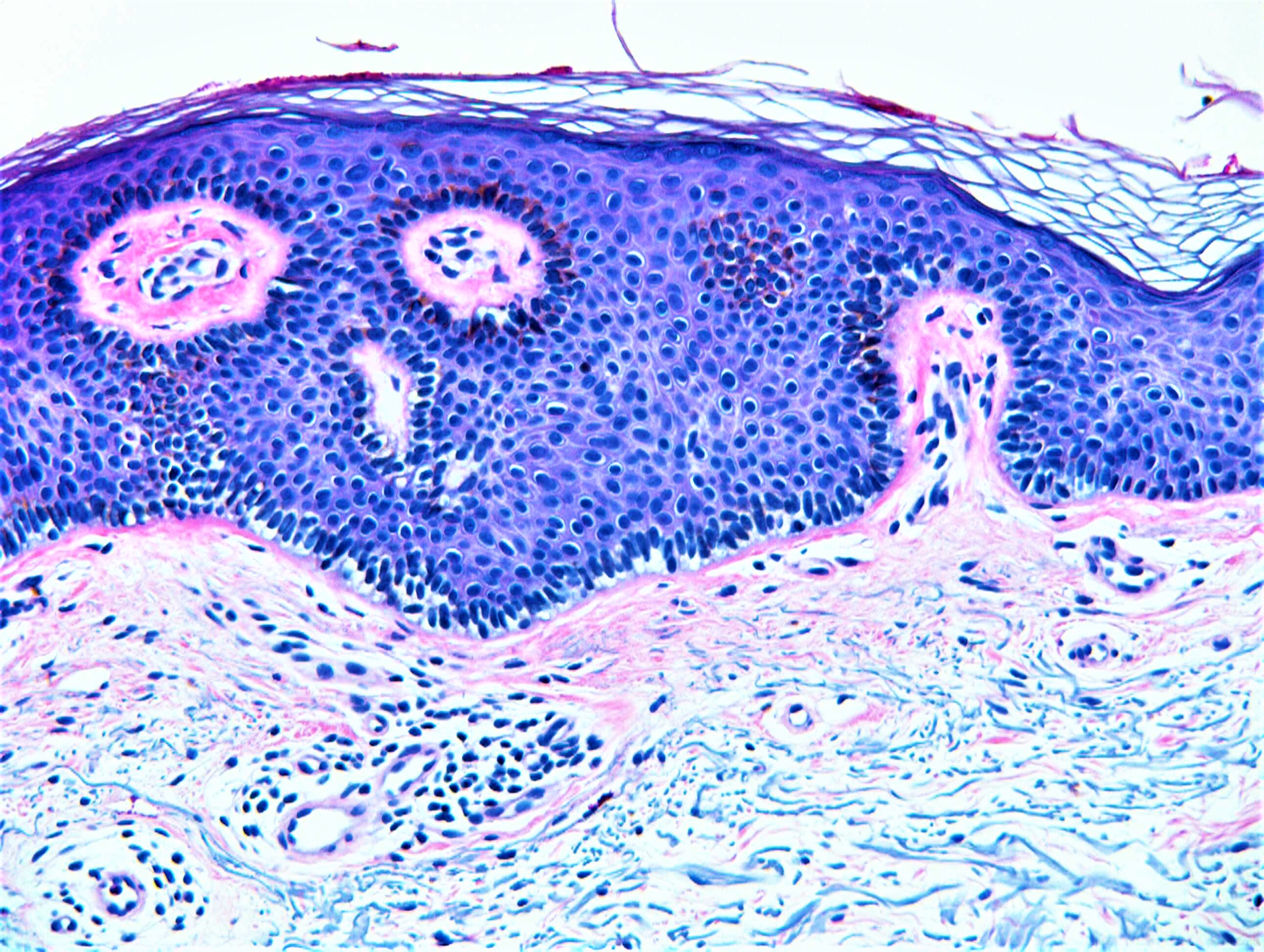
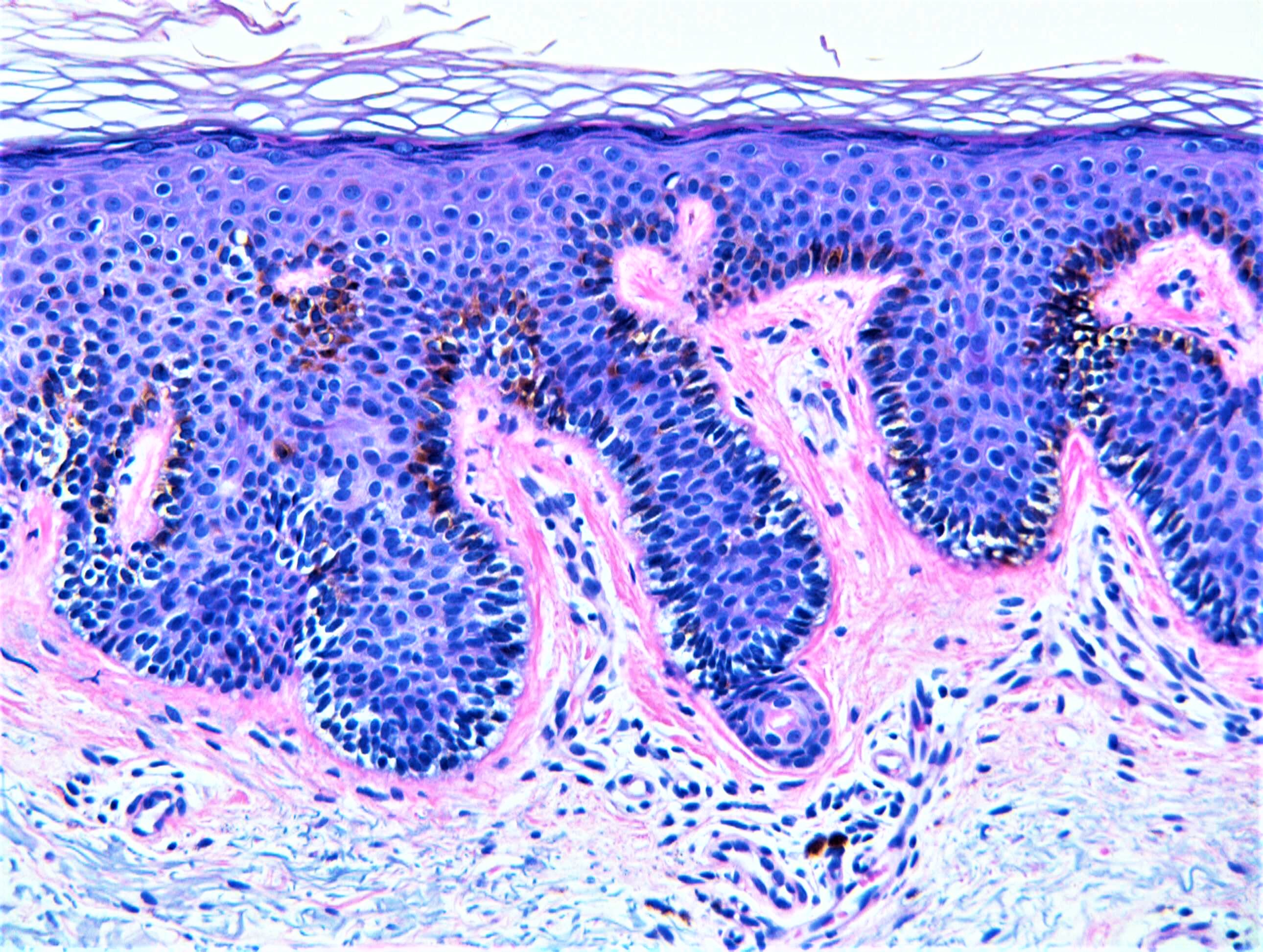
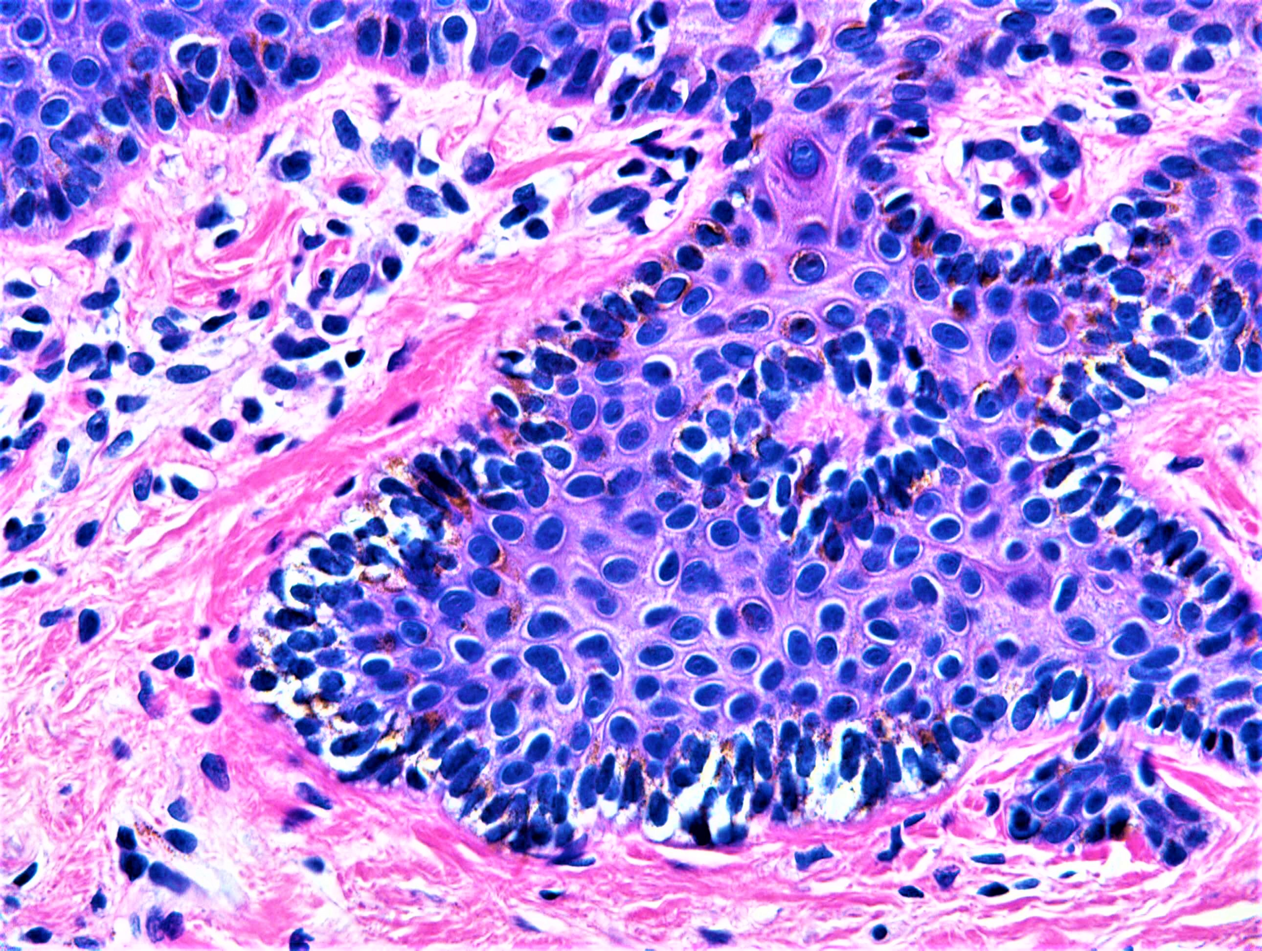
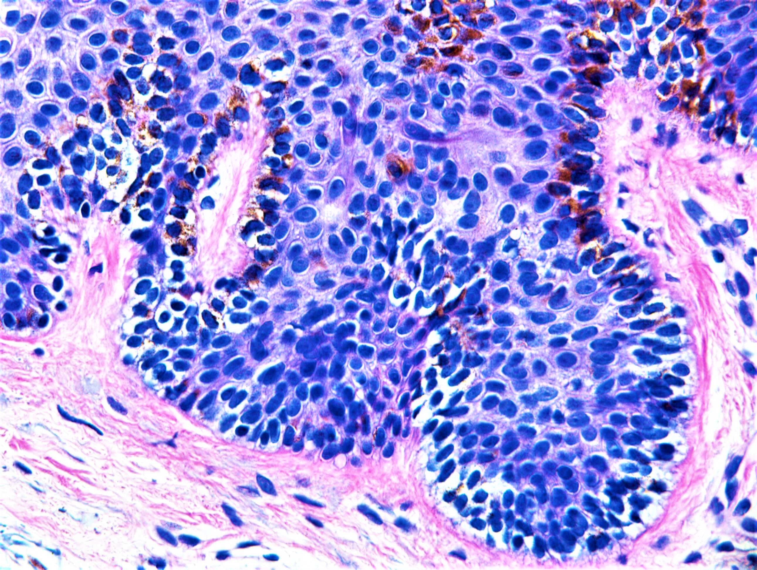
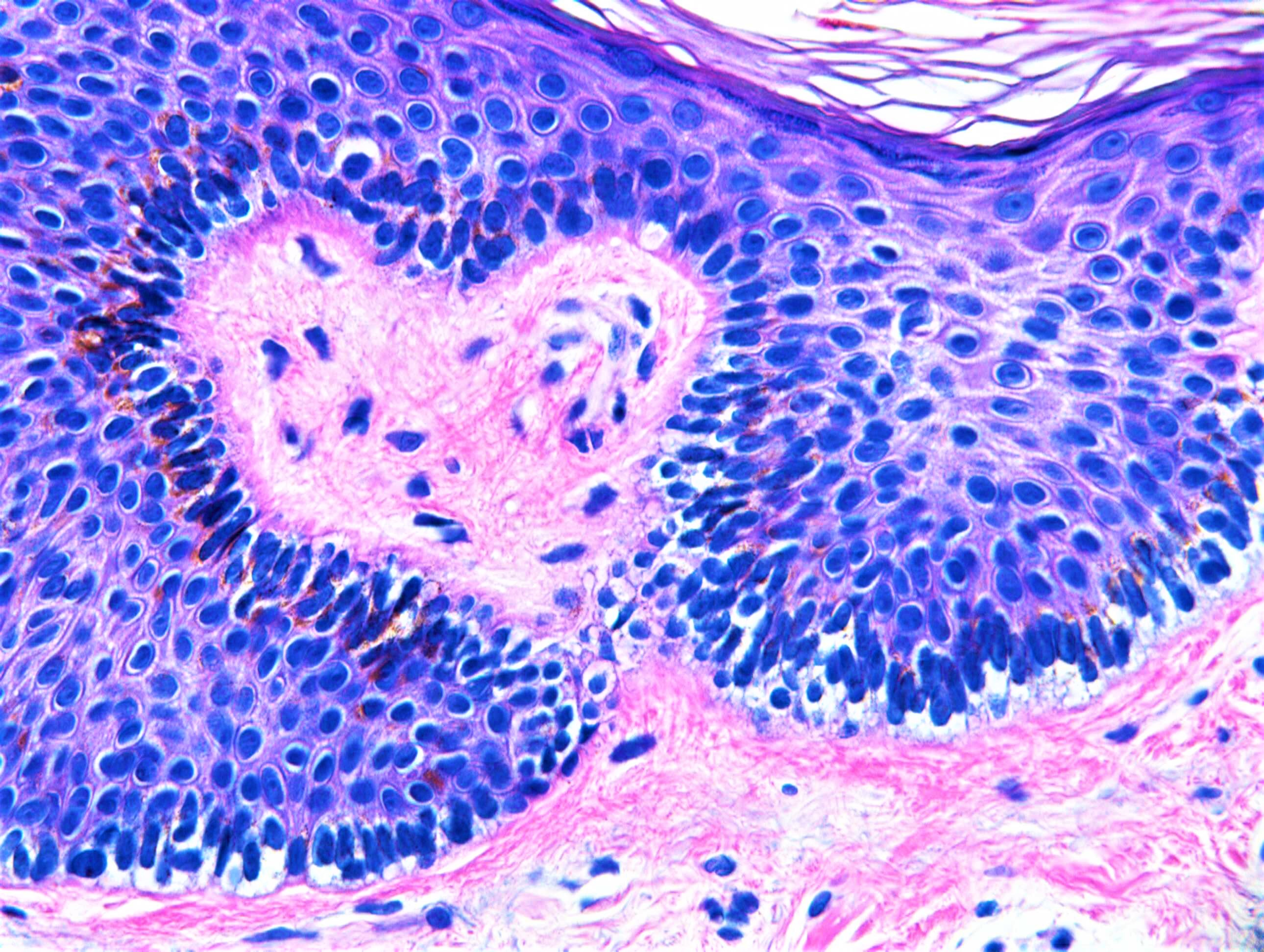
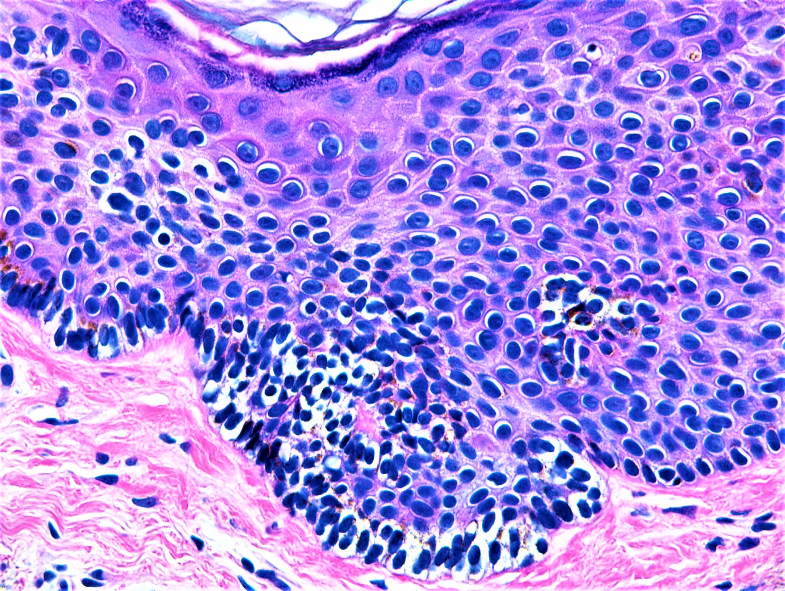
Clinical Details: M65. Forearm. ?Melanoma


  Report Record

User Feedback


Admin_Dermpath

Posted

Dear All 


Richard has posted his diagnosis on:


Share this comment


Link to comment
share_externally

vincenzo polizzi

Posted

Inflammatory Vitiligo or Hypopigmented Mycosis Fungoides?

The thick intrapapillary dermal collagen bands and some cytology always details make me favor HMF. But there is an odd atypical palisaded layer in basal epidermis: lymphocytes? Melanocytes? So many doubts...

Share this comment


Link to comment
share_externally

Rete ridge hyperplasia with increased basal melanocytes with vacuolation in some. Few melanophages in dermis. Solar lentigo or PUVA lentigo

Share this comment


Link to comment
share_externally

Saman Fatah

Posted

With my very limited knowledge on non-inflammatory dermpath, this lesion looks melanocytic, immuno may highlight the true nature of junctional activites and upward spread which is very hard to appreciate on H&E.

I thought about the possibility of lentiginous dyspalstic naevus of elderly or lentiginous melamona (if one regard them separate). 

Share this comment


Link to comment
share_externally

Raul Perret

Posted

As Saman mentioned, I would be careful with the dx. Clinical impression is worrisome, there is a filiform continuous hyperplasia of melanocytes and the dermis has extensive solar damage.

Share this comment


Link to comment
share_externally

Admin_Dermpath

Posted

Dr Richard Carr: Diagnosis 

Seborrheoic keratosis with basal clear cells. Case c/o Dr Philip Shapiro.


Share this comment


Link to comment
share_externally

vincenzo polizzi

Posted

So I was barking up the wrong tree...Shame on me!

Share this comment


Link to comment
share_externally


Join the conversation

You can post now and register later. If you have an account, sign in now to post with your account.

Guest
Add a comment...

×   Pasted as rich text.   Paste as plain text instead

  Only 75 emoji are allowed.

×   Your link has been automatically embedded.   Display as a link instead

×   Your previous content has been restored.   Clear editor

×   You cannot paste images directly. Upload or insert images from URL.

Loading...

×
×
  • Create New...