Jump to content
In this section we have spot diagnoses posted on a daily basis since June 2010, now over 4000! You can review the archived cases and read the suggested diagnoses by users and the final comment by the contributors.
Case are uploaded each week day by 10 am UK time with the correct diagnosis will generally be posted at 8 pm UK time. Why not view the most recent spot diagnosis and proffer a diagnosis?

Case Number : Case 2126 - 2 August 2018 Posted By: Raul Perret

Please read the clinical history and view the images by clicking on them before you proffer your diagnosis.
Submitted Date :
   (0 reviews)

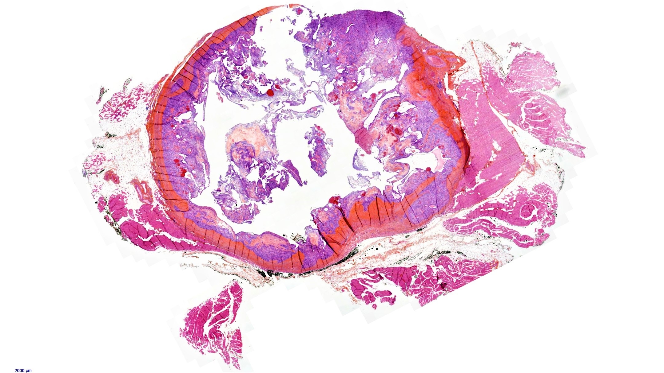
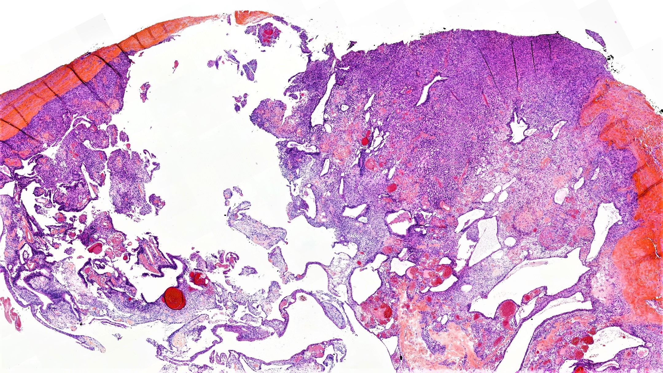
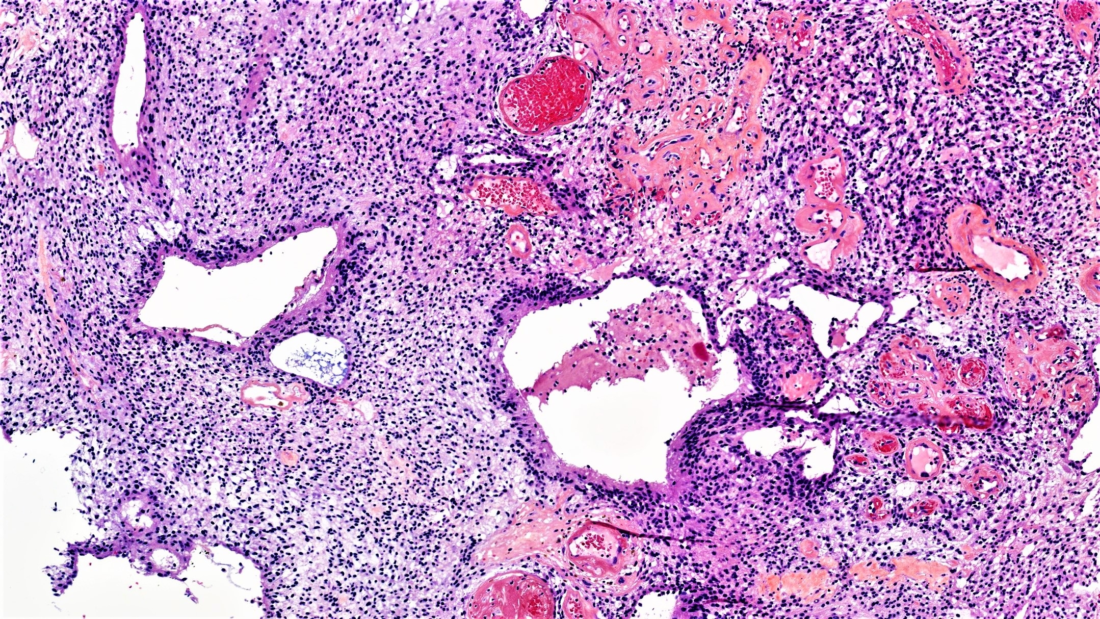
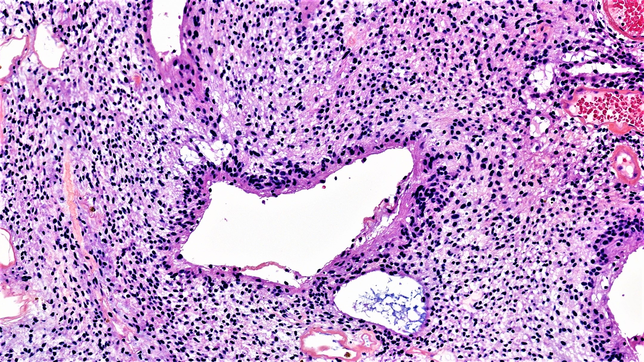
57 year old female with a 30 mm deep tumor localized in the buttock


  Report Record

User Feedback


Agreed with both although I like myxopapillary ependymoma a little bit better.

Share this comment


Link to comment
share_externally

Raul Perret

Posted

This is case of ancient change in a schwannoma. Important clues to consider this entity are the hyalinized blood vessels, the presence of prominent cleft artifact and in some cases the focal nuclear atypia (usually without mitotic activity). It is also useful to check for a capsule (quite prominent in this particular example. In addition, this case had prominent pseudoglandular structures that can be a potential pitfall. S100 was diffusely positive and ki66 was low. In regards to the differential with myxopapillary ependymoma, although s100 can be positive and may rarely arise on the subcutis of the lumbar region, in this case there were't true papillae centered by hyalinized blood vessels nor rosettes. Thank you for the comments

Share this comment


Link to comment
share_externally

Raul Perret

Posted

Sorry, forgot to mention that sox10 was diffusely positive also in this case, a feature that also helps in the distinction between ependymoma/myxopapillary ependymoma and schwannoma

Share this comment


Link to comment
share_externally

drkhaledhindi

Posted

Raul, i always get excited when i see your posts. Really awesome cases and info. Lucky to have you here.

Share this comment


Link to comment
share_externally


Join the conversation

You can post now and register later. If you have an account, sign in now to post with your account.

Guest
Add a comment...

×   Pasted as rich text.   Paste as plain text instead

  Only 75 emoji are allowed.

×   Your link has been automatically embedded.   Display as a link instead

×   Your previous content has been restored.   Clear editor

×   You cannot paste images directly. Upload or insert images from URL.

Loading...

×
×
  • Create New...