Jump to content
In this section we have spot diagnoses posted on a daily basis since June 2010, now over 4000! You can review the archived cases and read the suggested diagnoses by users and the final comment by the contributors.
Case are uploaded each week day by 10 am UK time with the correct diagnosis will generally be posted at 8 pm UK time. Why not view the most recent spot diagnosis and proffer a diagnosis?

Case Number : Case 2128 - 6 August 2018 Posted By: Limin Yu

Please read the clinical history and view the images by clicking on them before you proffer your diagnosis.
Submitted Date :
   (0 reviews)

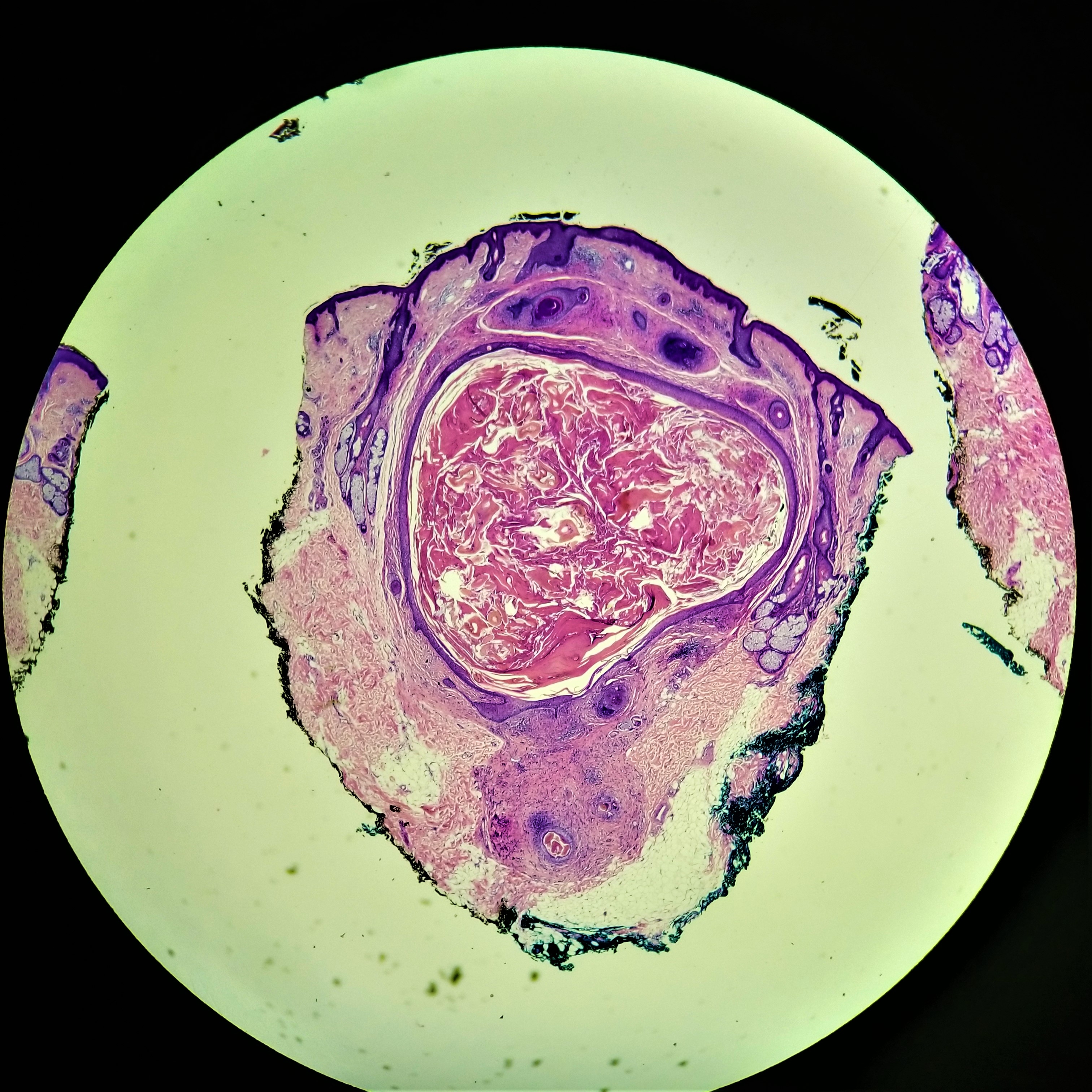
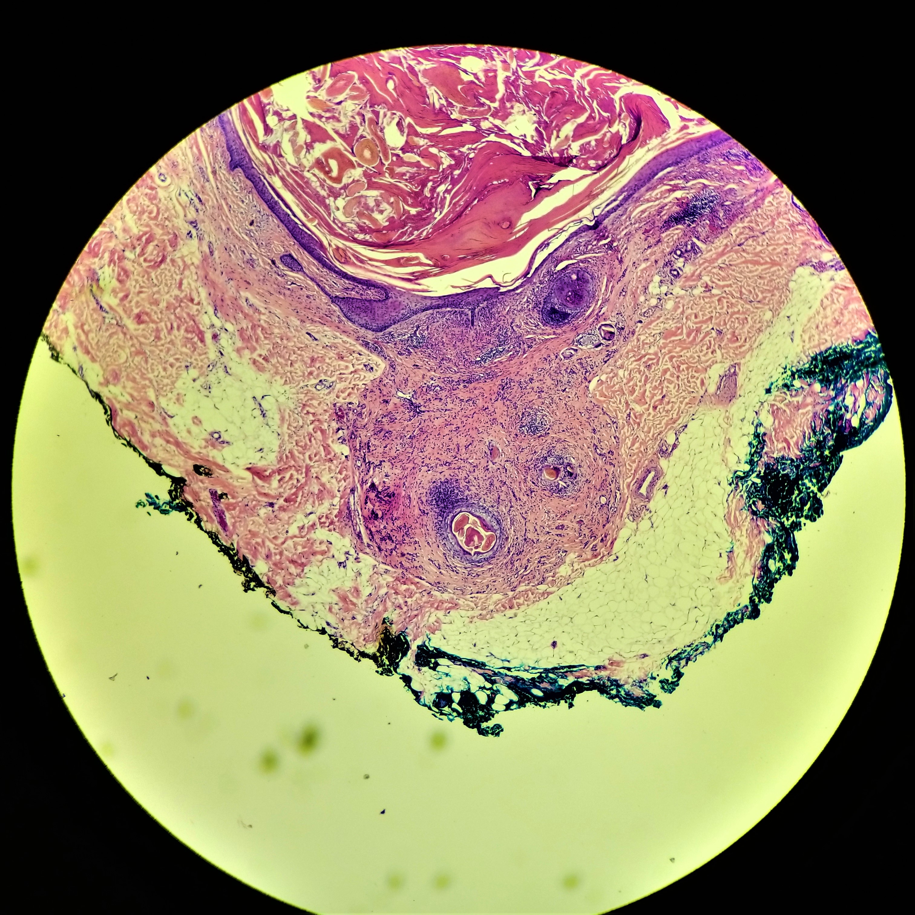
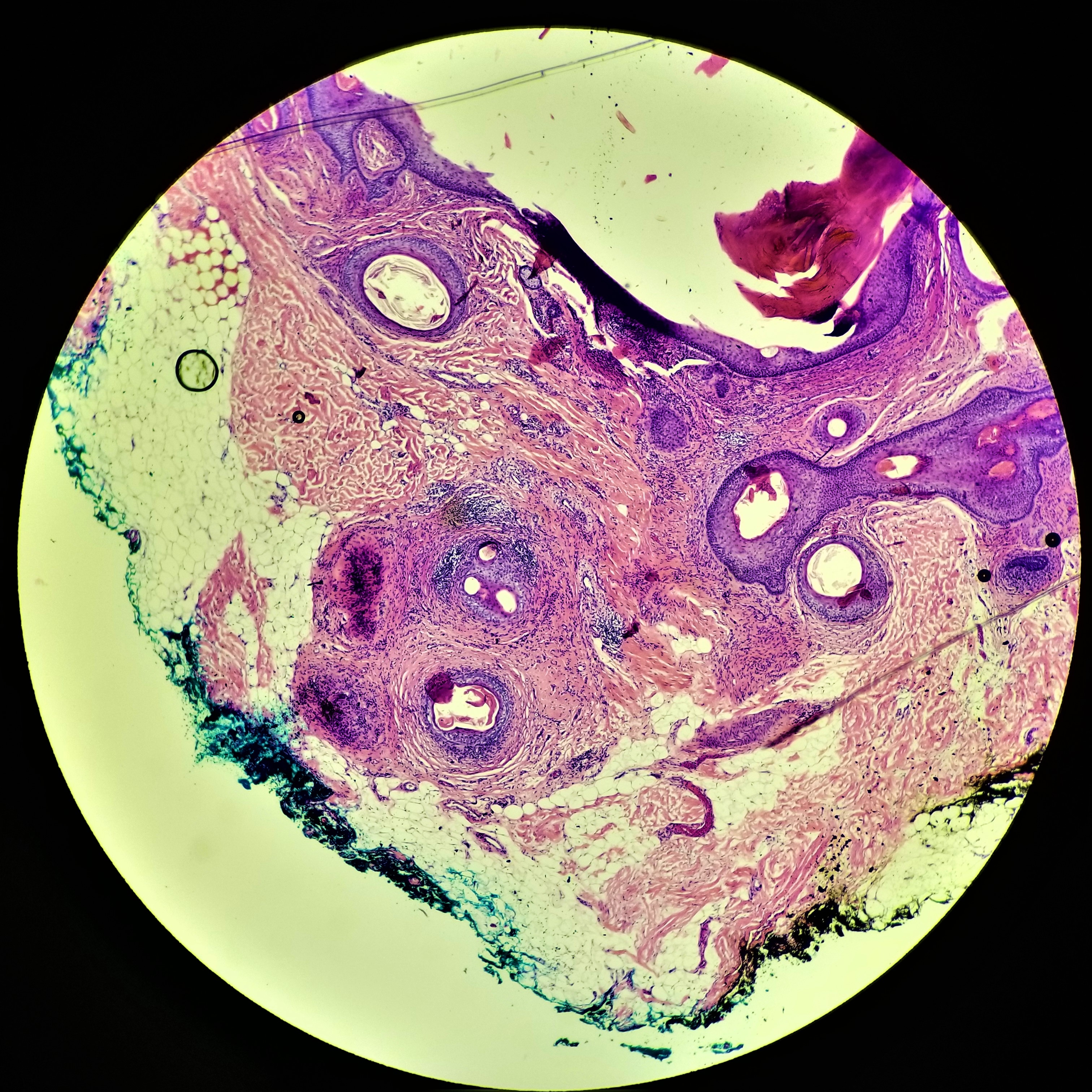
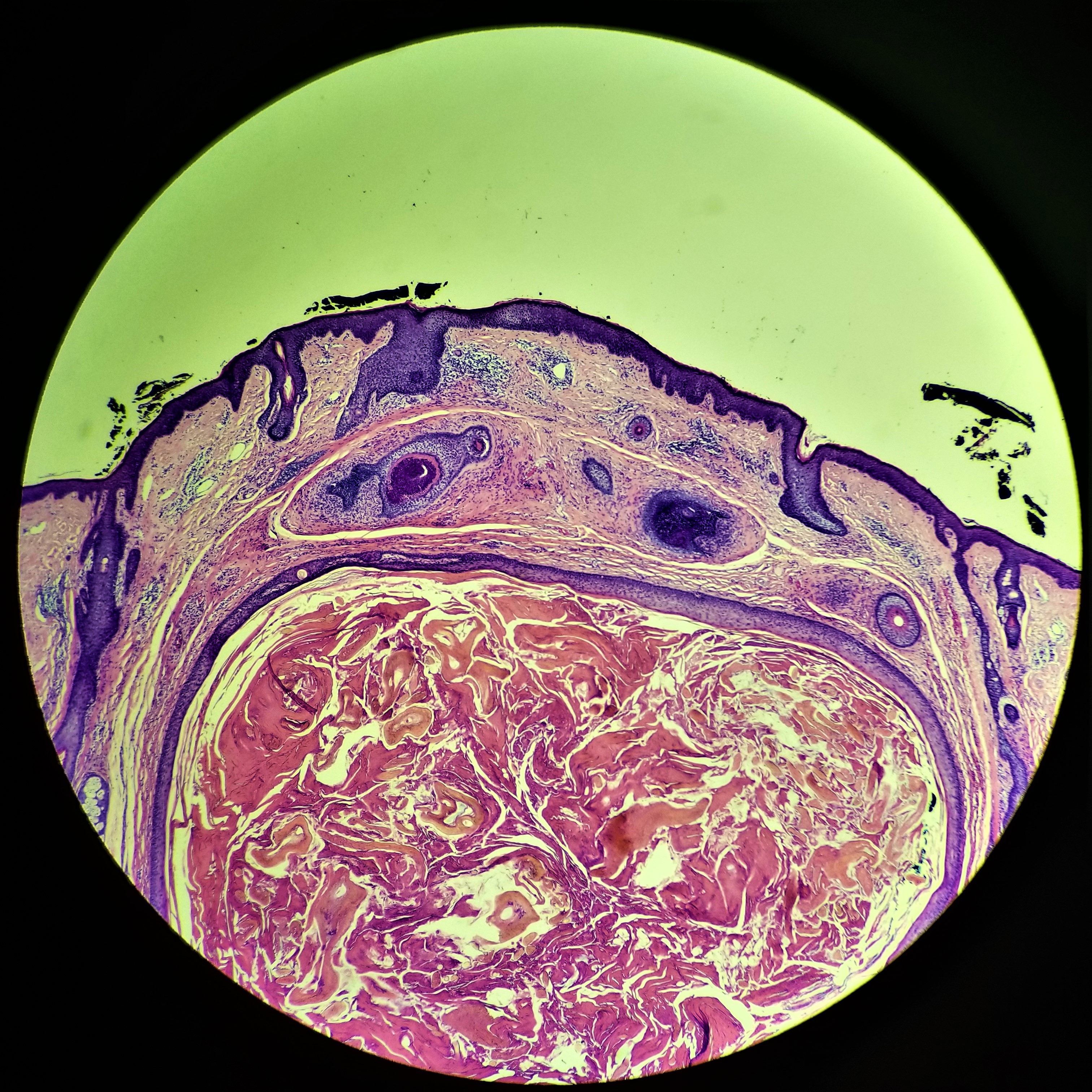
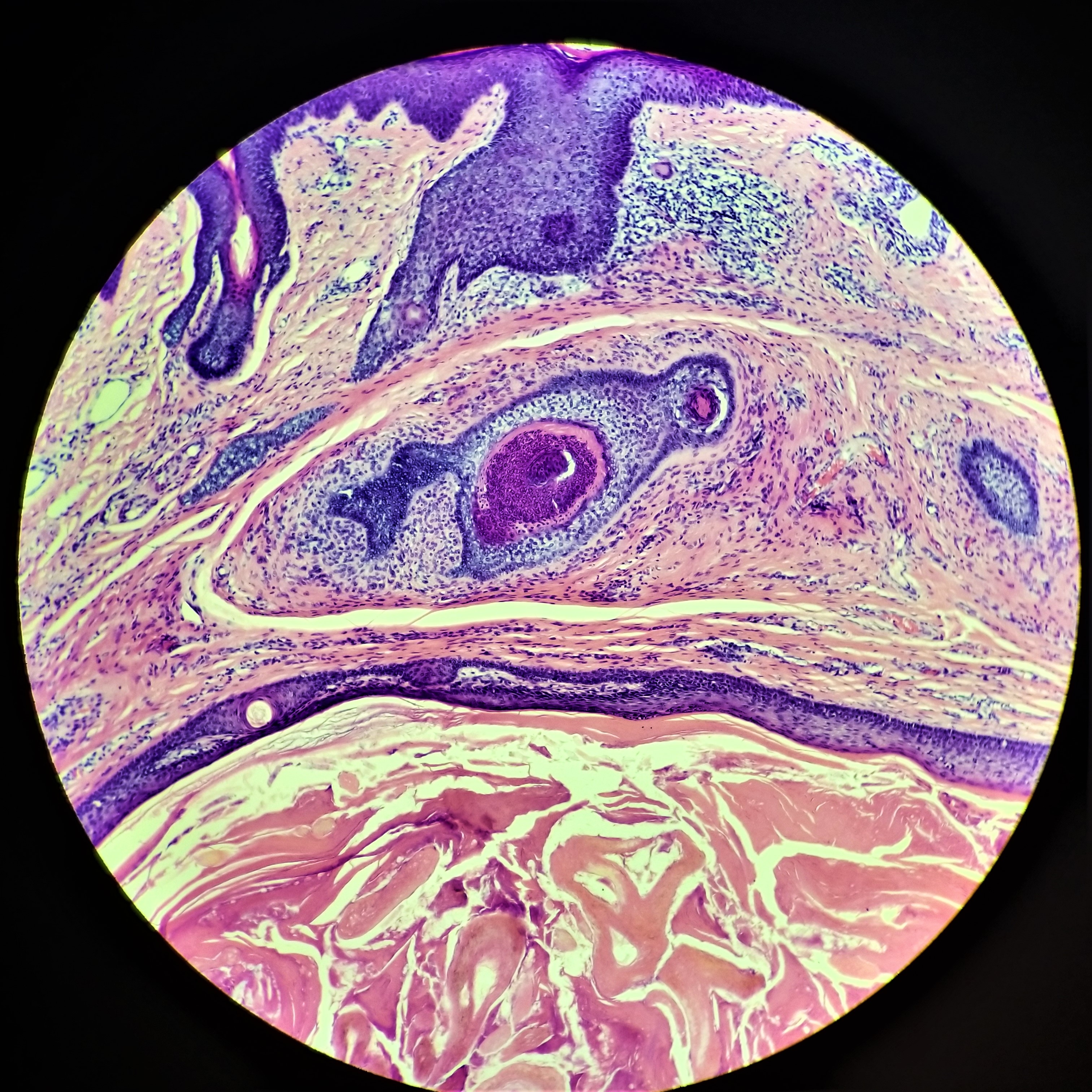
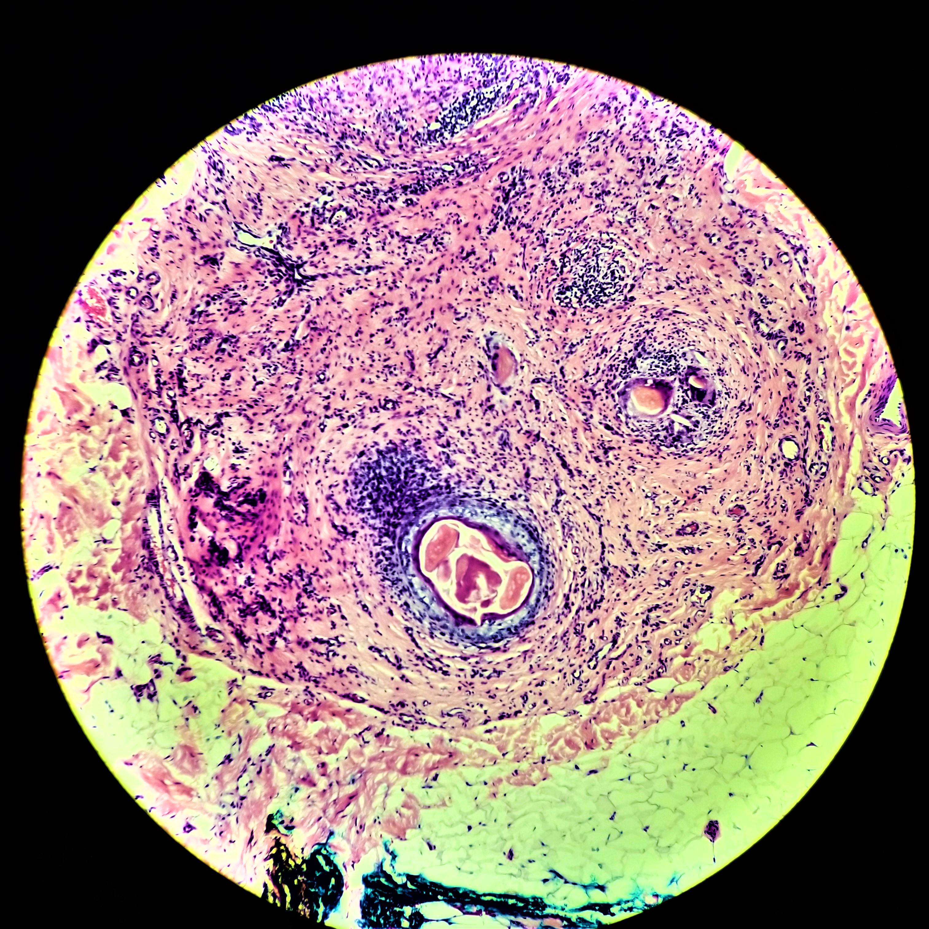
33F, lesion to face


  Report Record

User Feedback


Anil Patki

Posted

A keratinous cyst with trichilemmal keratinization. Around the main cyst are islands of basaloid cells with papillary mesenchymal bodies. I think this is a solitary trichoepitheioma. Alternative differential diagnoses are cutaneous keratocyst and milia.

Share this comment


Link to comment
share_externally

John Zhang

Posted

Central dilated hair follicle surrounded by many small hair follicles - trichofolliculoma. I think the last photo represents a reaction to ruptured hair follicles, rather than a fibrous and myxoid stromal component.

Share this comment


Link to comment
share_externally

This is a cystic lesion with epithelial and stromal components, sort of a biphasic cystic lesion. I usually put these types of cystic lesion in the category of fibrofolliculoma / trichodiscoma / folliculosebaceous cystic hamartoma. I do not see sebaceous units attached to the cyst, so i do not think it' s a FCH. The epithelial components look kind of mature so I favor trichodiscoma over fibrofolliculoma. But I think they all represent the opposite ends of the same type of lesion. I would agree with Vincenzo.

Share this comment


Link to comment
share_externally

Limin Yu

Posted

Thank everyone for the insightful discussion.  This is a trichofolliculoma for me -- a central cystic lesion with secondary radiating vellus hair follicles.  If this is more fibrous stromal changes, I would entertain the idea of fibrofolliculoma.  Thanks!

Share this comment


Link to comment
share_externally


Join the conversation

You can post now and register later. If you have an account, sign in now to post with your account.

Guest
Add a comment...

×   Pasted as rich text.   Paste as plain text instead

  Only 75 emoji are allowed.

×   Your link has been automatically embedded.   Display as a link instead

×   Your previous content has been restored.   Clear editor

×   You cannot paste images directly. Upload or insert images from URL.

Loading...

×
×
  • Create New...