Jump to content
In this section we have spot diagnoses posted on a daily basis since June 2010, now over 4000! You can review the archived cases and read the suggested diagnoses by users and the final comment by the contributors.
Case are uploaded each week day by 10 am UK time with the correct diagnosis will generally be posted at 8 pm UK time. Why not view the most recent spot diagnosis and proffer a diagnosis?

Case Number : Case 2214 - 4 December 2018 Posted By: Uma Sundram

Please read the clinical history and view the images by clicking on them before you proffer your diagnosis.
Submitted Date :
   (0 reviews)

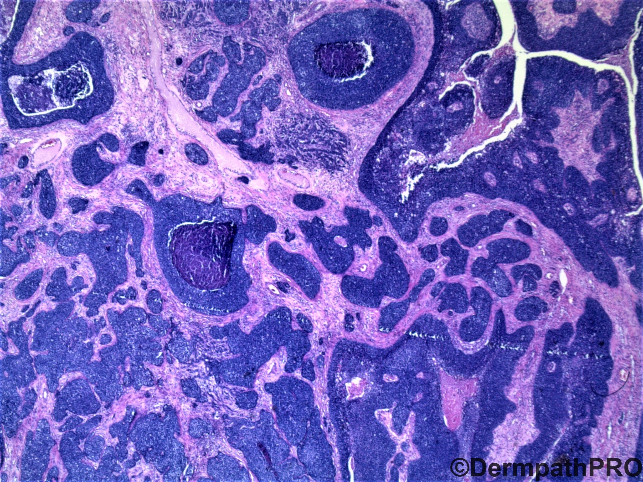
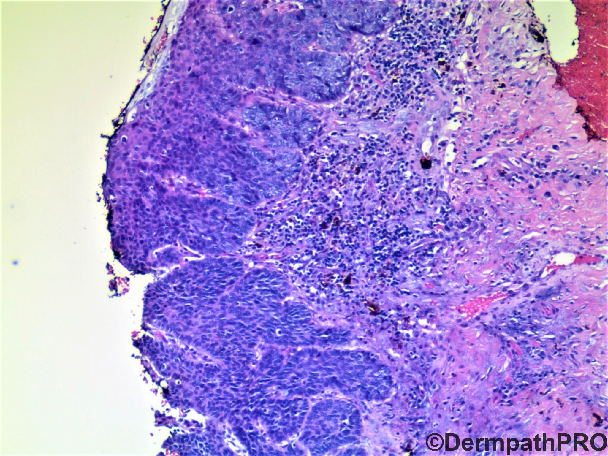
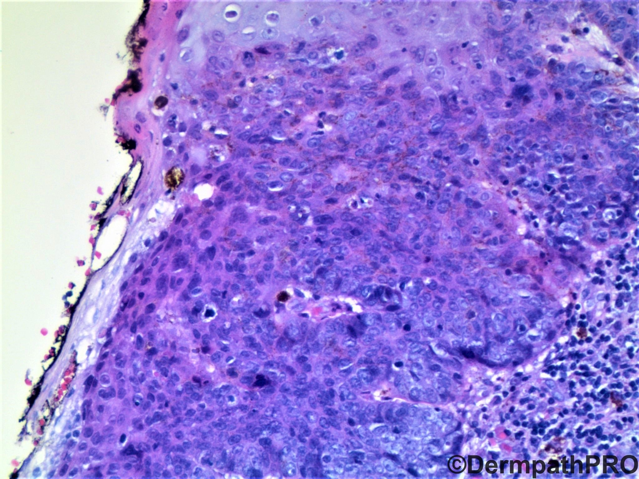
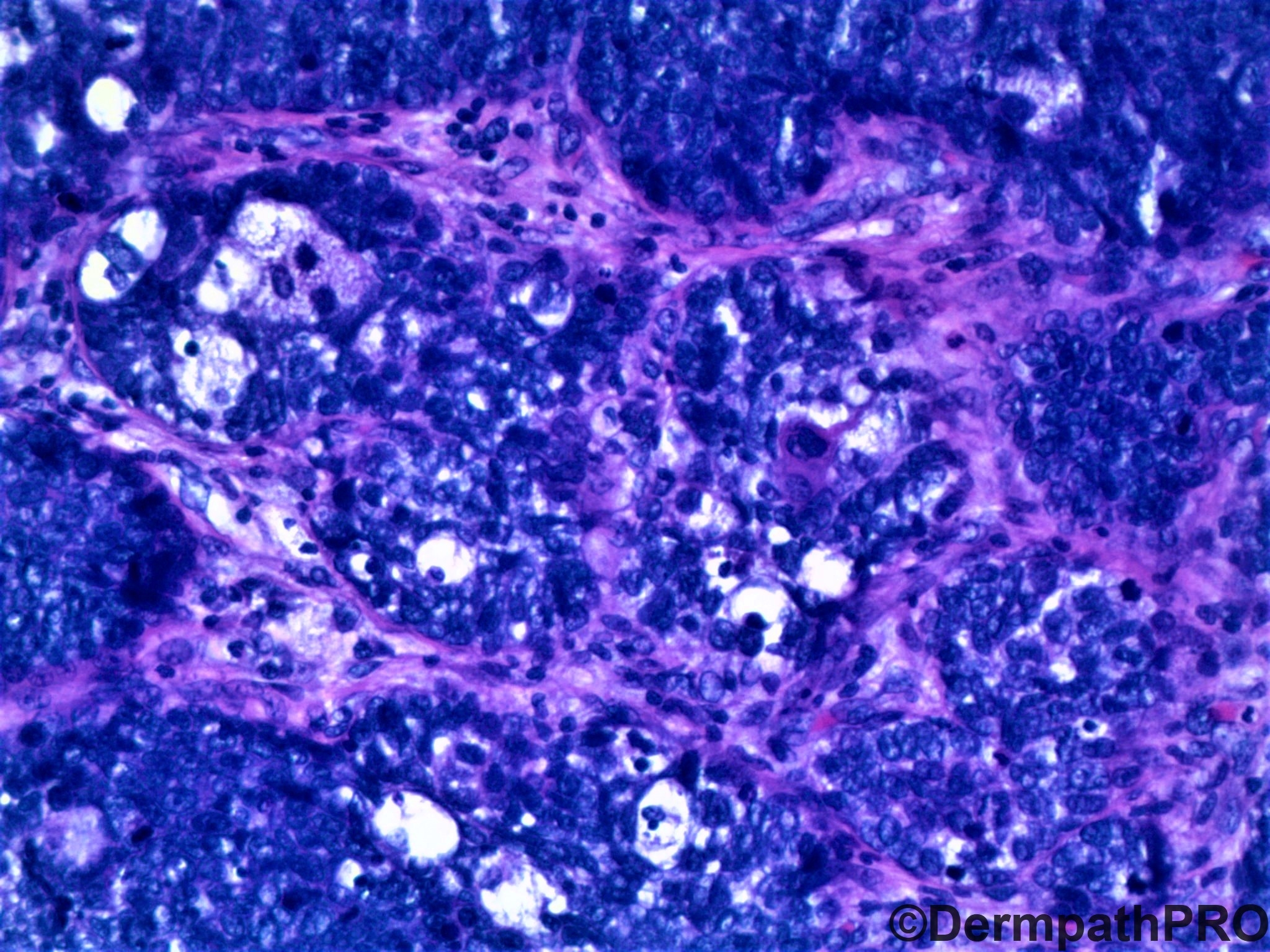
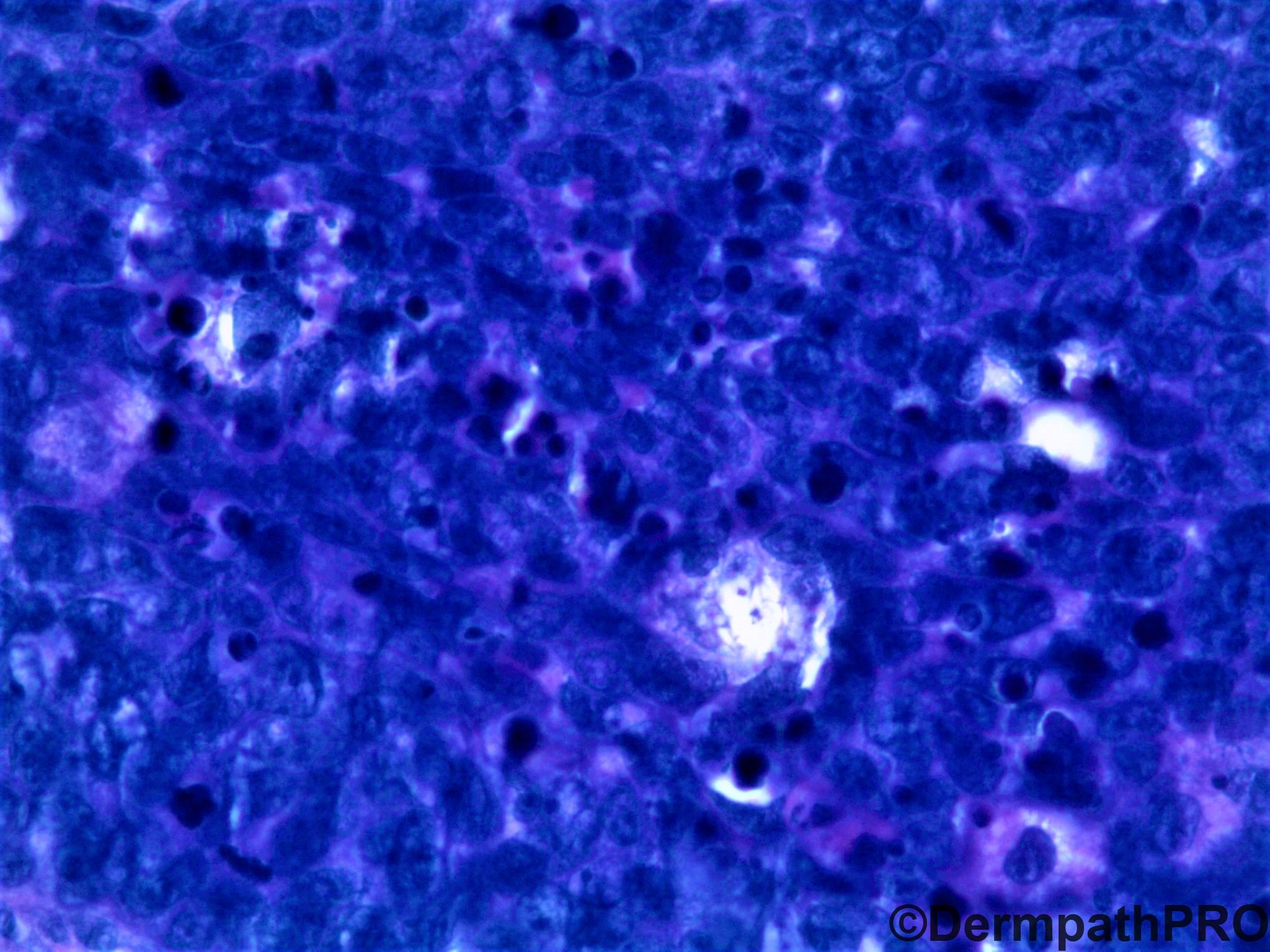
58 year old woman with development delay and a vulvar lesion.


  Report Record

User Feedback


Alex-Ventura-Leon

Posted

My impressions are Basalod SCC vs Sebaceous Carcinoma. I favor the first.

Share this comment


Link to comment
share_externally

Basal cell carcinoma.  Could be a manifestation of nevoid basal cell carcinoma syndrome as the site of the lesion and mental retardation of the patient suggest. 

Share this comment


Link to comment
share_externally

Krishnakumar subramanian

Posted

looks like sebaceous gland carcinoma with intraepithelial spread and dermal invasion. because of the basaloid component could be a sebaceous epithelioma change to adenocarcinoma. the tumor is in area other than head and neck and cystic changes are seen in tumor. I would like to rule out Muir torr syndrome, may be get some IHC done for loss of MSH-6 expression 


Share this comment


Link to comment
share_externally

Victor Delgado

Posted

I would like to favour Basaloid Squamous Cell Carcinoma. So, Dr. Uma Sundram wath was the final diagnosis?

Share this comment


Link to comment
share_externally

vincenzo

Posted (edited)

I favor extra ocular poorly differentiated sebaceous carcinoma.  Vulva is a well known extra-eyelids site.  This is a fully malignant neoplasia, instead BCC with sebaceous differentiation ( if does exist ), is a conventional BCC ( epithelial-stromal retraction, periepithelial mucin, peripheral palisading, mild/moderate atypia ). Muir-Torre syndrome?

Edited by vincenzo

Share this comment


Link to comment
share_externally

My first impression was a basaloid SCC, but the last 2 pictures show sebaceus differentiation that make us suspect of sabaceous carcinoma. IHQ with adipophilin and Factor XIIIa can be of great help. If we confirm the diagnosis, investigation of Muir Torre syndrome is necessary.

Share this comment


Link to comment
share_externally

Uma Sundram

Posted

Apologies, fellow spot diagnostic friends, for the delay in posting. An excellent discussion.

We too were between basaloid SCC and sebaceous carcinoma. Adipophilin was positive only in the sebocyte areas, leading us to favor this less. It may have been involvement of a pre existing sebaceous unit by carcinoma. All 4 MSI proteins were expressed with no loss. I did not favor BCC for the reasons already elaborated by the other wonderful posts here.

Share this comment


Link to comment
share_externally

Uma Sundram

Posted

Stay tuned for next weeks post -- I promise to be on top of it this time! The holiday rush in the US is leading to a LOT of skin biopsies :)

Share this comment


Link to comment
share_externally


Join the conversation

You can post now and register later. If you have an account, sign in now to post with your account.

Guest
Add a comment...

×   Pasted as rich text.   Paste as plain text instead

  Only 75 emoji are allowed.

×   Your link has been automatically embedded.   Display as a link instead

×   Your previous content has been restored.   Clear editor

×   You cannot paste images directly. Upload or insert images from URL.

Loading...

×
×
  • Create New...