Jump to content
In this section we have spot diagnoses posted on a daily basis since June 2010, now over 4000! You can review the archived cases and read the suggested diagnoses by users and the final comment by the contributors.
Case are uploaded each week day by 10 am UK time with the correct diagnosis will generally be posted at 8 pm UK time. Why not view the most recent spot diagnosis and proffer a diagnosis?

Case Number : Case 2215 - 5 December 2018 Posted By: Dr. Hafeez Diwan

Please read the clinical history and view the images by clicking on them before you proffer your diagnosis.
Submitted Date :
   (0 reviews)

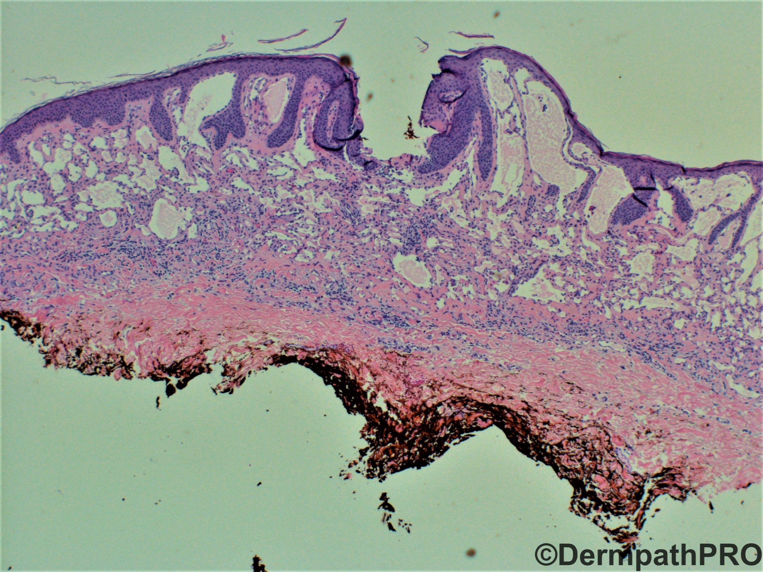
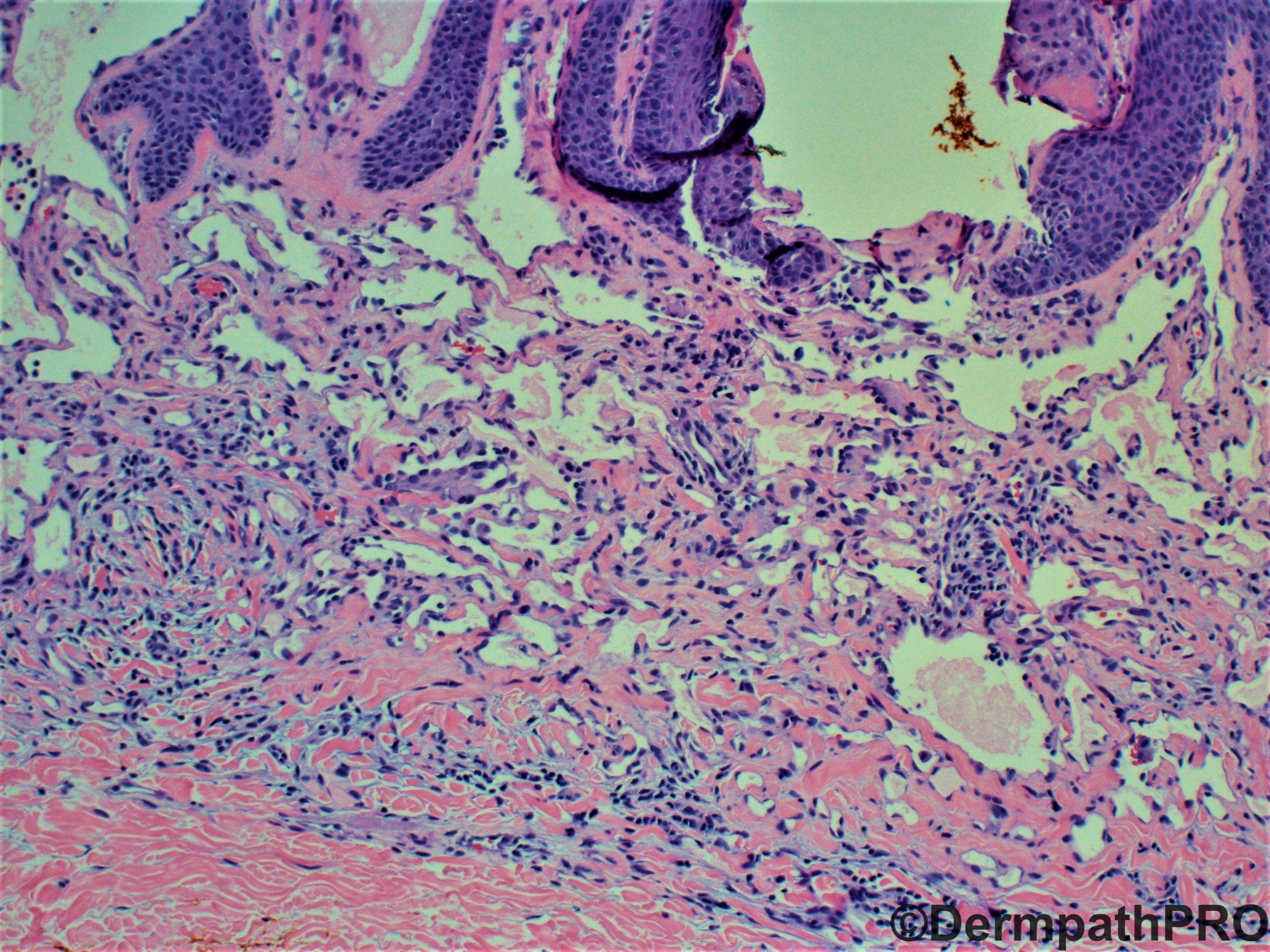
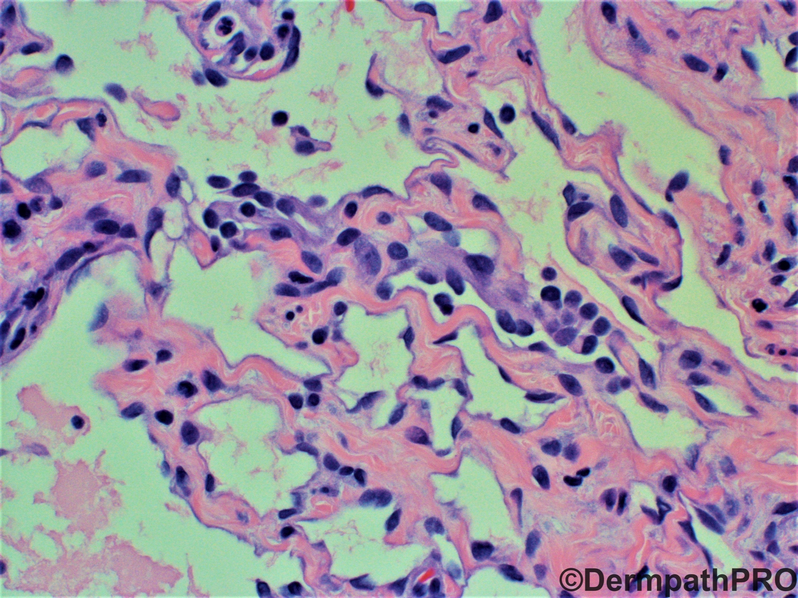
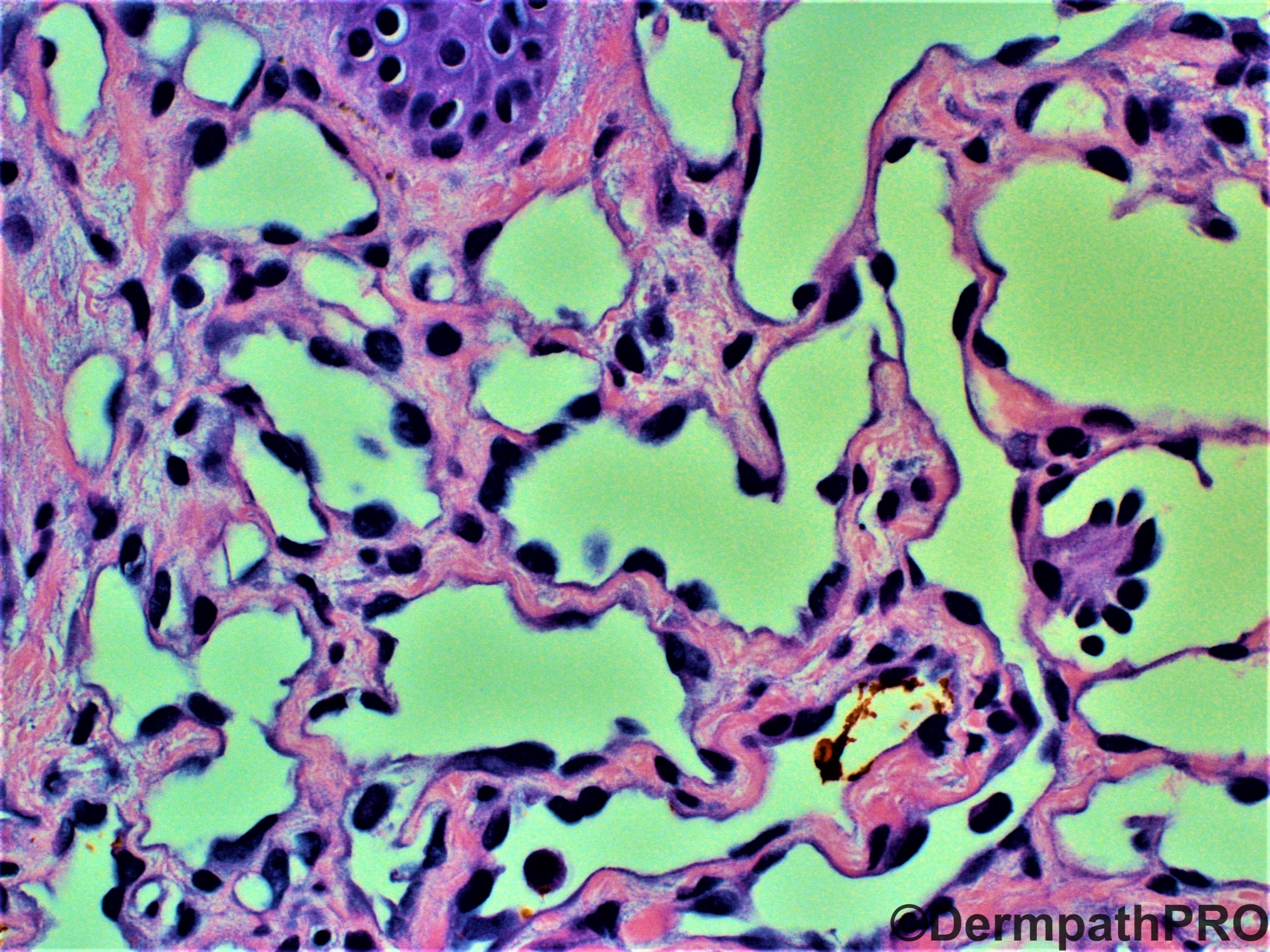
58 year-old female with right upper chest lesion. She has a history of inflammatory breast carcinoma. Clinical differential diagnosis: nevus versus seborrheic keratosis versus other.


  • Like 1
  Report Record

User Feedback


Hobnail hemangioma. I sometimes run HHV8 to make sure it is not Kaposi, because the lower portion of hobnail hemangiomas show slit-like vascular channels, almost spindle looking.

Share this comment


Link to comment
share_externally

Victor Delgado

Posted

Hobnail Hemangioma (targetoid hemosiderotic hemangioma). Dilated superficial vessels with hobnail endothelial cells, papillary tufts in superficial vessels, CD31 and D2-40 positive, CD34 negative.

DD. K. Sarcoma and Retiform Hemangioendothelioma.

Share this comment


Link to comment
share_externally

Krishnakumar subramanian

Posted

I would like to have some clinical information. whether any local radiotherapy was given and if so are the lesions occurring at the site of radiotherapy

If so these could be vascular malformations post radiotherapy and I would be care fully not to miss angiosarcoma which could be  deceptive

may be a Ki 67 can done to know the proliferation kinetics


Share this comment


Link to comment
share_externally

Alex-Ventura-Leon

Posted

Yes. Hobnail hemangioma is a good thought. But I'm kind of concerned about the possibility of Atypical Vascular Lesión in the setting of prior radiotherapy.

Share this comment


Link to comment
share_externally

Yes, agree with HH!!! Nice biphasic pattern: superficial areas with dilated irregular vessels, some of these with papillary endoluminal Dabska-like projections.  Deep areas with compressed spindle cell bounding slit-like vessels. Nice case!

Share this comment


Link to comment
share_externally

Raul Perret

Posted (edited)

For me first hypothesis = atypical vascular lesion. Was the patient treated with radiotherapy? We do MYC by immunohistochemistry routinely in this kind of cases. 

Edited by Raul Perret

Share this comment


Link to comment
share_externally

Dr. Hafeez Diwan

Posted

Yes, the patient received radiation therapy.  Atypical vascular lesion.

Share this comment


Link to comment
share_externally

Excellent point and excellent case! Upon further study it appears that atypical vascular lesion is known to sometimes show biphasic pattern as seen in hobnail hemangioma. The lack of hemosiderin, the lack of extravasated erythrocytes and the radiation history would favor atypical vascular lesion.

Share this comment


Link to comment
share_externally

Yes. I was wrong. After Raul’s comment, I was reconsidering the clinical-pathological features and changing my diagnosis!!!

Share this comment


Link to comment
share_externally


Join the conversation

You can post now and register later. If you have an account, sign in now to post with your account.

Guest
Add a comment...

×   Pasted as rich text.   Paste as plain text instead

  Only 75 emoji are allowed.

×   Your link has been automatically embedded.   Display as a link instead

×   Your previous content has been restored.   Clear editor

×   You cannot paste images directly. Upload or insert images from URL.

Loading...

×
×
  • Create New...