Jump to content
In this section we have spot diagnoses posted on a daily basis since June 2010, now over 4000! You can review the archived cases and read the suggested diagnoses by users and the final comment by the contributors.
Case are uploaded each week day by 10 am UK time with the correct diagnosis will generally be posted at 8 pm UK time. Why not view the most recent spot diagnosis and proffer a diagnosis?

Case Number : Case 2217 - 7 December 2018 Posted By: Dr. Richard Carr

Please read the clinical history and view the images by clicking on them before you proffer your diagnosis.
Submitted Date :
   (0 reviews)

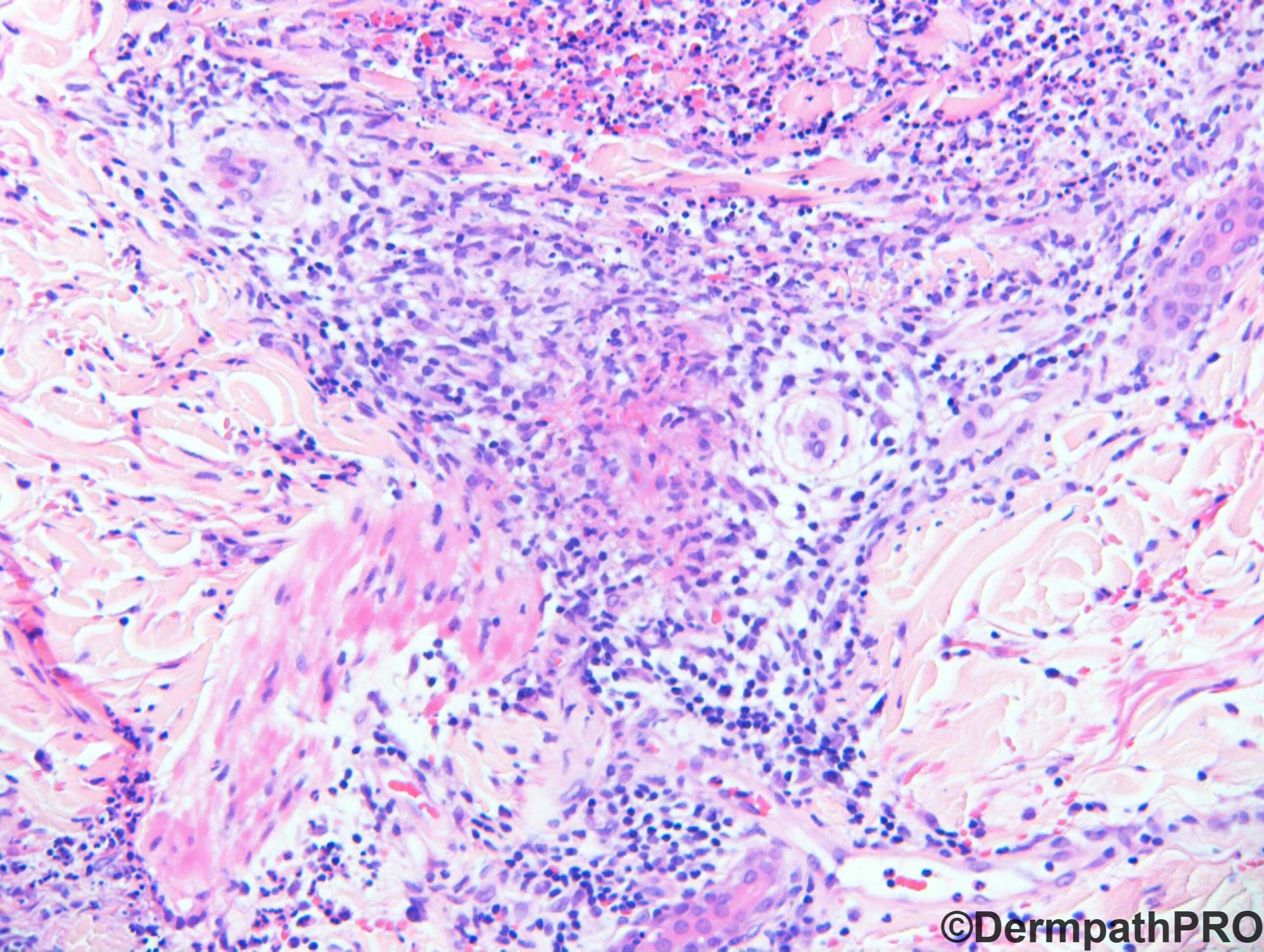
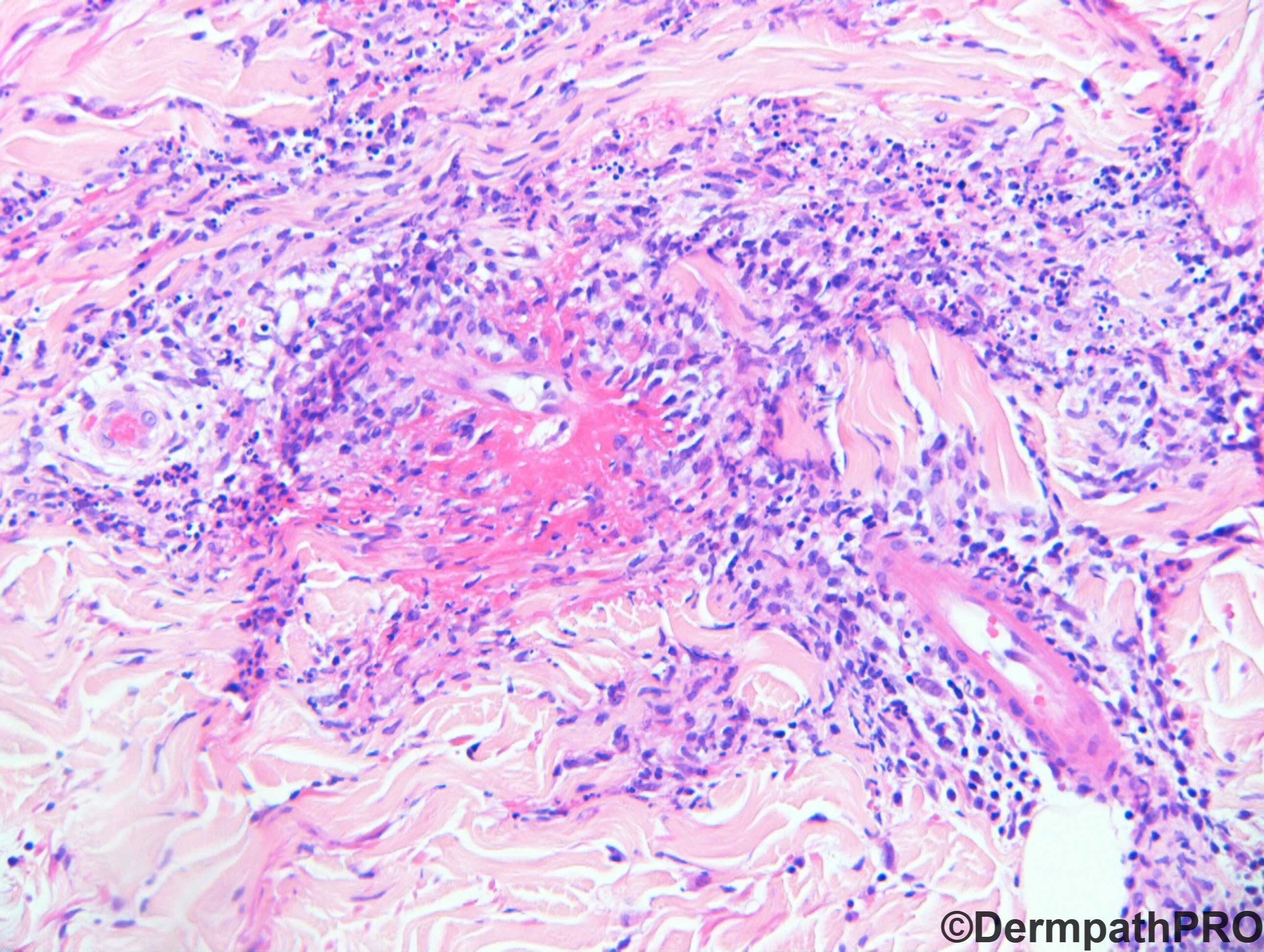
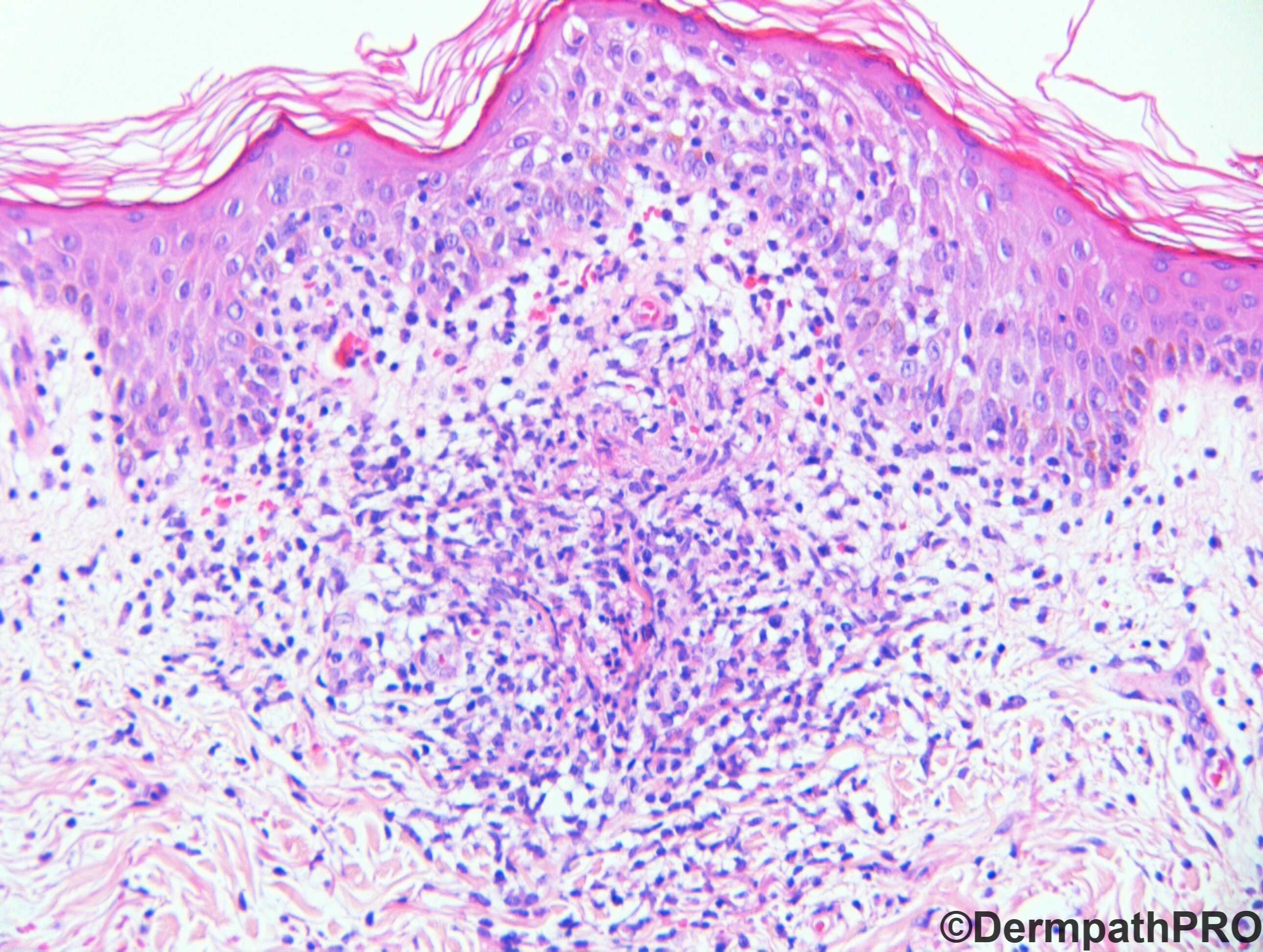
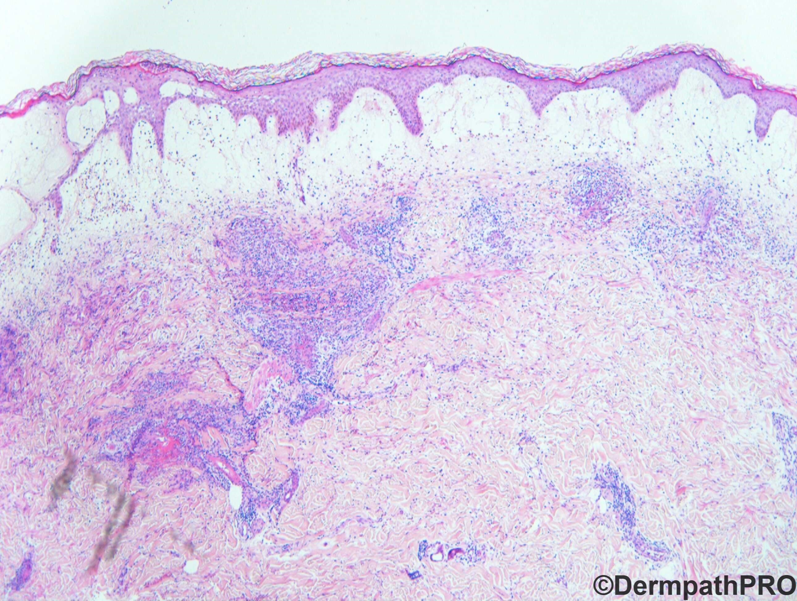
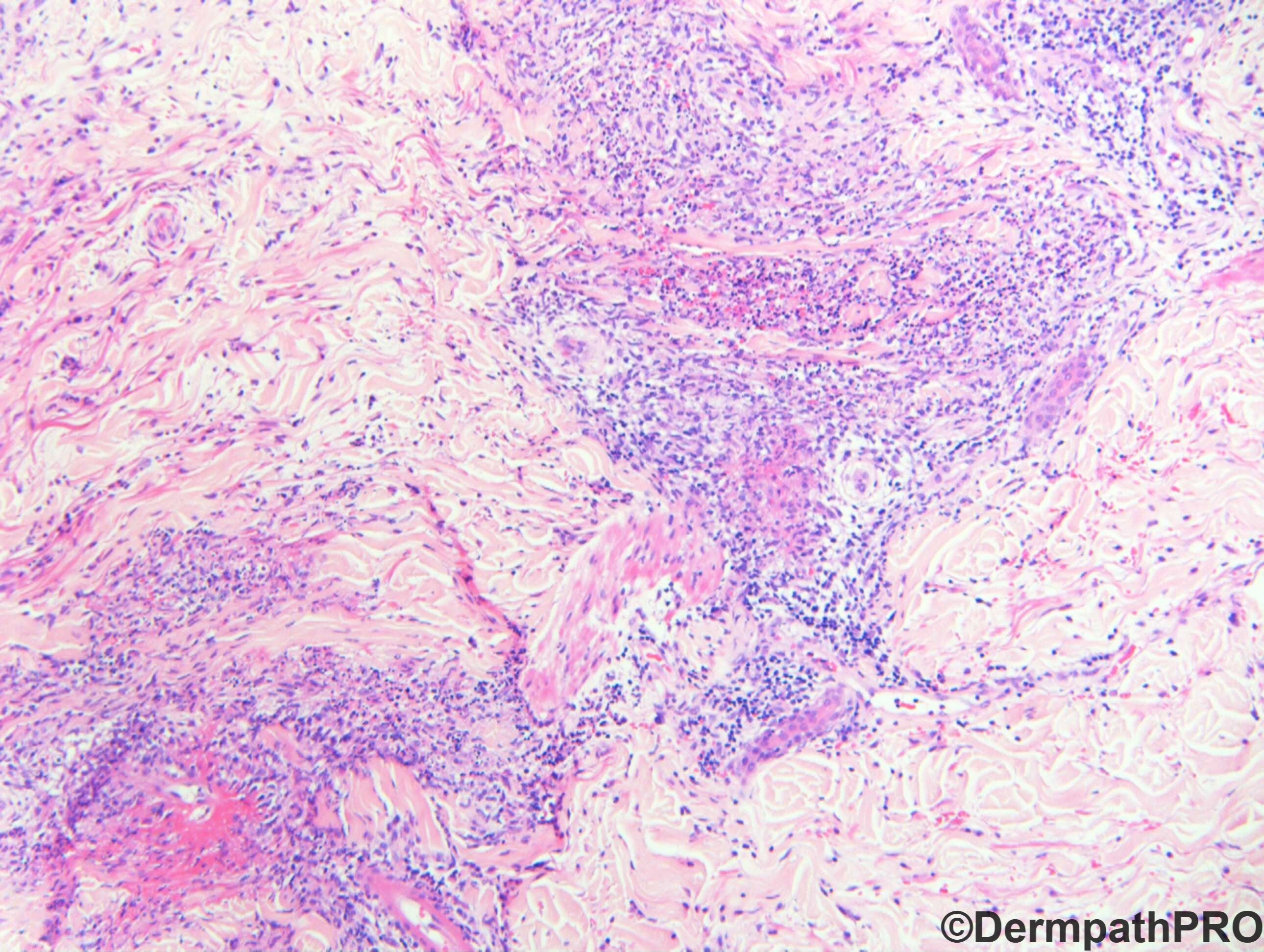
Presented with flu-like symptoms, sore throat, developed acute symmetrical rash over face, hands and forearms. Immunofluorescence negative. CRP176, WCC 6.5 Hb133 ANA negative, Blood cultures negative.

  Report Record

User Feedback


Krishnakumar subramanian

Posted

fever with rash and biopsy shows dermal edema with small vessel vasculitis with mixed inflammation with lymphocytes and a few eosinophils. fibrin deposition seen around vessels

Possible infective cause due to bacteria/rickettsial infections possibly

Share this comment


Link to comment
share_externally

vincenzo

Posted (edited)

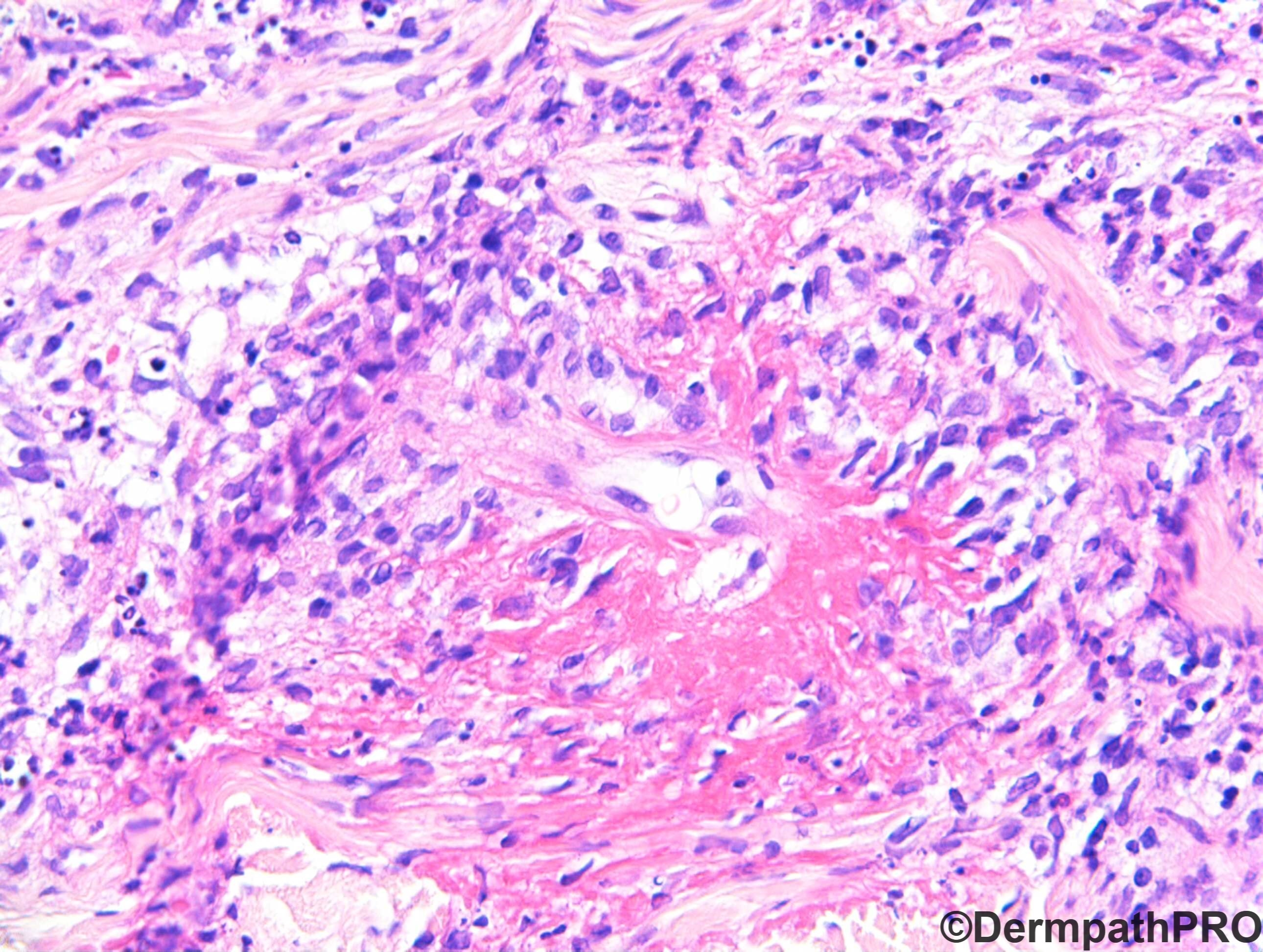
Difficult for me!  It seems a Sweet Syndrome , but Vasculitis doesn’t fit well. Eosinophils and vasculitis would be more in keeping with Churg Strauss but no Asthma.  Flames figures would be in keeping with Wells syndrome but with CS disease also.  Favore a Sweet syndrome with eosinophils and vasculitis for silhouette and other histological findings but not sure. 

Edited by vincenzo

Share this comment


Link to comment
share_externally

Alex-Ventura-Leon

Posted

Humm. It seem like a vasculitis whit granulomas (my impression) and flame figures (in keep whit eosinophils i think) so my interpretation is Eosinophilic granulomatosis with Polyangitis. But Im agree that is a difficult case.

Share this comment


Link to comment
share_externally

vincenzo

Posted (edited)

Maybe the secret to find our rosebud is in the last picture : cutaneous Well's syndrome in visceral larva migrans??? But doesn't fit clinically ( why symmetrical? )...No. SS is yet my favorite spot.

Edited by vincenzo

Share this comment


Link to comment
share_externally

There are a few necrotic keratinocytes. This could be drug induced erythema multiforme. IF can be negative in EM

Share this comment


Link to comment
share_externally

vincenzo

Posted (edited)

Found this:

Flame figures in linear IgA bullous dermatosis: a novel histopathologic finding.

Fulton E, et al. Dermatol Online J. 2017.
what do you thing guys?  Of course The IF would have been positive....I don’t Know!!! Waiting for the answer.  
Edited by vincenzo

Share this comment


Link to comment
share_externally

Saman Fatah

Posted

Acute Febrile Neutrophilic Dermatosis - Sweet’s Syndrome (SS). Without the clinical information it would be difficult to suspect this with confident.

Vasculitis can be seen as an epiphenomenon or a secondary process in SS and one should not exclude SS if other features consist with this diagnosis.

Some of the mononuclear cells which may look like histiocytes “histiocytoid” can be immature granulocyte precursors, MPO and other markers can guide their lineage in doubtful cases.

Share this comment


Link to comment
share_externally

Krishnakumar subramanian

Posted

looking at the marked dermal edema, it could be sweet syndrome

Share this comment


Link to comment
share_externally

Dr. Richard Carr

Posted

This case was from an external quality assurance scheme and had to be removed from scoring due to lack of consensus.

The slightly favoured response was leucocytoclastic vasculitis (44%) with Sweet's syndrome / neutrophilic dermatosis a close second (40%), and Vasculitis (26%) a moderate 3rd.

Other suggestions (all <1% unless stated, probably differentials) included: Sweet's with LCV (1.9%); Granulomatous vasculitis; Necrotising vasculitis; Erysipleas/cellulitis; Palisaded neutrophilic granulomatous dermatitis; EM, DH, Kikuchi, suppurative granulomatous; TEN, Stevens-Johnson; EBA; pyoderma gangrenosum; lupus; BP; fixed drug; Cowpox; EED; eosinophilic cellulitis.

My personal response: Leucocytoclastic vasculitis (Sweet's variant), commenting: Clinical sounds like Sweet's, histology is more LCV! I guess it's academic. 

Certainly Bernhard Zelger a mentor for vasculitis/vasuolopathy considers Sweet's as a type of LCV.

Regarding the pathology as Sweet's evolves it becomes vaguely granulomatous. This is fibrin (not a flame figure, eosinophils are not seen to any degree). I don't think this is the histiocytoid variant (which we have seen in dermpathpro).

Share this comment


Link to comment
share_externally

Alex-Ventura-Leon

Posted

Nice Case and great discussion by Dr. Carr and the other colleagues.

I learn more of Sweet Syndrome today.

Share this comment


Link to comment
share_externally

Nice case. My first and last impression is hypersentivity leucocytoclastic vasculitis due to previous infection.

Share this comment


Link to comment
share_externally


Join the conversation

You can post now and register later. If you have an account, sign in now to post with your account.

Guest
Add a comment...

×   Pasted as rich text.   Paste as plain text instead

  Only 75 emoji are allowed.

×   Your link has been automatically embedded.   Display as a link instead

×   Your previous content has been restored.   Clear editor

×   You cannot paste images directly. Upload or insert images from URL.

Loading...

×
×
  • Create New...