Jump to content
In this section we have spot diagnoses posted on a daily basis since June 2010, now over 4000! You can review the archived cases and read the suggested diagnoses by users and the final comment by the contributors.
Case are uploaded each week day by 10 am UK time with the correct diagnosis will generally be posted at 8 pm UK time. Why not view the most recent spot diagnosis and proffer a diagnosis?

Case Number : Case 1998 - 1 Feb 2018 Posted By: Iskander H. Chaudhry

Please read the clinical history and view the images by clicking on them before you proffer your diagnosis.
Submitted Date :
   (0 reviews)

68M;

Biopsy 1) Mid back incision biopsy. Red rash. 18/12 can be itchy/ indurated plaques.
Biopsy 2) Abdomen Incision biopsy. Annular scaly part to rash

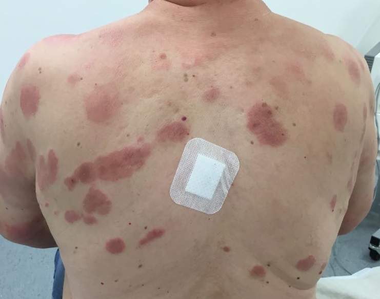
Edited by Admin_Dermpath


  Report Record

User Feedback


Erythematous pruritic plaques on the trunk and those on the dorsa of the hands show symmetry. Epidermis shows psoriasiform hyperplasia with patchy spongiosis and eosinophils. Dermis shows perivascular infiltrate with eosinophils. These features suggest a psoriasiform drug rash. Has the patient received beta blockers or terbinafine?

Share this comment


Link to comment
share_externally

Dr. Mona Abdel-Halim

Posted

Spongiotic Psoriasiform reaction pattern with eosinophils, not worrisome of CTCL on pathological level. I agree with the possibility of drug reaction. 

Share this comment


Link to comment
share_externally

Nitin Khirwadkar

Posted

Yes, spongiosis and psoriasiform. Eosinophils++. So, a drug reaction remains a distinct possibility. Otherwise, psorisiform eczema. No features of MF.

Share this comment


Link to comment
share_externally

Admin_Dermpath

Posted

Dr Richard Carr's comments:


Agree clinical could be CTCL (although hands look quite lichenified). The pathology is eczematous and not worrying for CTCL. Agree drug induced worth considering. If you have more than one biopsy PCR can be helpful and might allay clinical colleagues worries.

Share this comment


Link to comment
share_externally

vincenzo polizzi

Posted

Agree with above comments

But sincerely I was thinking of something like erythema chronicum migrans/Lyme disease...This is a very hard case for me.

Share this comment


Link to comment
share_externally

Saman Fatah

Posted

Congratulation again to the admin team for adding clinical photos when appropriate consent level allows. The images are of excellent quality and makes a significant difference especially in an inflammatory Dermpath case such as this one.

1. I would consider urticarial/pre-bullous/prodromal phase of an immunobullous disease especially bullous pemphigoid first. The protraced course of 18 month without expression of blisters is a bit unusual in my view and DIF can support or exclude this easily.

2. The reactive annular erythemas group is worth consdering if DIF is negative including Eosinophilic Annular Erythema. The gross spongiosis, eosinophil exocytosis and mild superficial perivascular lymphotic infiltrate will be atypical for conventional figurate erythemas including the superficial variant.

3. Drug is possible on histology basis alone but I am not aware of this clinical pattern of cutaneous drug eruption and it is very unlikely in this context.

4. Again neither clinically nor histologically is fits with an insect bite or anthopode assault.

5. Rare causes of other eosinophil rich dermatosis one need to be aware are Wells Syndrome and Eosinophilic Dermatosis of Haematological Malignancy (AKA insectbite-like reactions especially in those with CLL but certaingly not exclusive to this hence renamed lately EDHM). The infiltrate is probably too sperficial for the latter on histology alone. 


Share this comment


Link to comment
share_externally

Admin_Dermpath

Posted

From Dr Saleem Taibjee, Dermatologist and Dermatopathologist, U.K. Clinically it doesn’t look like to eczema to me.
I can understand why CTCL was considered. Would be an unusual drug reaction, but worth considering.

As we know, treatment can modify histology, so may be worthwhile repeating a biopsy in view of the clinical.

The previous posted suggestion of ruling out pre-bullous pemphigoid is also not unreasonable.


Share this comment


Link to comment
share_externally

Admin_Dermpath

Posted

Dr Chaudhry's diagnosis:

Based on clinical differentials working diagnosis is cutaneous drug eruption 

Share this comment


Link to comment
share_externally


Join the conversation

You can post now and register later. If you have an account, sign in now to post with your account.

Guest
Add a comment...

×   Pasted as rich text.   Paste as plain text instead

  Only 75 emoji are allowed.

×   Your link has been automatically embedded.   Display as a link instead

×   Your previous content has been restored.   Clear editor

×   You cannot paste images directly. Upload or insert images from URL.

Loading...

×
×
  • Create New...