Jump to content
In this section we have spot diagnoses posted on a daily basis since June 2010, now over 4000! You can review the archived cases and read the suggested diagnoses by users and the final comment by the contributors.
Case are uploaded each week day by 10 am UK time with the correct diagnosis will generally be posted at 8 pm UK time. Why not view the most recent spot diagnosis and proffer a diagnosis?

Case Number : Case 1999 - 2 Feb 2018 Posted By: Dr. Richard Carr

Please read the clinical history and view the images by clicking on them before you proffer your diagnosis.
Submitted Date :
   (0 reviews)

RAC7818

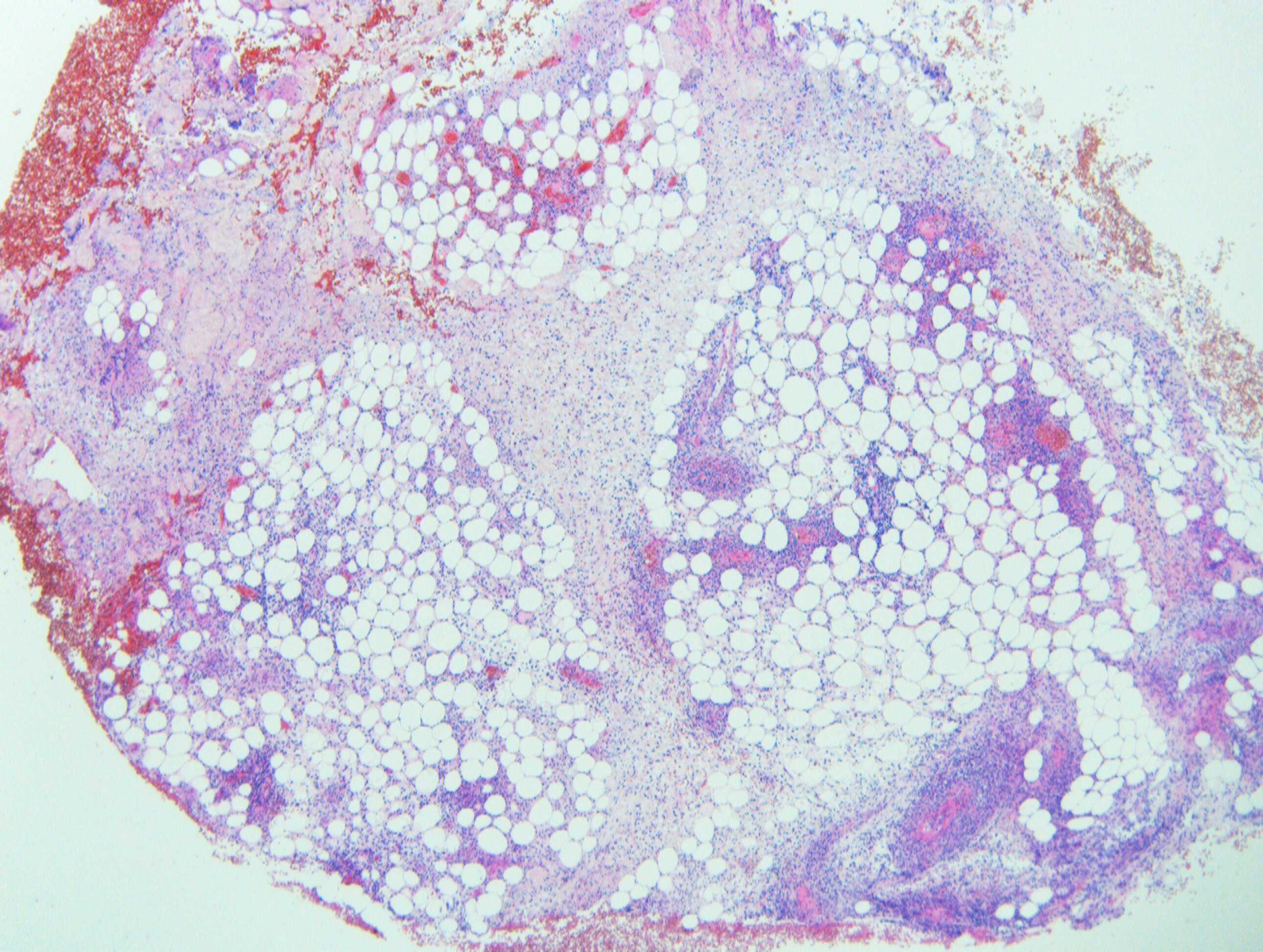
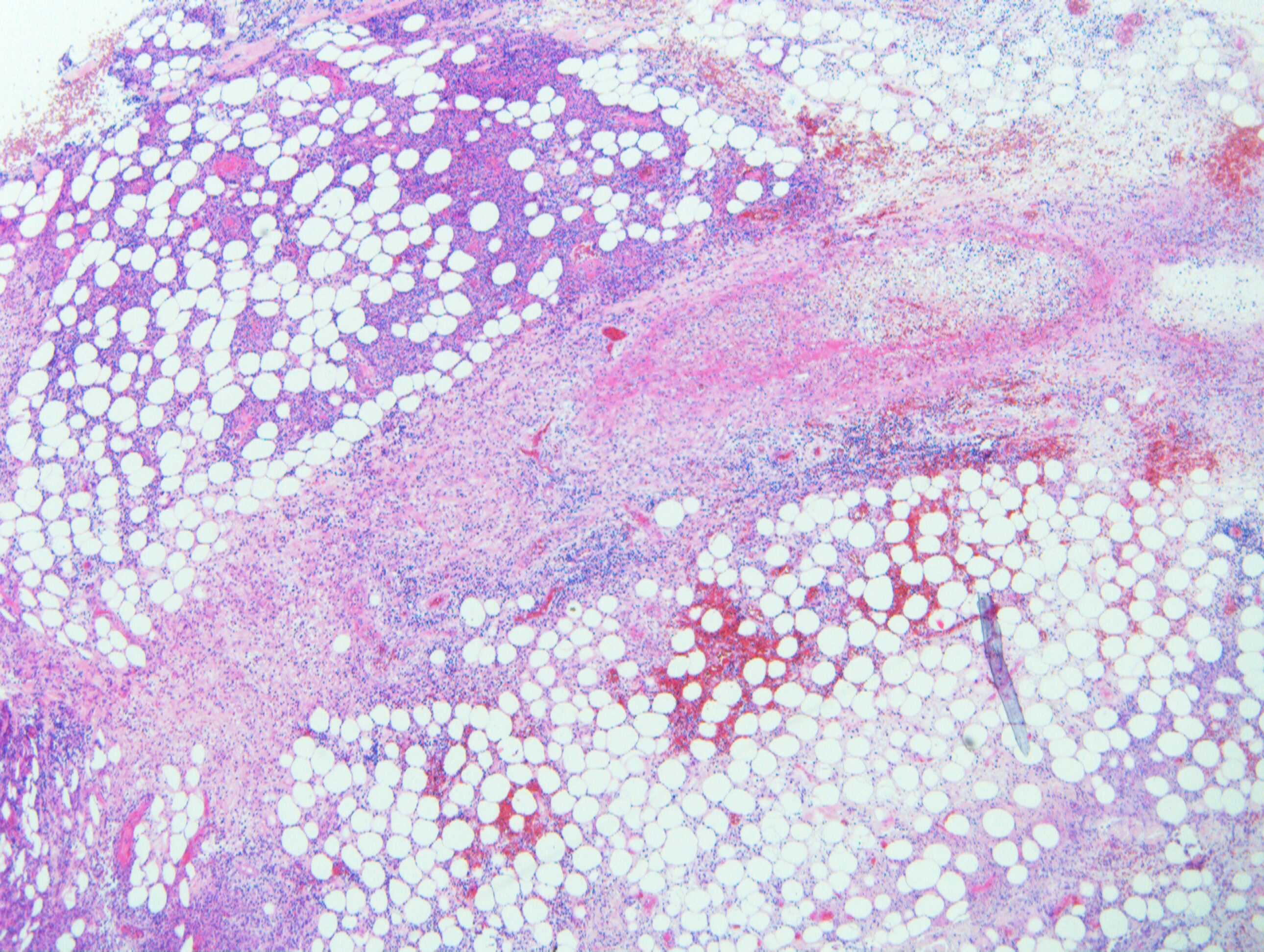
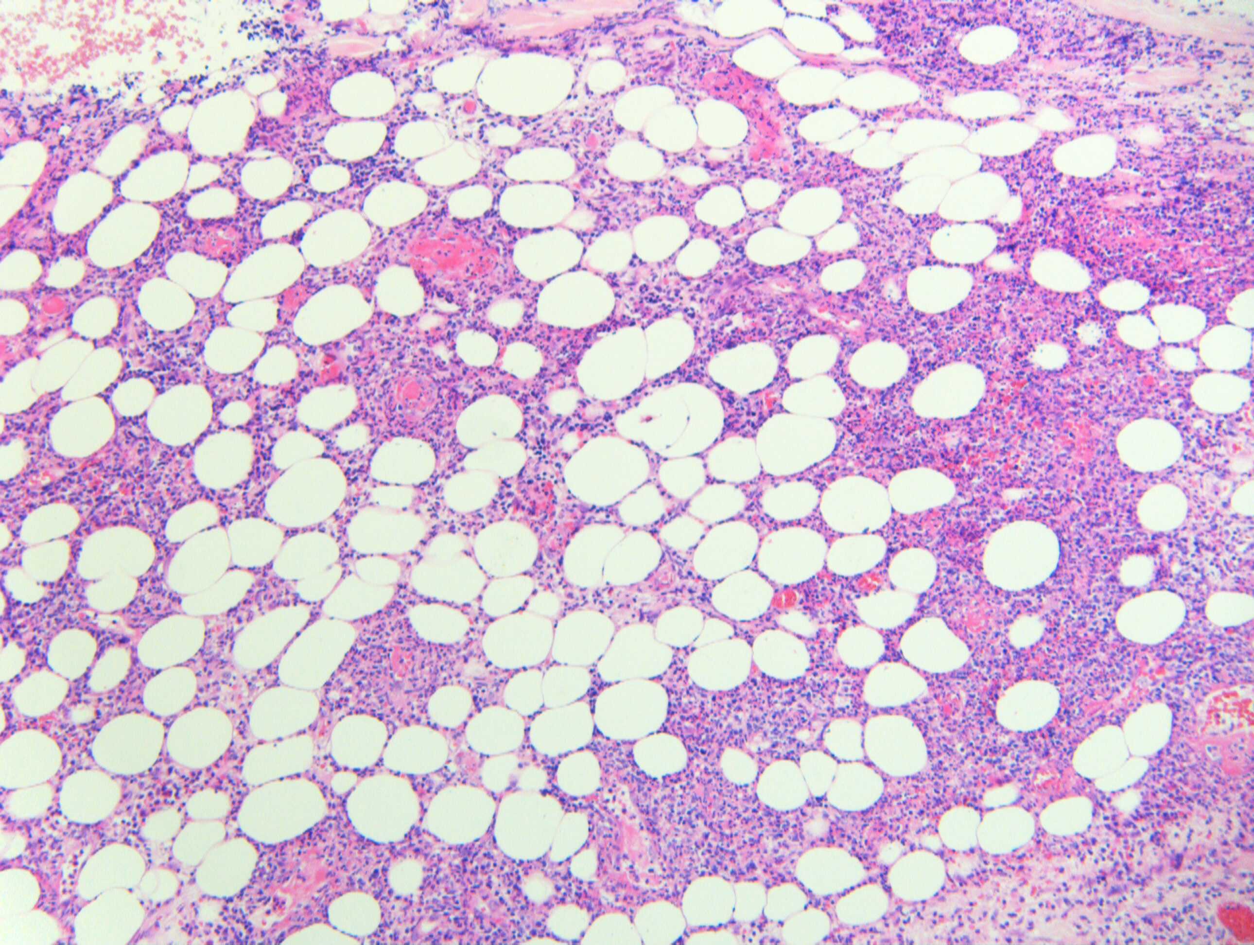
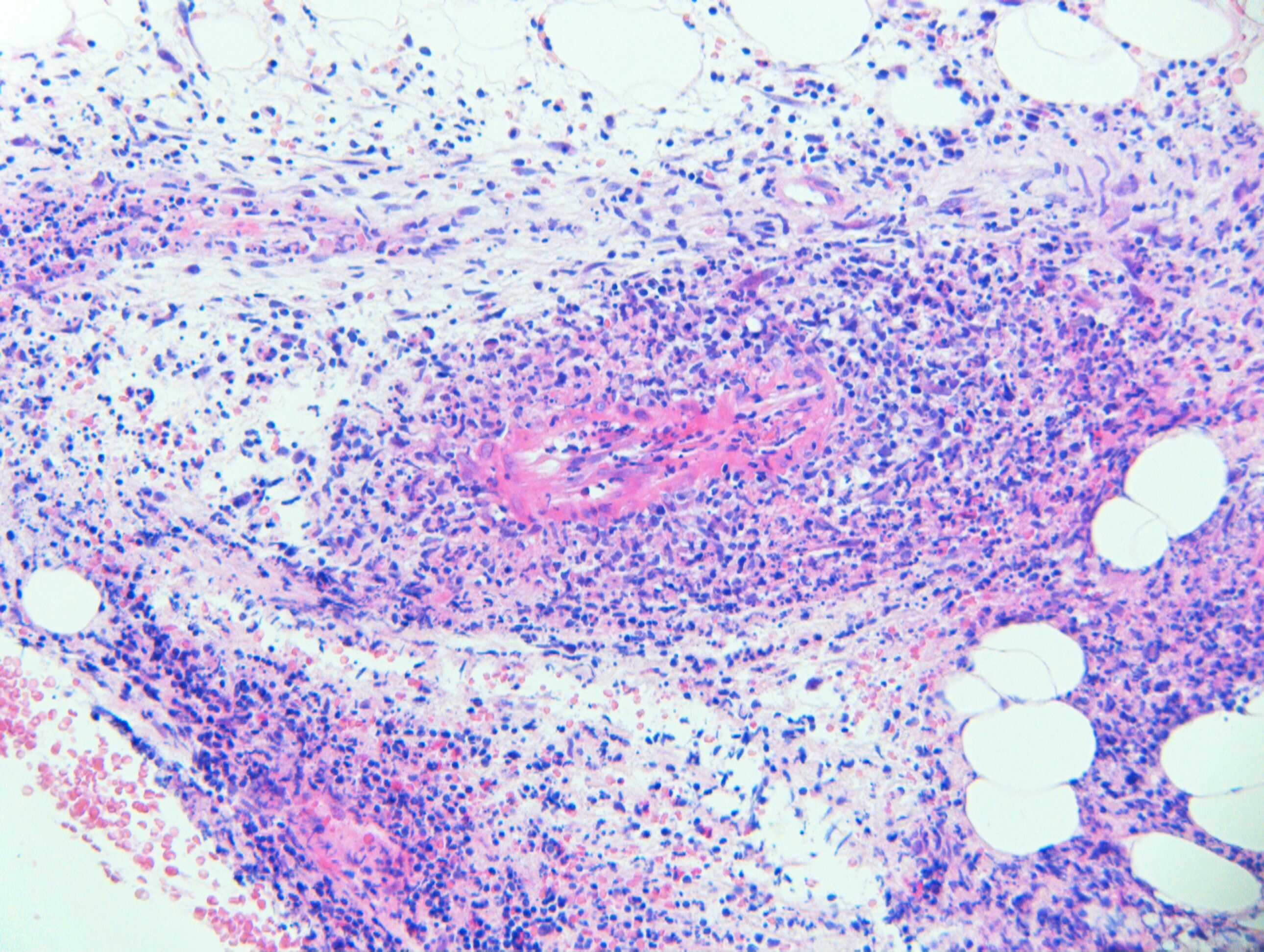
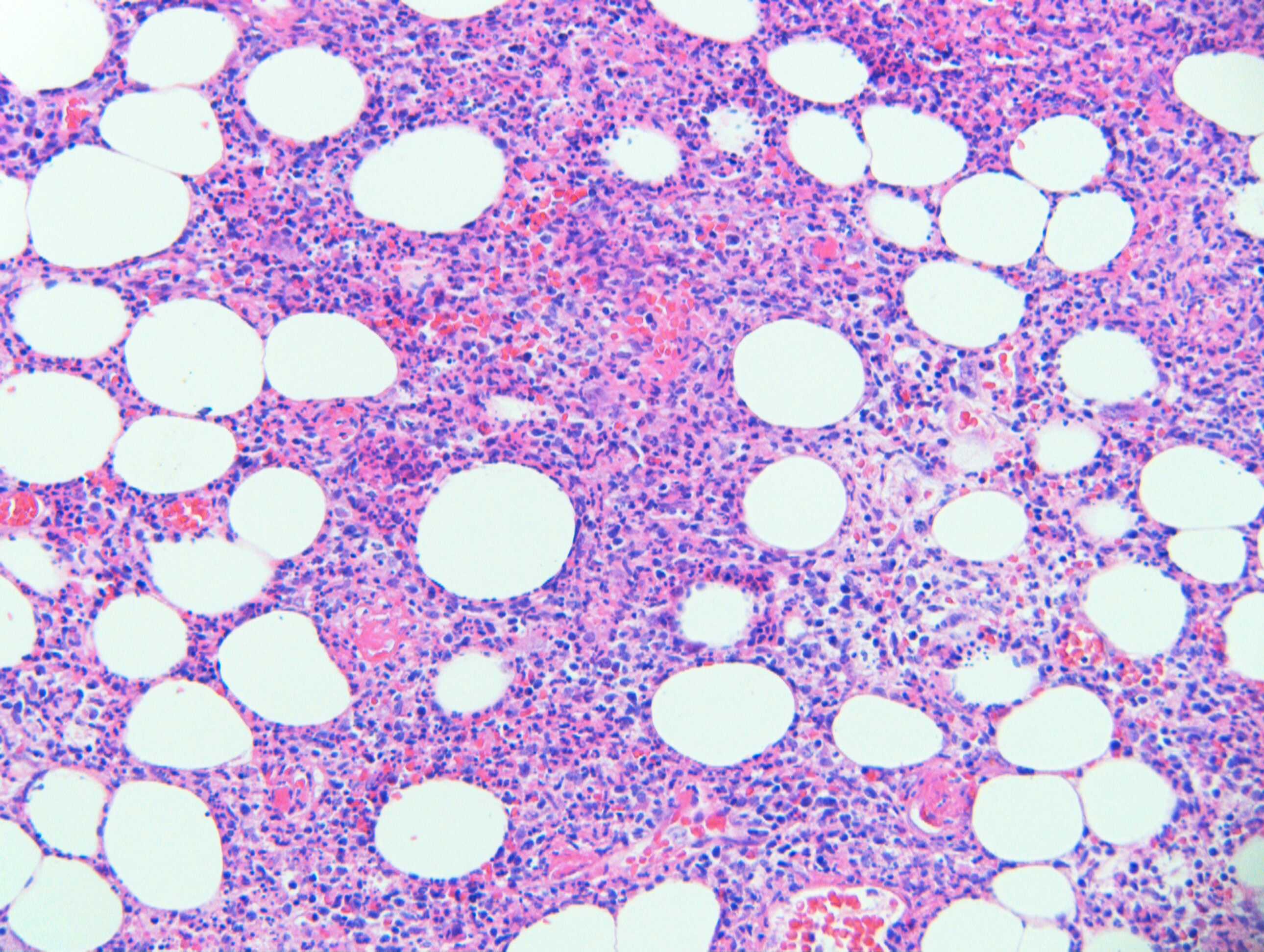
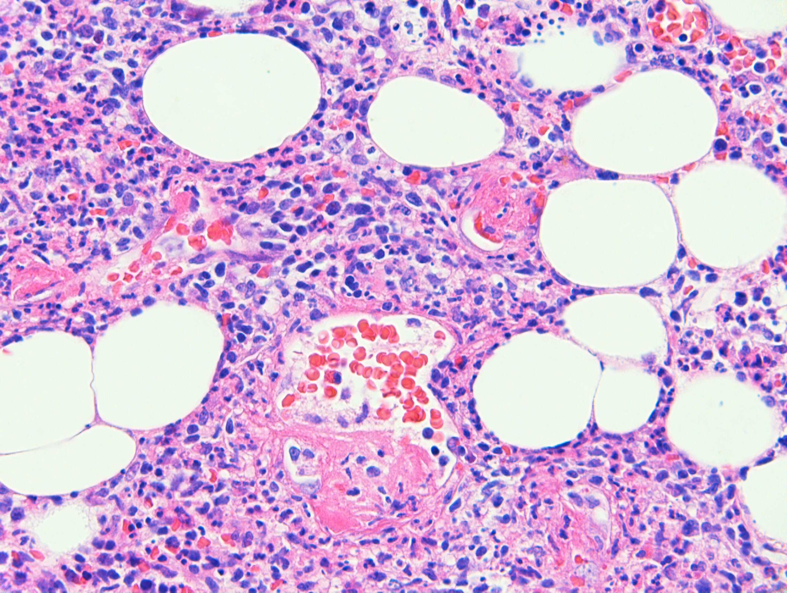
Clinical Details: F40. Occipital lump. ?Lymph node. Case c/o Dr Yee Wah Tsang

Edited by Admin_Dermpath


  Report Record

User Feedback


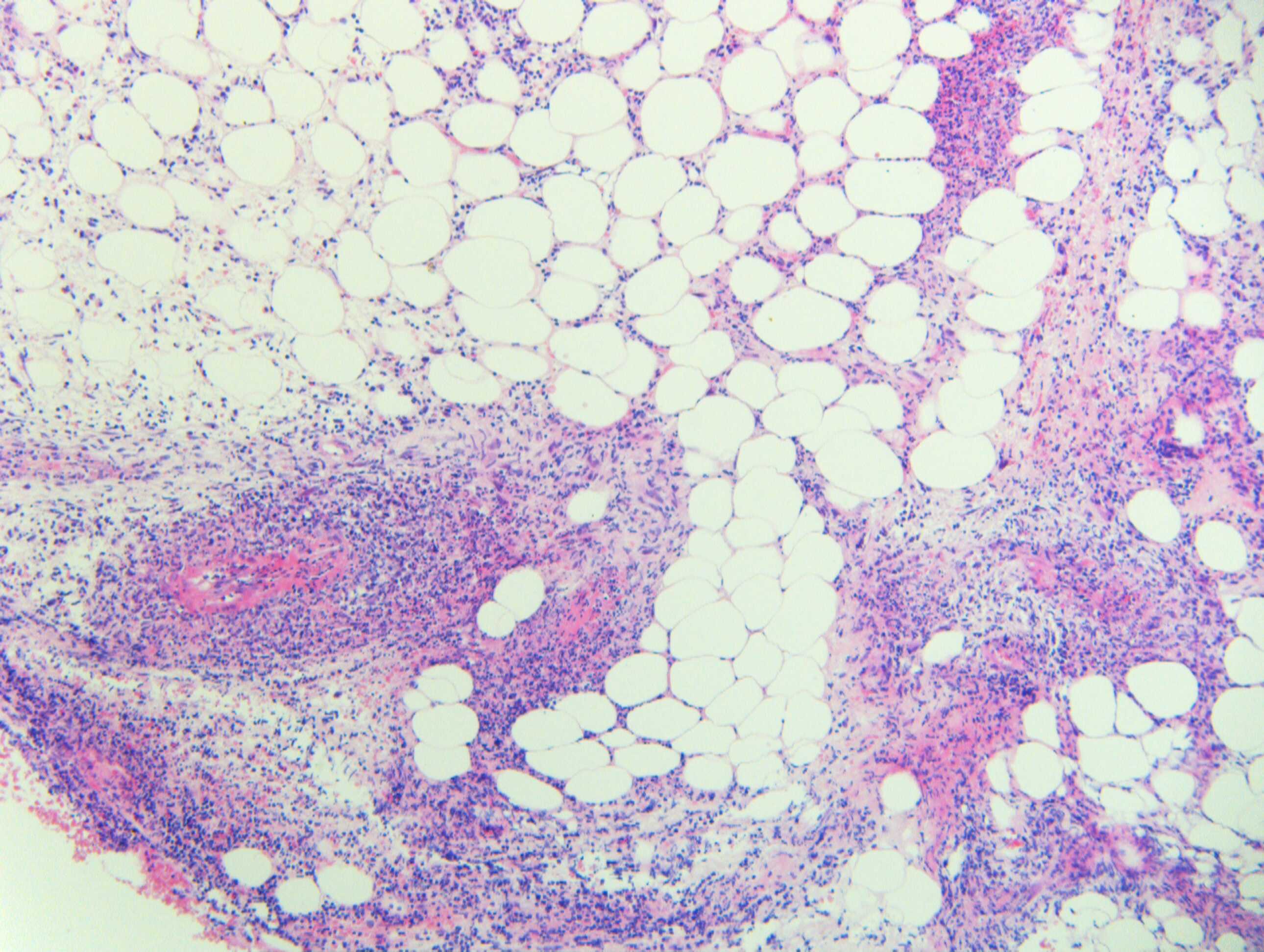
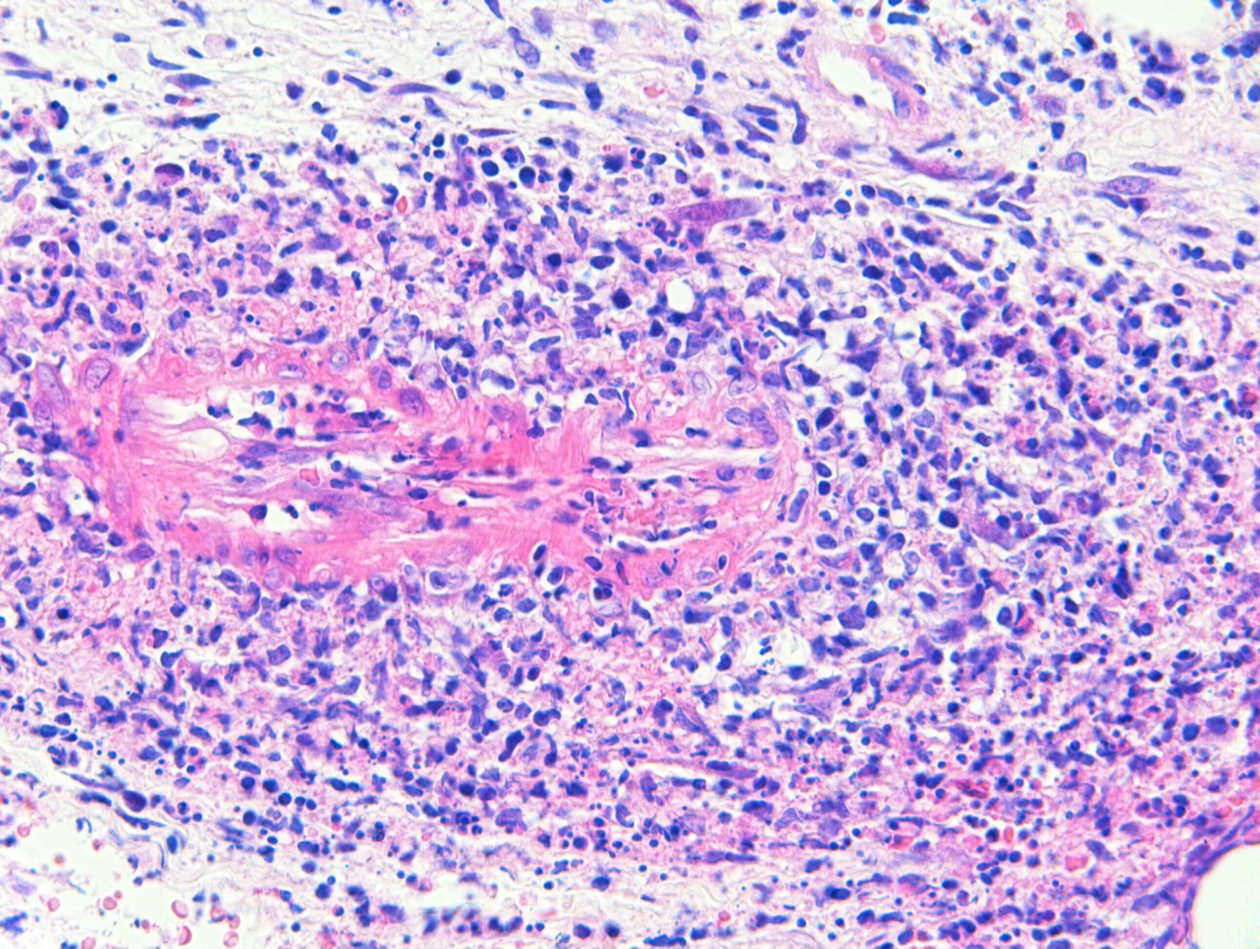
This is a neutrophilic lobular panniculitis with spillover into the septa. It's very difficult to give etiologic diagnosis without clinical details and correlation. It could be infectious, factitious, due to alpha 1 antitrypsin deficiency or due to myelodysplastic syndromes. Could not identify the eosinophilic structure in the last image. It could be a foreign body or trophozoit form of some parasite like acanthameba. Another interesting possibility is that of acute infectious id panniculitis ( Magro et al J Cut Path 2008;35:941-46). Would be interesting to know the details from Dr Carr!

Share this comment


Link to comment
share_externally

To add to the above comment, PAS and Fite Faraco stains would give some more clues

Share this comment


Link to comment
share_externally

Dr. Mona Abdel-Halim

Posted

Thinking also of infectious panniculitis. Suspecting fungal structures in the last image. Special stains are needed. 

Share this comment


Link to comment
share_externally

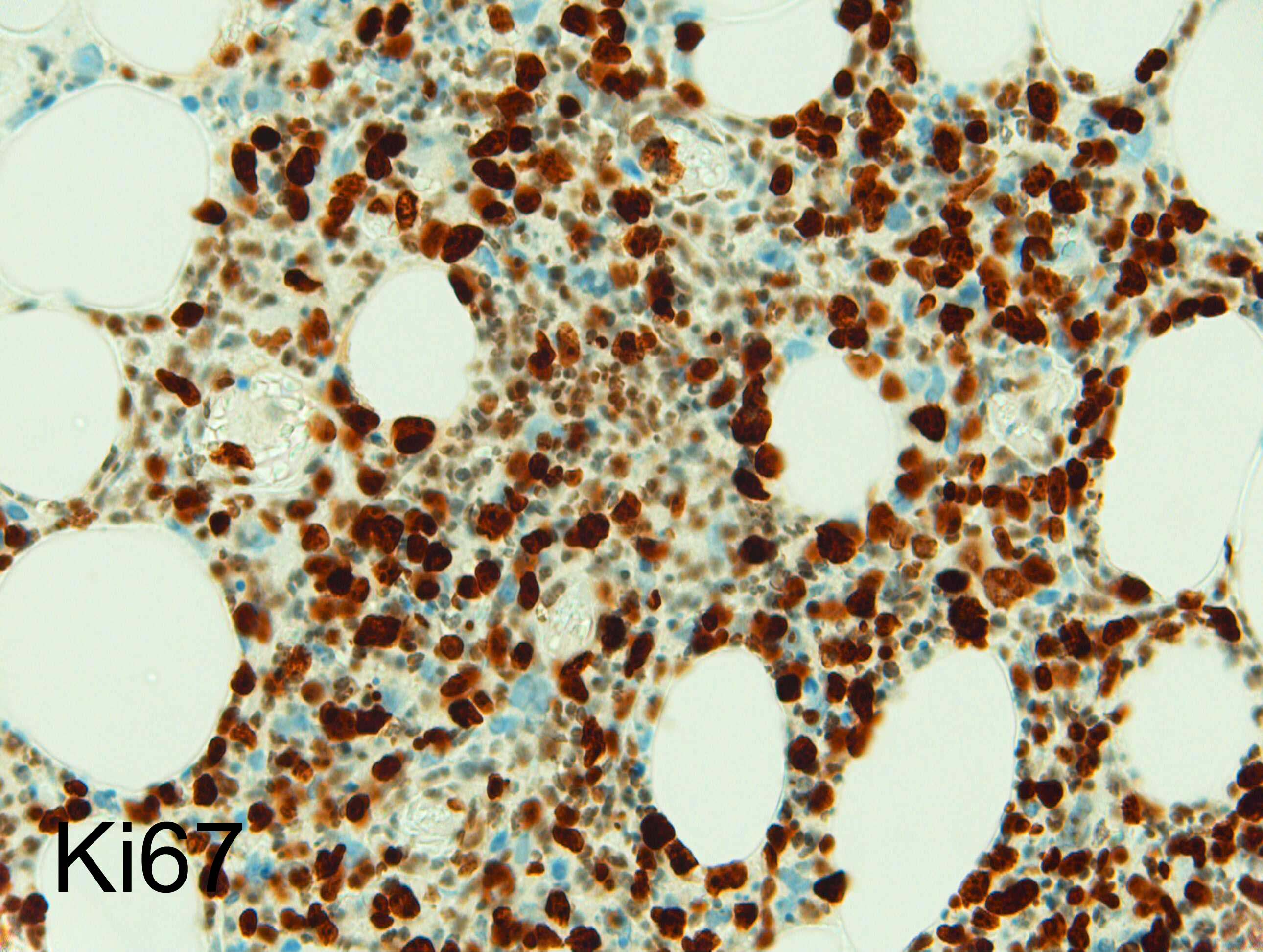
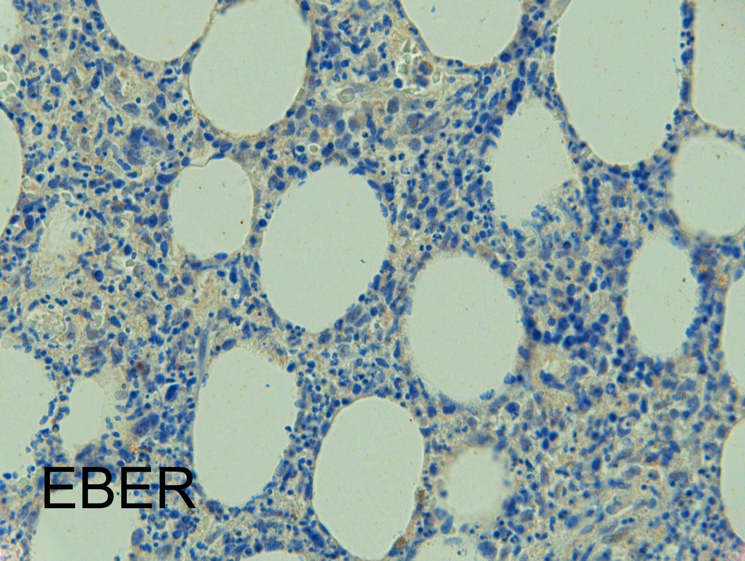
acute panniculitis pattern but also angiocentric. Agree with all above comments. The last figure show lot of neutrophilic debris but also some clusters of quite atypical cells with irregular hyperchromatic nuclei and clear cytoplasm on the right side of the vessel. So would like to rule out a NK/-T or T cell lymphoma. EBV stain?

Share this comment


Link to comment
share_externally

Saman Fatah

Posted

Cutaneous lymphomas with a panniculitic presentation, angiocentricity/angiodestruction is striking in this case. Main players are Subcutaneous Panniculitis-like T-Cell Lymphoma (SPTCL) and Gamma Delta T-Cell Lymphoma (GDTCL).

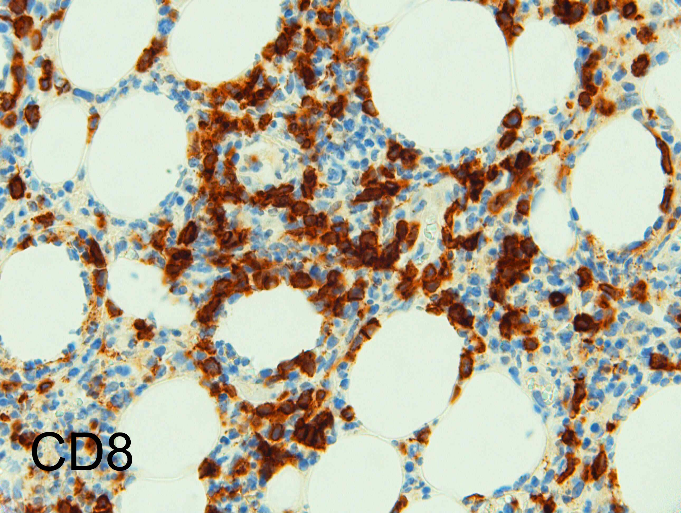
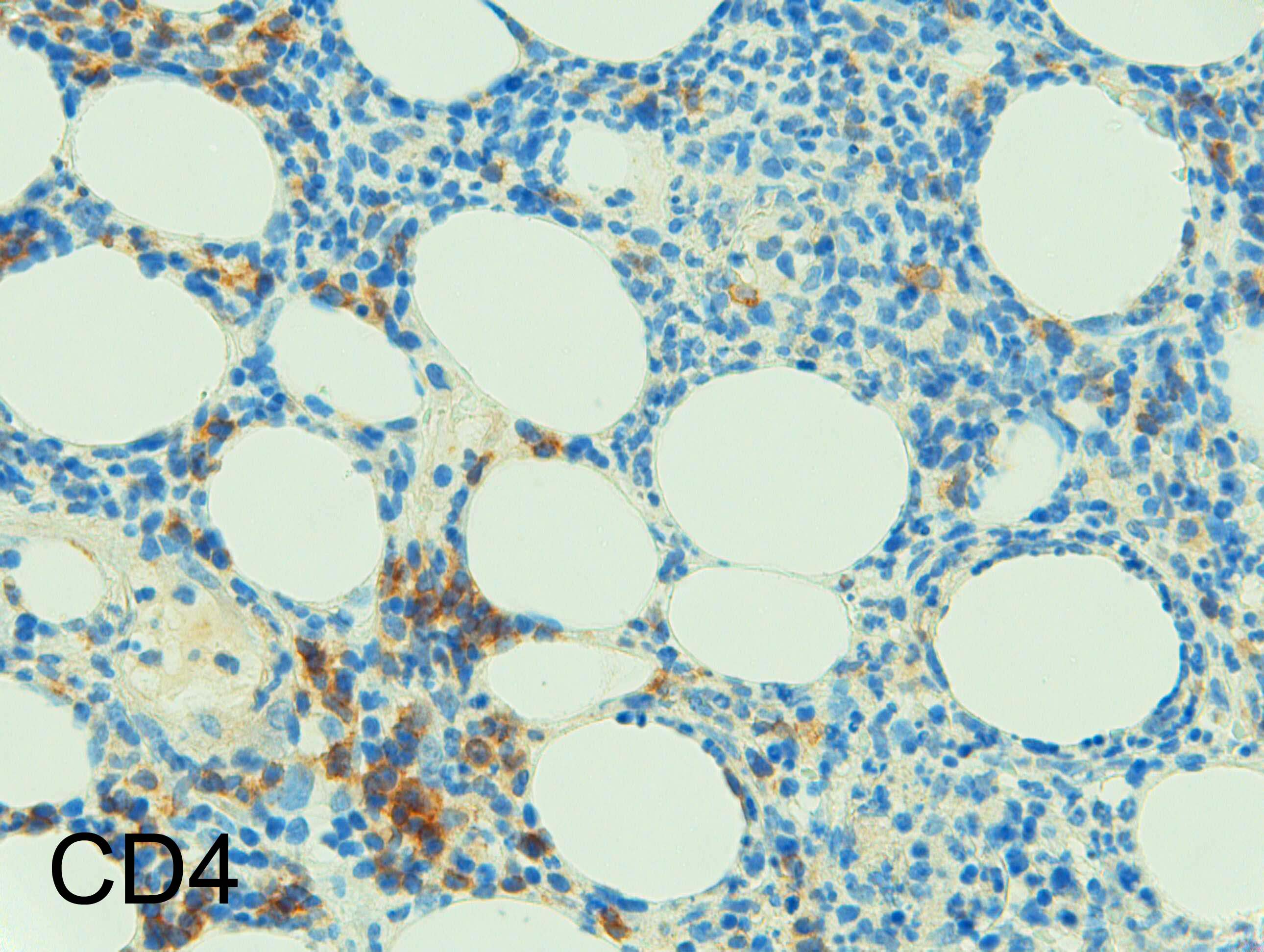
Needs complete immunophenotyping including Beta F1 and proper correlation with the clinical features to finalise the diagnosis. Based on what is availabe so far and CD8+ cells, SPTCL slightly favoured though the degree of the vessle destruction is a bit unusual for a classic SPTCL. 

Share this comment


Link to comment
share_externally

Dr. Mona Abdel-Halim

Posted

Interesting!

so it is a subcutaneous panniculitis like T cell lymphoma

Share this comment


Link to comment
share_externally

Dr. Richard Carr

Posted

We reviewed this histology because the patient presented with a new rash (that was eczematous). This case had been reported initially rather cryptically and referred for a specialist opinion to a national haematological centre who confirmed T-cell receptor gene re-arrangements. It was reported as a T-cell lymphoma CD4+ve (unusual for panniculitis-like T-cell lymphoma). The original IHC was confusing so we repeated it and the larger cells are CD8+ve. We don't have Beta-F1 so cannot confirm but given that the patient presented with a solitary lesion and is alive and well some 9 years later we concluded it was in keeping with a panniculitis-like T-cell lymphoma.

Share this comment


Link to comment
share_externally

Thanks for the illustrative case! We've seen cases where the initial diagnosis of SPLTCL was quite difficult as the infiltrate was rather subtle in the first biopsy.

Wonder if there was any erythrophagocytosis in this case.

Anyone encountered cases with initial lupus-like morphology - with interface reaction pattern and increased mucin? Seems to be reported that sometimes there might be SPLTCL on subsequent biopsy.

Share this comment


Link to comment
share_externally

Dr. Richard Carr

Posted

No erthyrophagocytosis seen Elaine. Yes I've seen rare cases of lupus changing to SPLTCL.

Share this comment


Link to comment
share_externally


Join the conversation

You can post now and register later. If you have an account, sign in now to post with your account.

Guest
Add a comment...

×   Pasted as rich text.   Paste as plain text instead

  Only 75 emoji are allowed.

×   Your link has been automatically embedded.   Display as a link instead

×   Your previous content has been restored.   Clear editor

×   You cannot paste images directly. Upload or insert images from URL.

Loading...

×
×
  • Create New...