Jump to content
In this section we have spot diagnoses posted on a daily basis since June 2010, now over 4000! You can review the archived cases and read the suggested diagnoses by users and the final comment by the contributors.
Case are uploaded each week day by 10 am UK time with the correct diagnosis will generally be posted at 8 pm UK time. Why not view the most recent spot diagnosis and proffer a diagnosis?

Case Number : Case 2005 - 12 Feb 2018 Posted By: Limin Yu

Please read the clinical history and view the images by clicking on them before you proffer your diagnosis.
Submitted Date :
   (0 reviews)

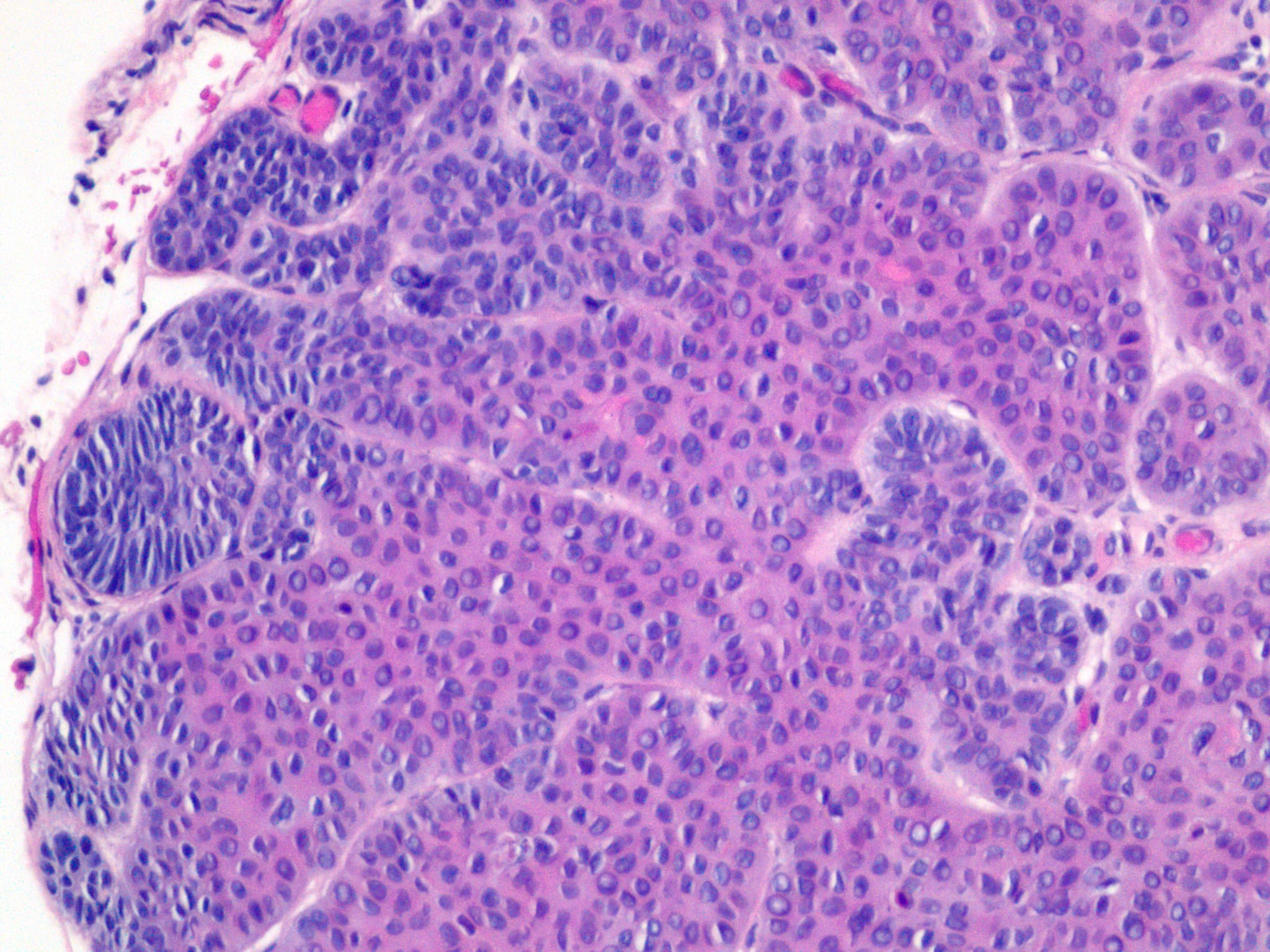
80 yo, M, right upper eyelid


  Report Record

User Feedback


Dr. Richard Carr

Posted

Agree. Strongly in favour of a rather indolent appearing nodular BCC (trichoepithelioma-like). CD10 (epithelial dominant staining), CK20 (reactive Merkel cells limited to trapped follicles only) and BerEP4 (diffuse & strong throughout sparing only central keratocysts) can be helpful.

Share this comment


Link to comment
share_externally

Infundibulocystic BCC? Kind of big to consider basaloid follicular hamartoma.

Share this comment


Link to comment
share_externally

What do the pale pink cells suggest? Can we call it a BCC with isthmic differentiation?

Share this comment


Link to comment
share_externally

Thank everyone!  I signed out this case as an Infundibulocystic BCC. TE is a close ddx.

Share this comment


Link to comment
share_externally

Mani Makhija

Posted

 Yes agree. I call them Infundibulocytic BCC (when tumorous and large as pointed above). There are others who shy away from the term as the number of infundibular cysts are variable and sometimes scant but the circumscription, central eosinophilic cords and peripheral basaloid nubbins are suggestive (for me). If I remember correctly Dr Ackerman's in his book on follicular tumors says these can even have papillary mesenchymal bodies and absence of clefts. 
In smaller lesions basaloid follicular hamartomas is a d/d. 
Infundibulocystic BCC are most often sporadic; however its is a variant associated with Gorlin syndrome or the multiple hereditary infundibulocytic basal cell carcinoma syndrome.  

Share this comment


Link to comment
share_externally

Dr. Richard Carr

Posted

On ‎14‎/‎02‎/‎2018 at 06:55, Anil Patki said:

What do the pale pink cells suggest? Can we call it a BCC with isthmic differentiation?

Dear Anil,

I don't see why not.

More generally I personally prefer nodular ("infundibulo-cystic" or "TE-like" I only mention to my friends as I prefer not confuse the clinical colleagues) BCC's and have observed that they are very much more commonly located in the nasolabial and delicate areas of the face. I have seen several examples mis-reported as trichoepitheliomas. Remember I have said previously every time you want to diagnose a TE in a patient >50y on the face 19 of 20 cases will be BCC. I don't think I have seen convincing papillary mesenchymal cells in this variant of BCC and a big clue is that they are almost always >80% epithelial compared with TE (that almost always do have PMCs) that are usually 50:50 stroma:epithelium (usually with stromal clefts). If in doubt have it excised with narrow margins. I've seen these wrongly reported very indolent lesions deep in the facial muscles by the time the penny dropped. Also stromal keratin derived amyloid is not stroma so watch out for that too in these lesions.

Share this comment


Link to comment
share_externally


Join the conversation

You can post now and register later. If you have an account, sign in now to post with your account.

Guest
Add a comment...

×   Pasted as rich text.   Paste as plain text instead

  Only 75 emoji are allowed.

×   Your link has been automatically embedded.   Display as a link instead

×   Your previous content has been restored.   Clear editor

×   You cannot paste images directly. Upload or insert images from URL.

Loading...

×
×
  • Create New...