Jump to content
In this section we have spot diagnoses posted on a daily basis since June 2010, now over 4000! You can review the archived cases and read the suggested diagnoses by users and the final comment by the contributors.
Case are uploaded each week day by 10 am UK time with the correct diagnosis will generally be posted at 8 pm UK time. Why not view the most recent spot diagnosis and proffer a diagnosis?

Case Number : Case 2012 - 21 Feb 2018 Posted By: Iskander H. Chaudhry

Please read the clinical history and view the images by clicking on them before you proffer your diagnosis.
Submitted Date :
   (0 reviews)

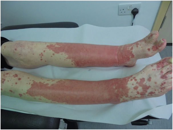
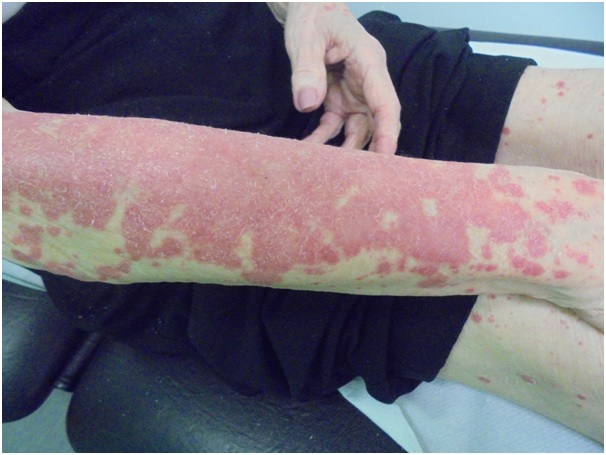
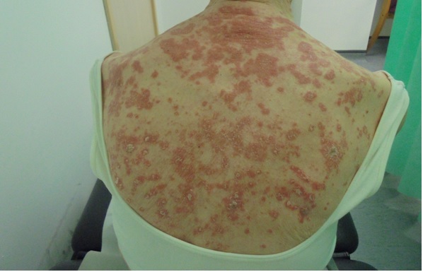
85 year old female. Right upper arm incisional biopsy. ? extensive erythematous rash. Known thyroid Ca with poor prognosis


  Report Record

User Feedback


Thanks for sharing clinical photographs. The epidermis shows parakeratosis with deficient granular layer, acanthosis, altered architecture of keratinocytes and a few apoptotic keratinocytes. Dermis shows edema, dilated vessels and perivascular lymphocytic infiltrate. Clinical photographs show a symmetric erythematous papulosquamous rash. This looks like a psoriasiform drug eruption.

Share this comment


Link to comment
share_externally

Saman Fatah

Posted

Nice example of PRP, not sure labelling it as paraneoplastic just because of the background is justified. 

Share this comment


Link to comment
share_externally

Dr. Richard Carr

Posted

I agree drugs and psoriasiform/PRP-like drug or just PRP. I was not actively aware PRP could be paraneoplastic (cue a google!). I'd also add in some other "histological" differentials such as the nutritional deficiency group here (including necrolytic migratory erythema, Zinc deficiency etc). I thought there might be some neutrophils. I'd would do a PAS for fungi in routine sign-out..

Share this comment


Link to comment
share_externally

One clinical point against PRP is the absence of follicular keratotic papules. Secondly, PRP's association with neoplasia could be fortuitous as Dr Saman has pointed out. It doesn't fulfill Curth's criteria.

Share this comment


Link to comment
share_externally

Admin_Dermpath

Posted

Dr Chaudhry's Final diagnosis: PRP as it fitted with the clinical - differential was a PRP like drug eruption. Interestingly the typical alternating para and hyperkeratosis are not clearly seen - is that other people's experience ? 

Share this comment


Link to comment
share_externally

Dr. Richard Carr

Posted

An older version of Weedon says it's one of the diagnoses he missed most frequently. We rarely see it biopsied here because clinicians don't necessarily biopsy clear-cut cases or it's just uncommon, so I tend to collect them in my slide collection (just searched the database and I have 9 collected, 4 photographed, only 3 cases suggested PRP on the histo request form clinically which is interesting). In my overall experience they don't tend to read the textbook! We usually need our clinical colleagues to give us this diagnosis although I have suggested it a few times with variable clinical correlation.

Share this comment


Link to comment
share_externally


Join the conversation

You can post now and register later. If you have an account, sign in now to post with your account.

Guest
Add a comment...

×   Pasted as rich text.   Paste as plain text instead

  Only 75 emoji are allowed.

×   Your link has been automatically embedded.   Display as a link instead

×   Your previous content has been restored.   Clear editor

×   You cannot paste images directly. Upload or insert images from URL.

Loading...

×
×
  • Create New...