Jump to content
In this section we have spot diagnoses posted on a daily basis since June 2010, now over 4000! You can review the archived cases and read the suggested diagnoses by users and the final comment by the contributors.
Case are uploaded each week day by 10 am UK time with the correct diagnosis will generally be posted at 8 pm UK time. Why not view the most recent spot diagnosis and proffer a diagnosis?

Case Number : Case 2119 - 20 July 2018 Posted By: Dr. Richard Carr

Please read the clinical history and view the images by clicking on them before you proffer your diagnosis.
Submitted Date :
   (0 reviews)

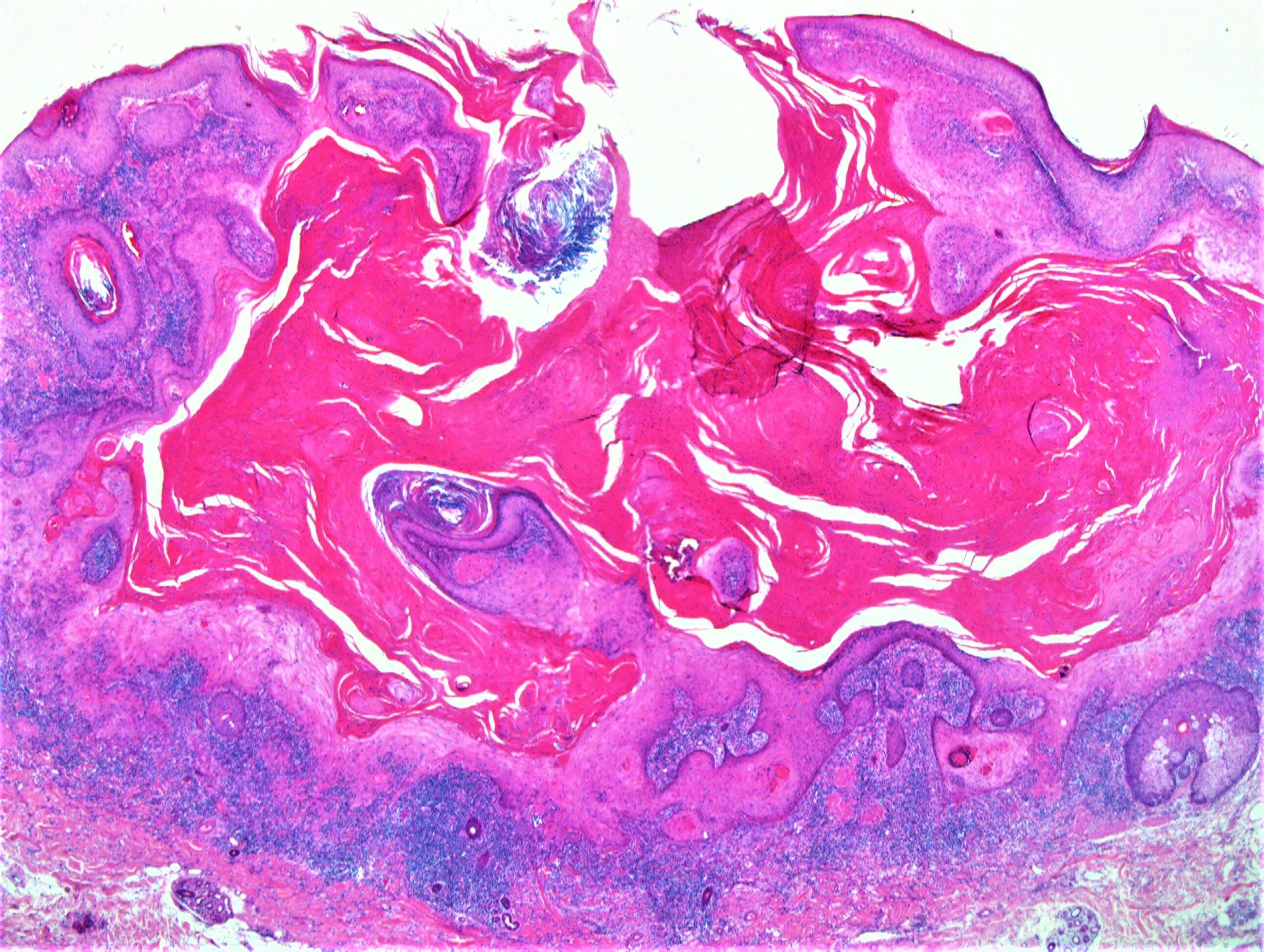
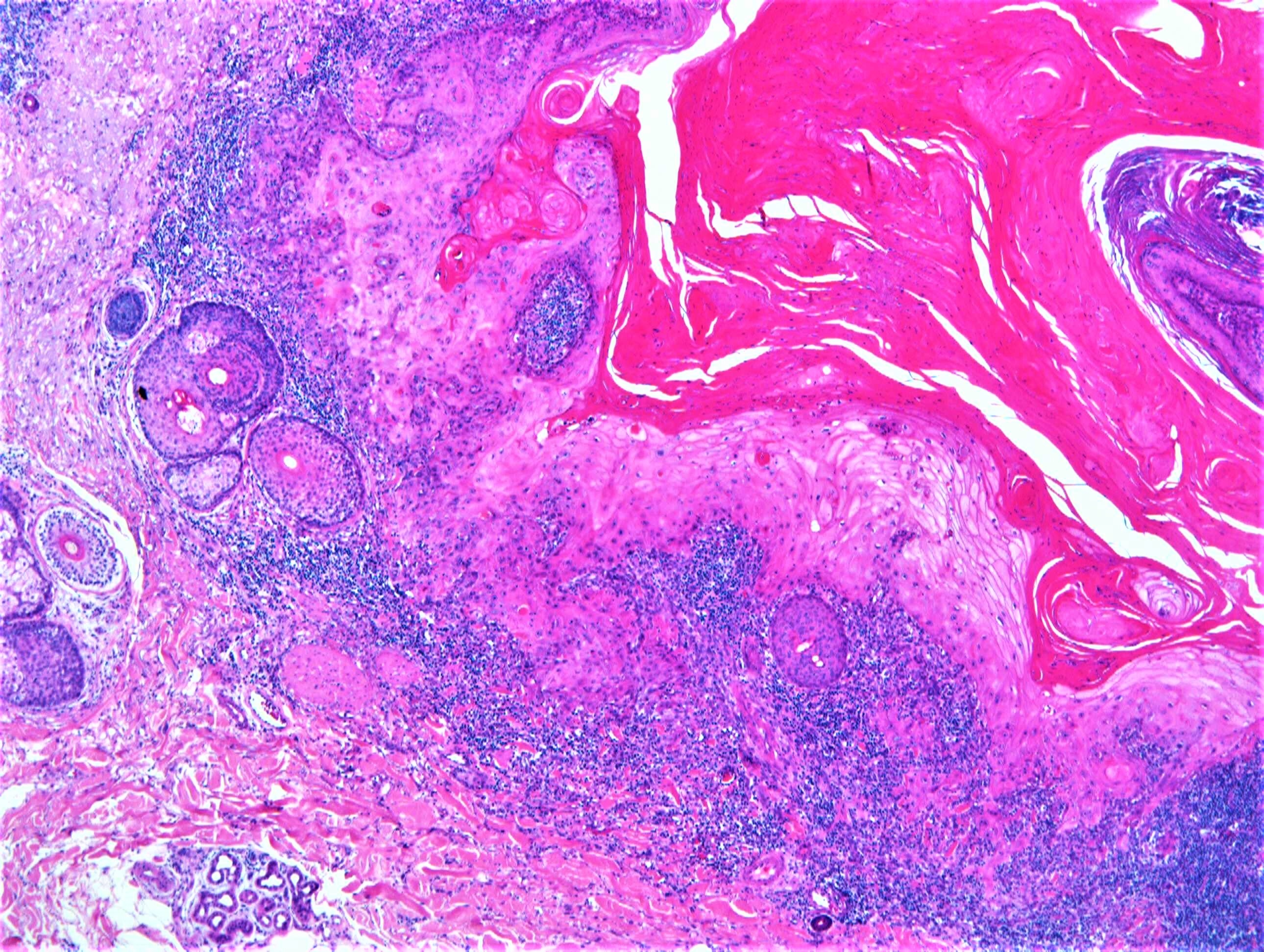
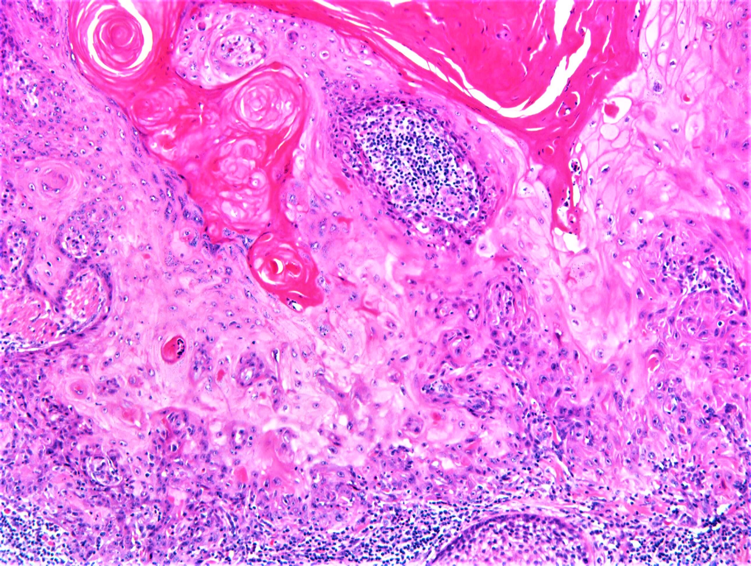
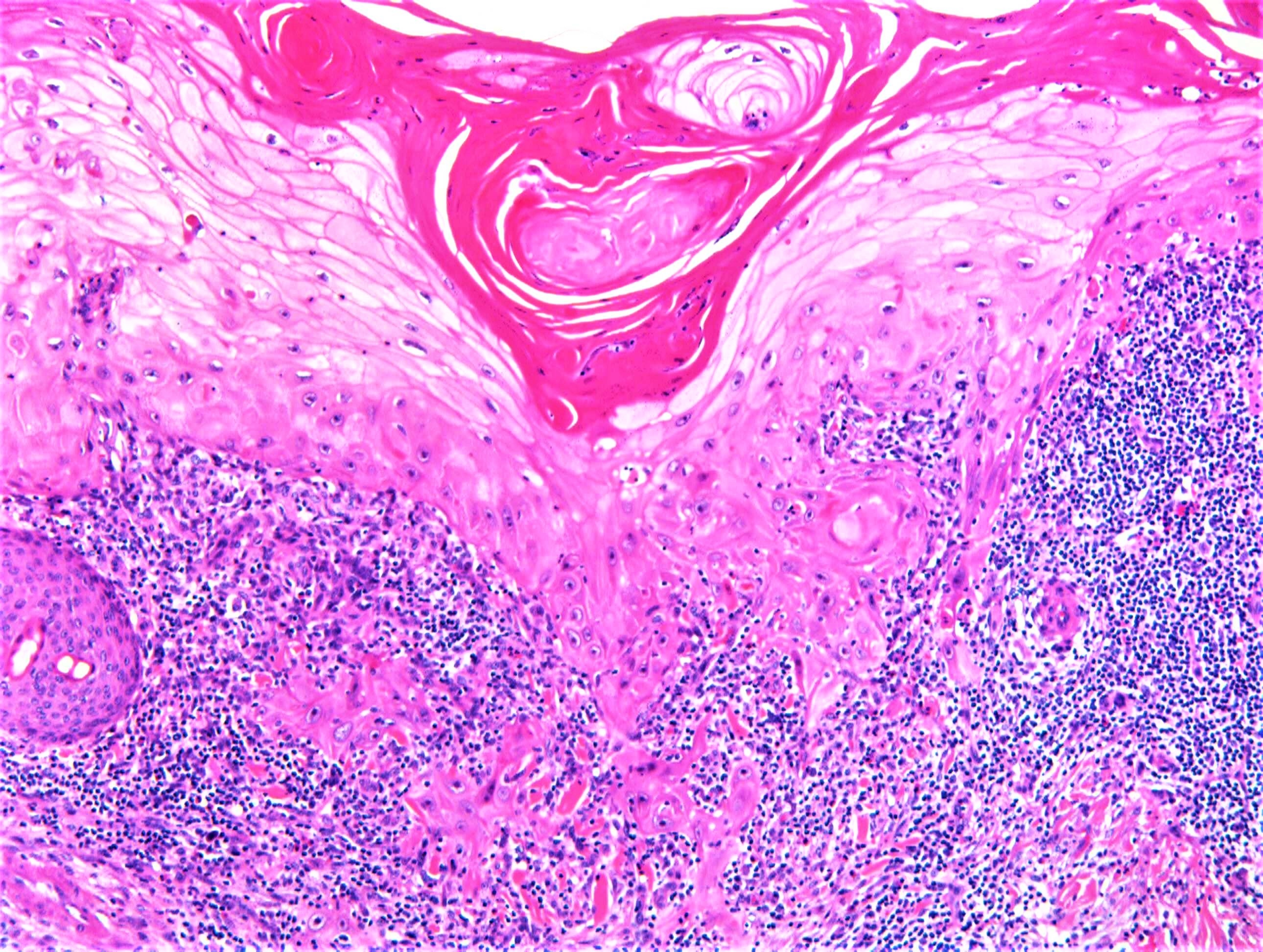
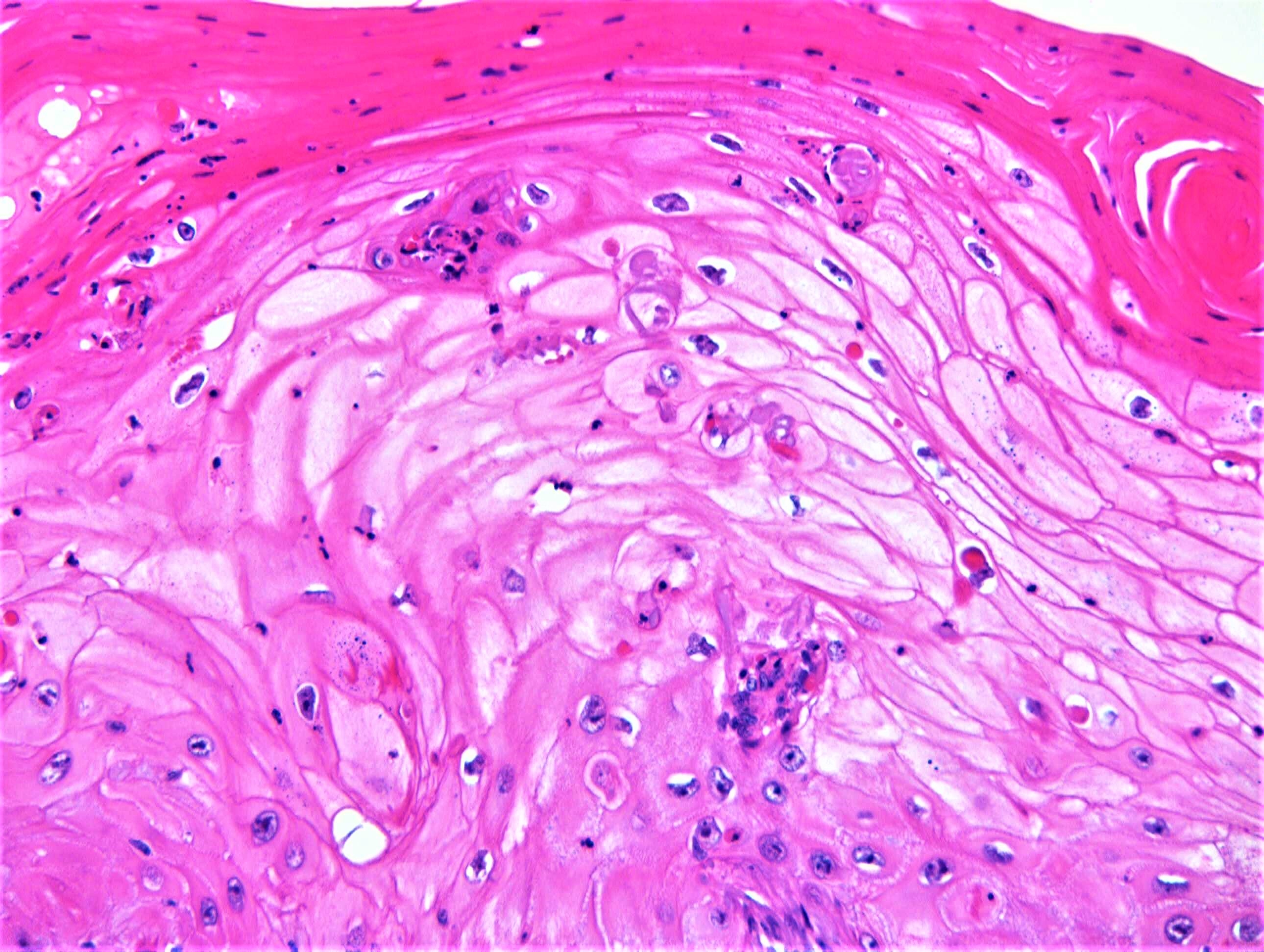
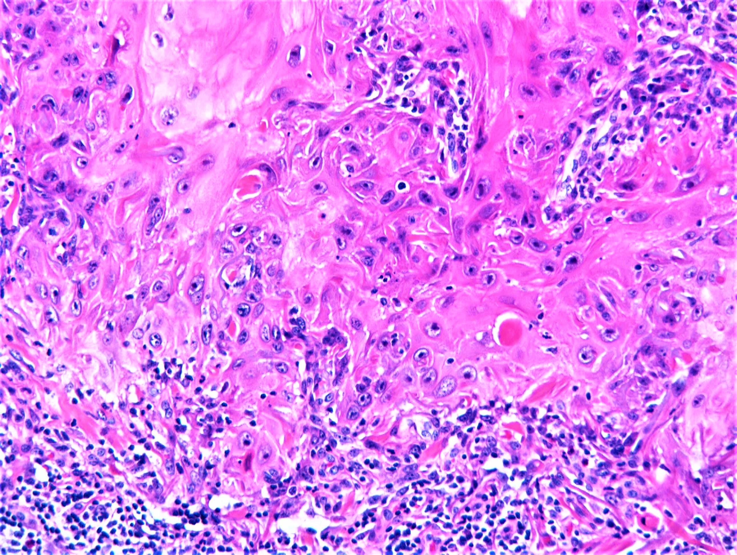
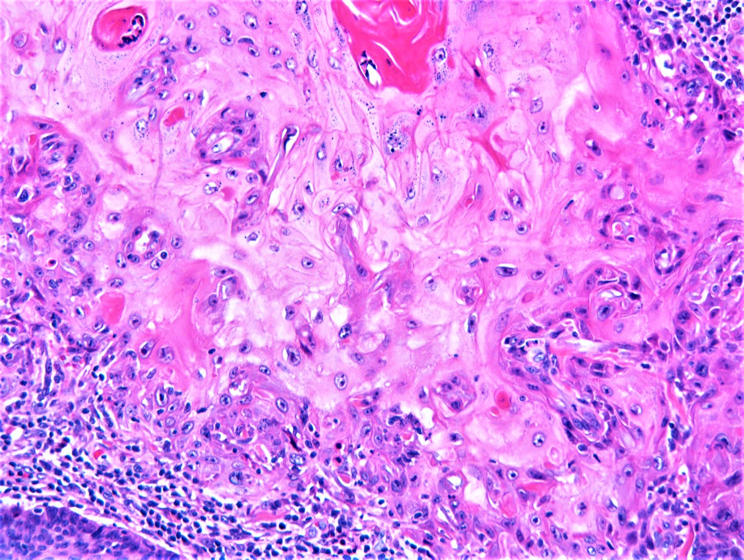
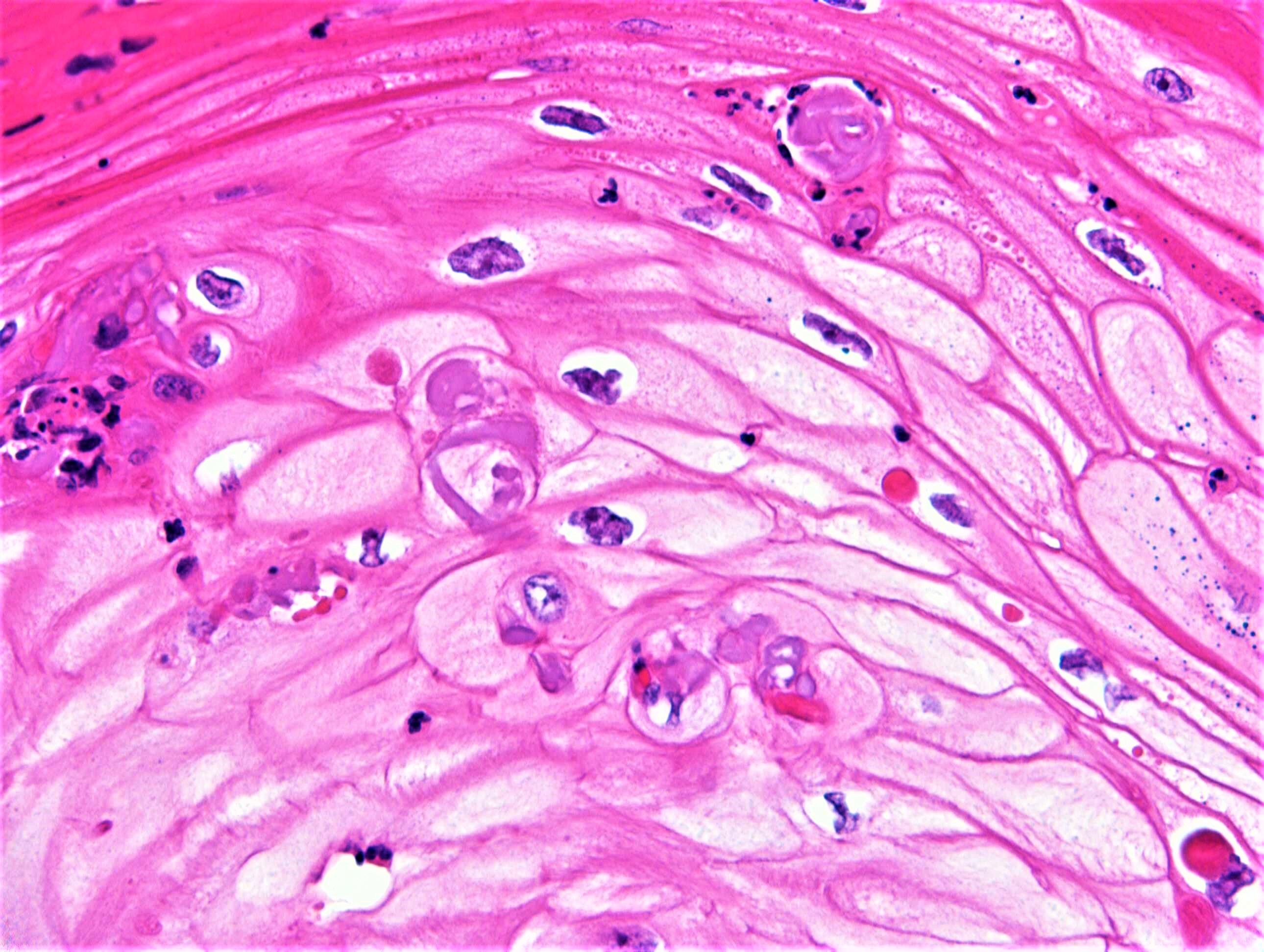
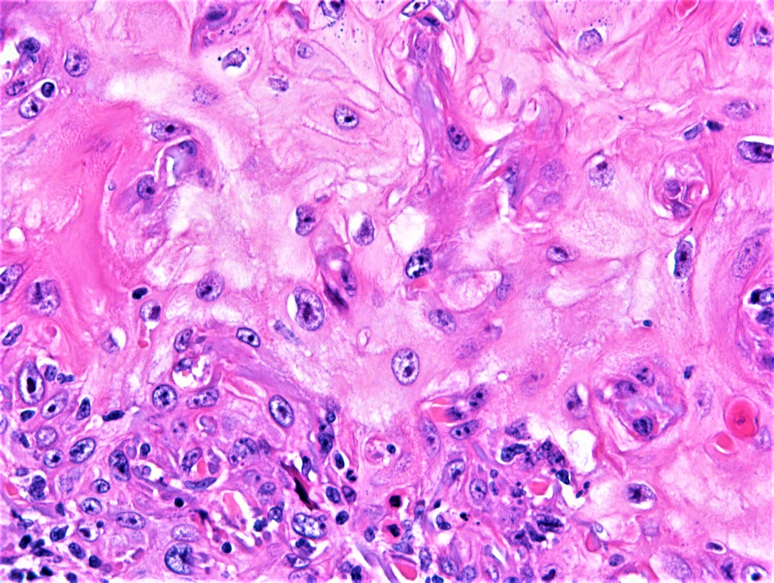
F72 Neck. 4-5 weeks h/o hyperkeratotic nodular lesion.


  • Like 1
  Report Record

User Feedback


Anil Patki

Posted

Keratin filled crater with epidermal buttresses. Epidermal invaginations show horn pearls and individual cell keratinization. Lymphocytic infiltrate at the base. I think this is a keratoacanthoma

Share this comment


Link to comment
share_externally

Agreed, particularly with the clinical history of 4-5 weeks. But in the real world of dermatopathology, we rarely get that kind of clinical history. Therefore, I have the tendency to just call these lesions crateriform well-differentiated keratinizing squamous cell carcinoma because I think it doesn't make a big difference to call it a SCC instead of a KA, but at least I'm protected if the lesion somehow does crazy things like metastasis. Would like to know what other colleagues handle these cases in practice (without good history).

Share this comment


Link to comment
share_externally

Yes. KA. Nice elastotic material engulfment. 

Share this comment


Link to comment
share_externally

John Zhang

Posted

Based on histology I think it is KA or KA-type SCC. I prefer KA given the fast growing history. I have a question for everyone: how specific is the elastotic material engulfment to differentiate KA from SCC? Every time we see engulfment, does it mean that it won't metastasis? Thanks!

Share this comment


Link to comment
share_externally

Raul Perret

Posted

15 hours ago, anh said:

Agreed, particularly with the clinical history of 4-5 weeks. But in the real world of dermatopathology, we rarely get that kind of clinical history. Therefore, I have the tendency to just call these lesions crateriform well-differentiated keratinizing squamous cell carcinoma because I think it doesn't make a big difference to call it a SCC instead of a KA, but at least I'm protected if the lesion somehow does crazy things like metastasis. Would like to know what other colleagues handle these cases in practice (without good history).

I think Richard may be the correct person for elaborating on this

Share this comment


Link to comment
share_externally

vincenzo

Posted (edited)

When we see some elastotic material entrapment,  but into a well differentiated squamous growth, it means  our current case is a quickly developed tumor and at the same time a well differentiated squamous tumor. KA is a good example of this odd behavior. But I think Richard is the most qualified expert to answer in this question. 

Edited by vincenzo

Share this comment


Link to comment
share_externally

Dr. Richard Carr

Posted

I don't think I could have given you a more "text-book" histological example with clinical corroboration of a keratoacanthoma (in my opinion a distinctive clinicopathological entity that requires more study - I don't think I've ever said KA is an entirely benign entity. My thinking currently KA sits in the grey zone with some cases of persistence, local recurrence and exceedingly rarely metastasis or perhaps slightly more often frank malignant transformation) but the vast majority having no adverse outcome. It may show venous and perineural invasion without any adverse outcome.  If we don't make the diagnosis we won't be able to study the cases and advance knowledge. Weedon communicated to me that he saved a youngish patient with KA and venous invasion from having a neck dissection. Also currently we don't know the best treatment for patients that might include watch & wait, curretage, excision etc - that is why we need more well planned clinicopatholoical studies. Regarding elastic entrapment it is highly characteristic especially when present in the mature central areas but is not specific to KA (can be seen in pseudoepitheliomatous hyperplasia - of many causes). In the higher grade follicular (infundibular tricholemmal) SCC you may see striking growth permeating through the dermis with striking elastic fibres but this highly infiltrative variant of FSCC characteristically lacks central maturation in all areas (see discussion below). I would add a caveat that the crateriform variants of FSCC can be awfully well differentiated superficially and this is a huge trap so I like always to see the base of the lesion and may have to admit uncertainty in superficial and partial biopsies. Again suggest checking out the YouTube, Dermtalks, Carr, Follicular squamous cell carcinoma.

Features I like to see in KA as follows:

Clinical correlation e.g. rapid growth

Symmetrical exo-endophytic lesion

Well-formed epidermal collarette

Uniform maturation from peripheral proliferative borders with collagen/elastic entrapment through to well-differentiated central areas with glassy eosinophilic cytoplasm

Neutrophil microabscesses

Signs of regression (fibrosis, lichenoid reaction, keratin granulomas).

I am pasting a section from our drafted paper on follicular (infundibular-tricholemmal) SCC: 

Perhaps the most difficult distinction is between FSCC and KA, also considered a tumour of infundibular origin. Diaz-Cascajo comment that KA is a more rapidly growing tumour*Diaz-Cascajo2004 but in our series a significant proportion of FSCC (24.4%) grew within 1 to 2 months. A key determinant of KA is subsequent stabilisation and regression in most instances. However, patients in the UK tend to be referred urgently and lesions often excised during the proliferative phase when regression may be minimal or absent. Our local practice has been to distinguish KA from SCC using a combination of features. In favour of KA is a symmetrical, exo-endophytic lesion with a rounded/arciform profile to the base, a well-formed epidermal collarette, an abrupt transition from basaloid peripheral cells to central cells with glassy eosinophilic cytoplasm and tricholemmal keratinisation, engulfed collagen and elastic fibres, a well-formed laminated keratin-filled crater, neutrophil microabscesses, and regressional features including lichenoid reaction, peripheral individual cell necrosis and fibrosis. Similarly to Weedon*Weedon2010, we allow infiltrative proliferative peripheries with cellular pleomorphism limited to the peripheral cell layers provided that central maturation is present in all areas of the tumour. In contrast, most FSCC had a distinctive circumscribed lobular profile with frequent presence of follicular mucin. We encountered a less common infundibular-cystic variant of FSCC with exo-endophytic profile and central keratin-filled crater which closely simulated KA, but this too lacked elastic and collagen entrapment and showed pushing lobular borders without regression. An additional subset of high risk FSCC, also illustrated by others, *Fitzpatrick1985,*Billingsley1999,*Kossard2008,*Misago2011 should be distinguished from KA in showing a poorly formed crater, high proliferation throughout the lesion, asymmetry and deeper invasion into subcutis in the absence of regression. Features also favouring SCC include prominent dyskeratosis, ‘dirty’ parakeratosis (hyperchromatic enlarged nuclei and prominent cellular debris), ulceration and zonal necrosis.

Share this comment


Link to comment
share_externally

Dr. Mona Abdel-Halim

Posted

Thanks Dr Carr for teaching us KA and fSCC.. 

Share this comment


Link to comment
share_externally


Join the conversation

You can post now and register later. If you have an account, sign in now to post with your account.

Guest
Add a comment...

×   Pasted as rich text.   Paste as plain text instead

  Only 75 emoji are allowed.

×   Your link has been automatically embedded.   Display as a link instead

×   Your previous content has been restored.   Clear editor

×   You cannot paste images directly. Upload or insert images from URL.

Loading...

×
×
  • Create New...