Jump to content
In this section we have spot diagnoses posted on a daily basis since June 2010, now over 4000! You can review the archived cases and read the suggested diagnoses by users and the final comment by the contributors.
Case are uploaded each week day by 10 am UK time with the correct diagnosis will generally be posted at 8 pm UK time. Why not view the most recent spot diagnosis and proffer a diagnosis?

Case Number : Case 2121 - 26 July 2018 Posted By: Raul Perret

Please read the clinical history and view the images by clicking on them before you proffer your diagnosis.
Submitted Date :
   (0 reviews)

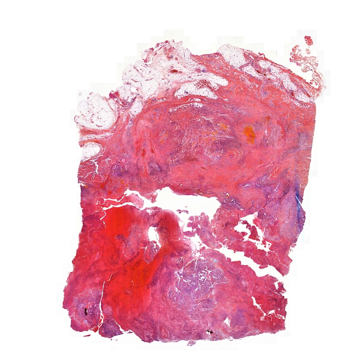
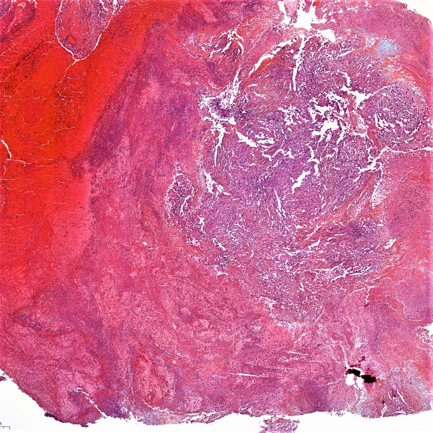
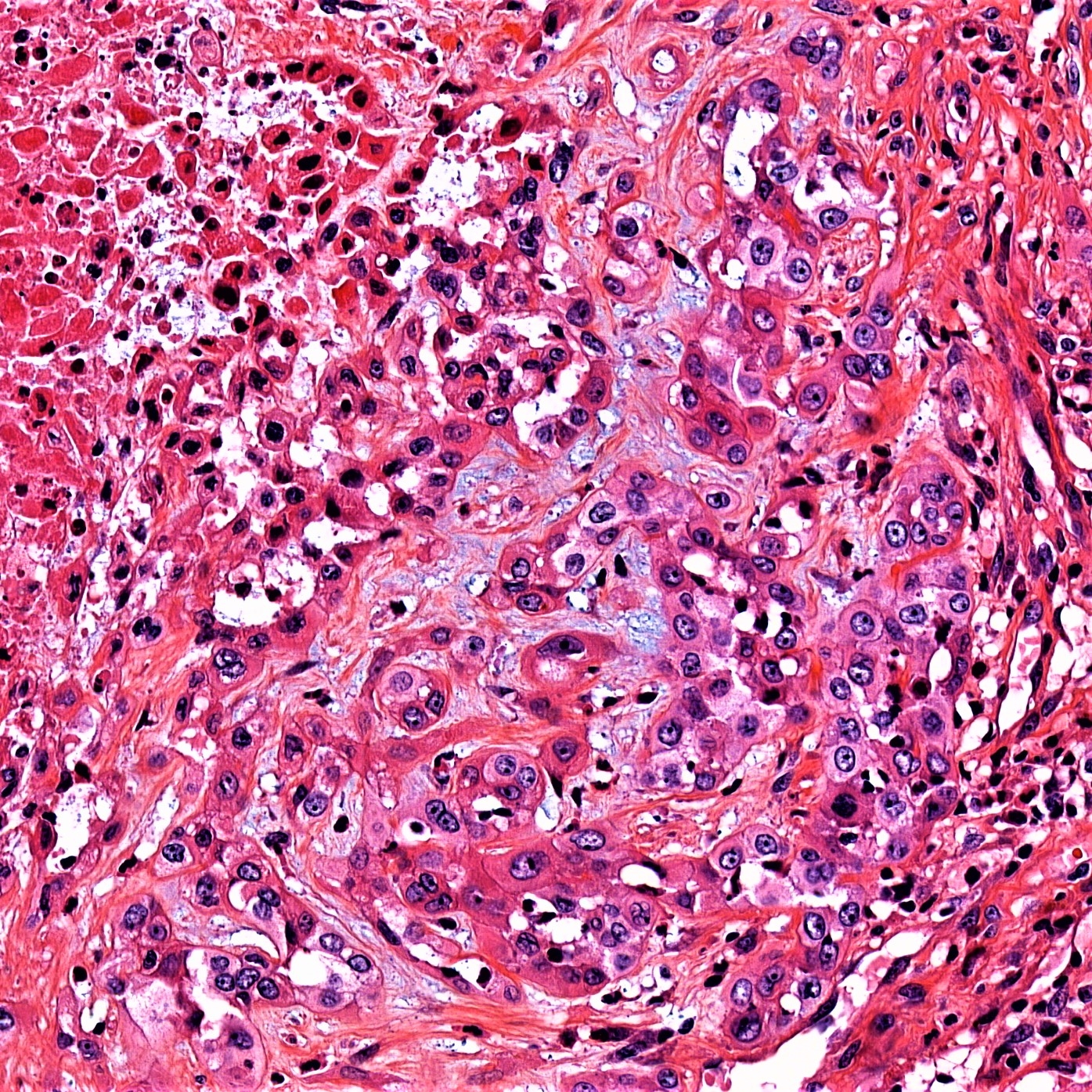
19 year old male with a 19 mm polypoid mass on the perineum.


  Report Record

User Feedback


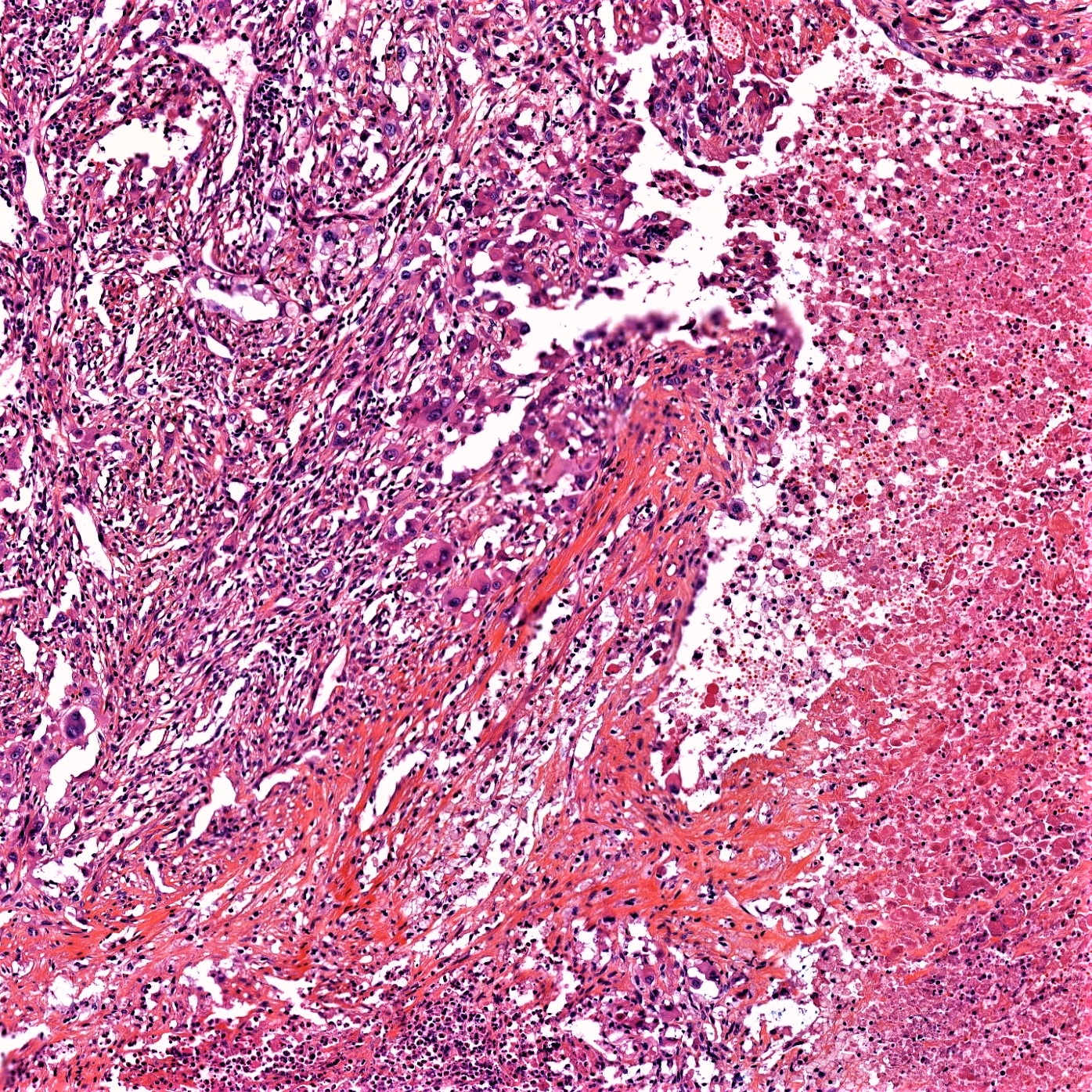
ATypical epithelioid lesion. Appears forming lumen. Some myxoid stroma. The question is whether the lumens represent glands or vascular lumens. Looks like the background is hemorrhagic too. So my guess is it an epithelioid vascular lesion. For now, I would do Cytokeratin, cd31, cd34 (or whateverendothelial markers available ). Then I will start thinking about classifying ths lesion once i know its differentiation.

Share this comment


Link to comment
share_externally

Anil Patki

Posted

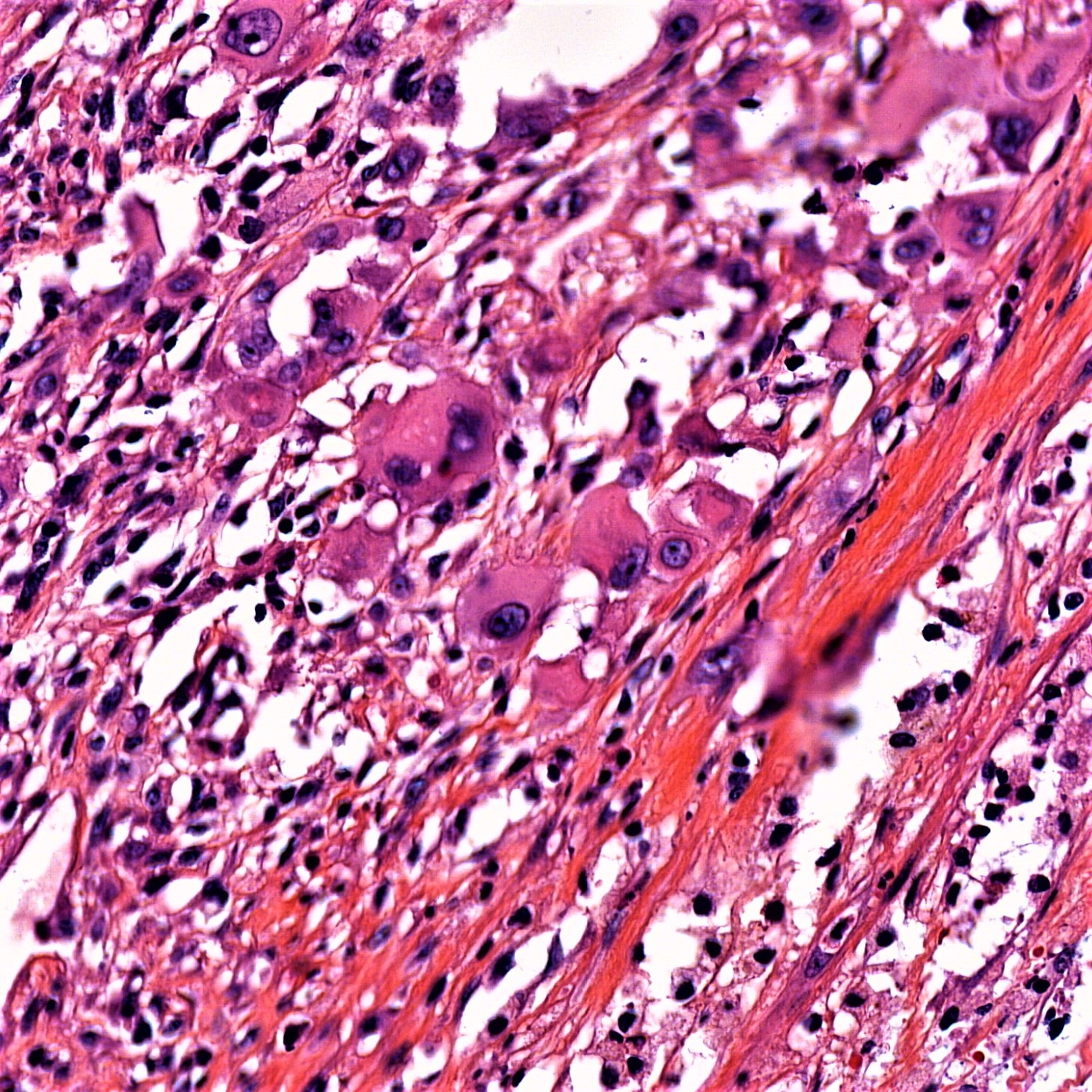
Stroma contains atypical and multinucleate cells. Is it a fibroepithelial polyp of the anus considering it's location on the perineum?

Share this comment


Link to comment
share_externally

vascular lumina possibility as mentioned by anh. will also consider a rhabdoid tumour and spread from testicular germ cell tumour (embryonal/yolk sac)

Share this comment


Link to comment
share_externally

John Zhang

Posted

Great discussions! Doesn't strike me as a vascular lesion as the spaces are too ill-formed but ruling them out with an endothelial marker why not! Rhabdoid tumor with muscle lineage and germ cell tumors are good thoughts would like to rule them out with IHC stains. I also want to add S100 to rule out clear cell sarcoma.

Share this comment


Link to comment
share_externally

Difficult. ?SCC arising in a teratoma?

Share this comment


Link to comment
share_externally

Alice Roberts

Posted

I’m going with metastatic lesion, rhabdoid or testicular germ cell tumor.  First off, check Clinical history.  For IHC Would start with S100, pan keratin, EMA and desmin.

Share this comment


Link to comment
share_externally

Raul Perret

Posted

This was a particularly interesting case of epithelioid sarcoma, proximal type. The tumor cells expressed CD34, EMA and CK AE1/AE3 diffusely and were negative for s100, CD31, ERG and desmin. INI-1 (BAF47) was loss in all cells. The presentation of this case was rather typical both clinically and morphologically. The presence of massive necrosis (tumor cells were hard to find on HE) and neoplastic cells with vesicular nuclei and prominent eosinophilic cytoplasm (rhabdoid like) are an almost constant finding in this variant of ES. It is nice that some of you noted the pseudovascular arrangement of cells a feature that has been previously described in the literature (and was the reason of ERG and CD31). Differential diagnosis between ES-proximal type and malignant extrarenal rhabdoid tumor is based on clinical grounds (MERT occurs almost exclusively in children less than 1 year old), and molecular findings (MER affected patients typically have homozygous deletions or mutations of BAF47). A recent article by Yoshida et al. also metioned that SALL4 expression by immunohistochemistry is frequently seen in MERT compared to ES, although I do not have experience on this matter. Finally, concerning the prognosis, ES are highly aggresive tumors which frequently metastasize, usually to the lungs. Hope you guys enjoy the case.

Share this comment


Link to comment
share_externally


Join the conversation

You can post now and register later. If you have an account, sign in now to post with your account.

Guest
Add a comment...

×   Pasted as rich text.   Paste as plain text instead

  Only 75 emoji are allowed.

×   Your link has been automatically embedded.   Display as a link instead

×   Your previous content has been restored.   Clear editor

×   You cannot paste images directly. Upload or insert images from URL.

Loading...

×
×
  • Create New...