Jump to content
In this section we have spot diagnoses posted on a daily basis since June 2010, now over 4000! You can review the archived cases and read the suggested diagnoses by users and the final comment by the contributors.
Case are uploaded each week day by 10 am UK time with the correct diagnosis will generally be posted at 8 pm UK time. Why not view the most recent spot diagnosis and proffer a diagnosis?

Case Number : Case 2124 - 31 July 2018 Posted By: Uma Sundram

Please read the clinical history and view the images by clicking on them before you proffer your diagnosis.
Submitted Date :
   (0 reviews)

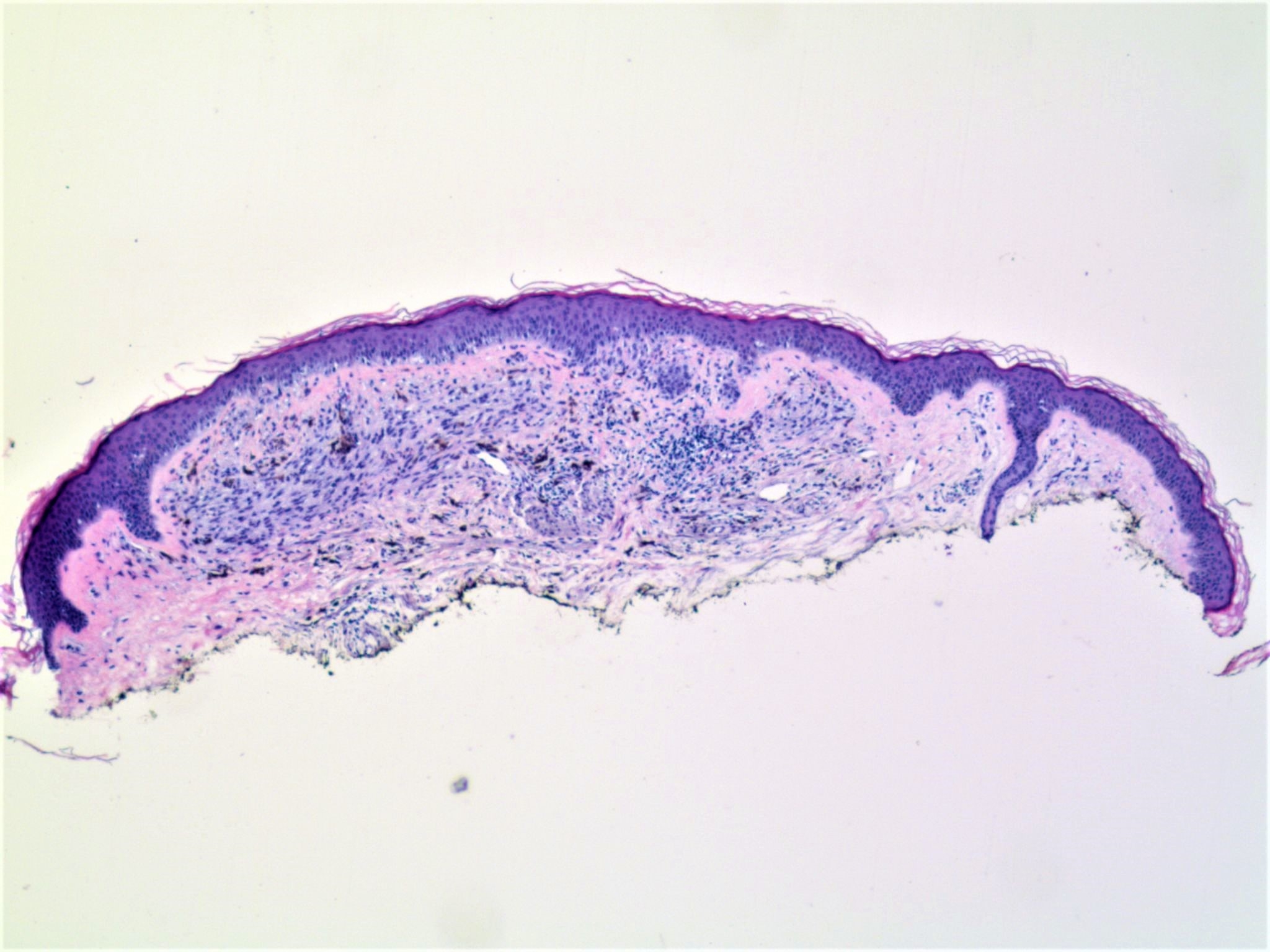
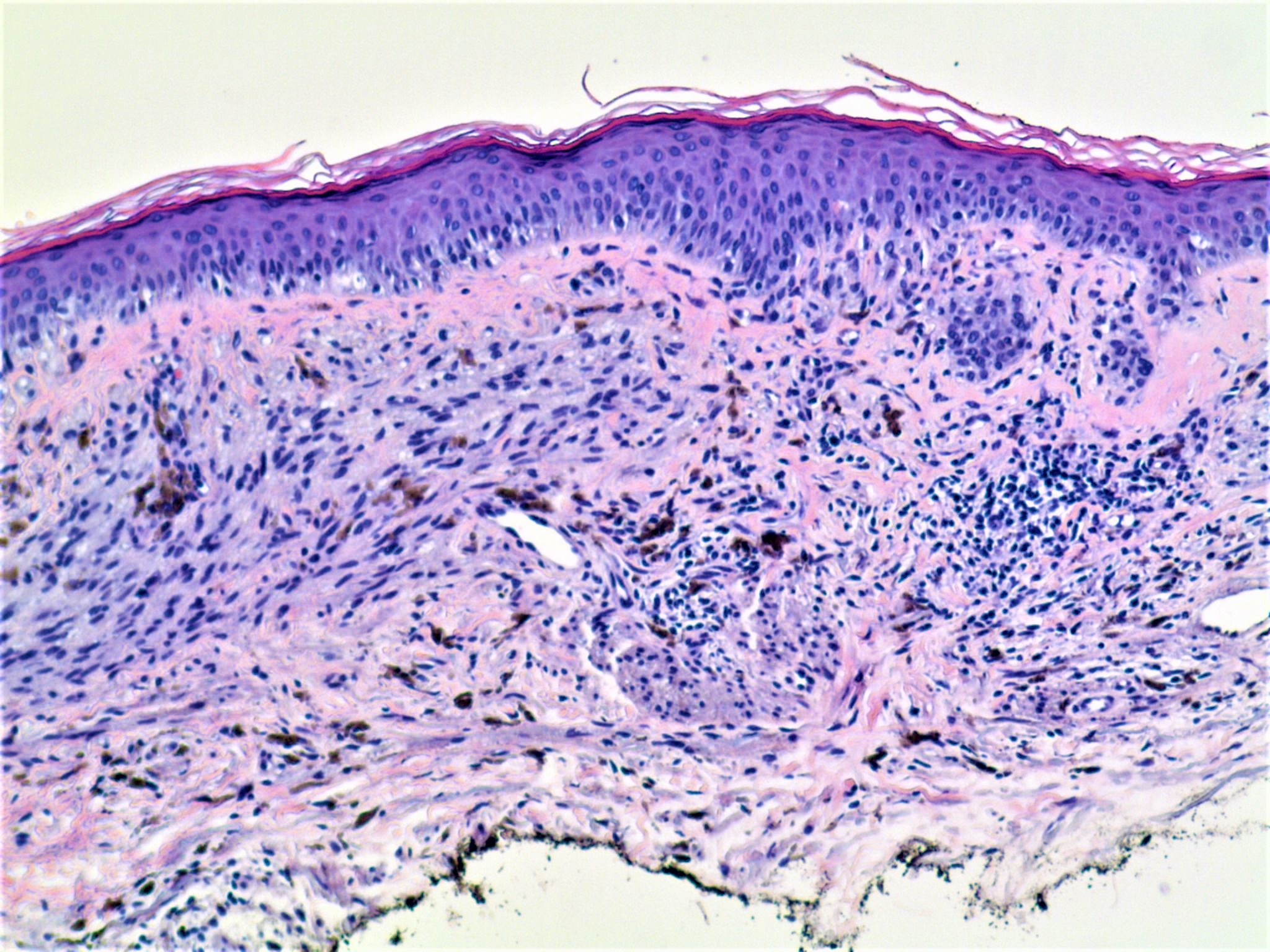
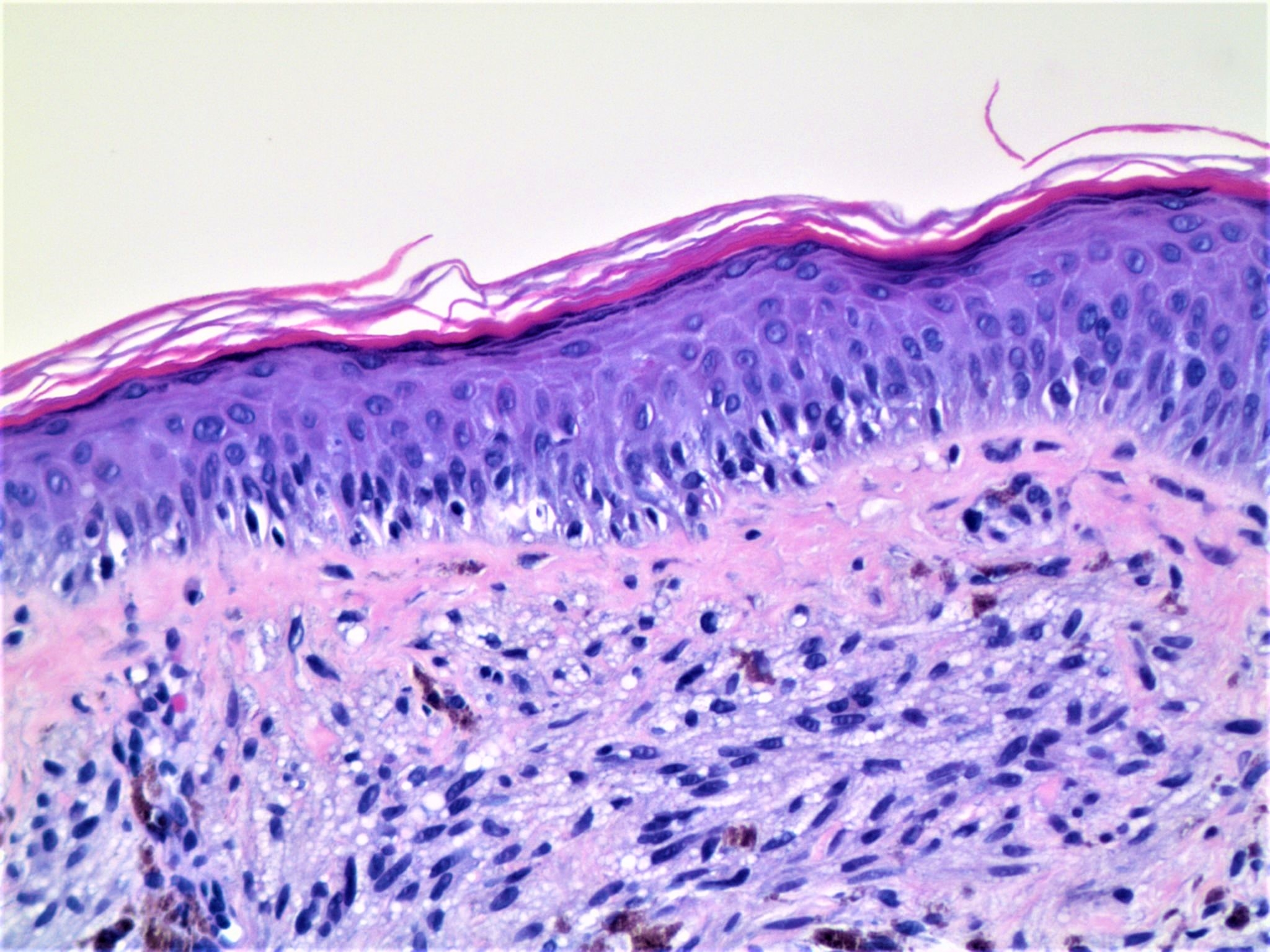
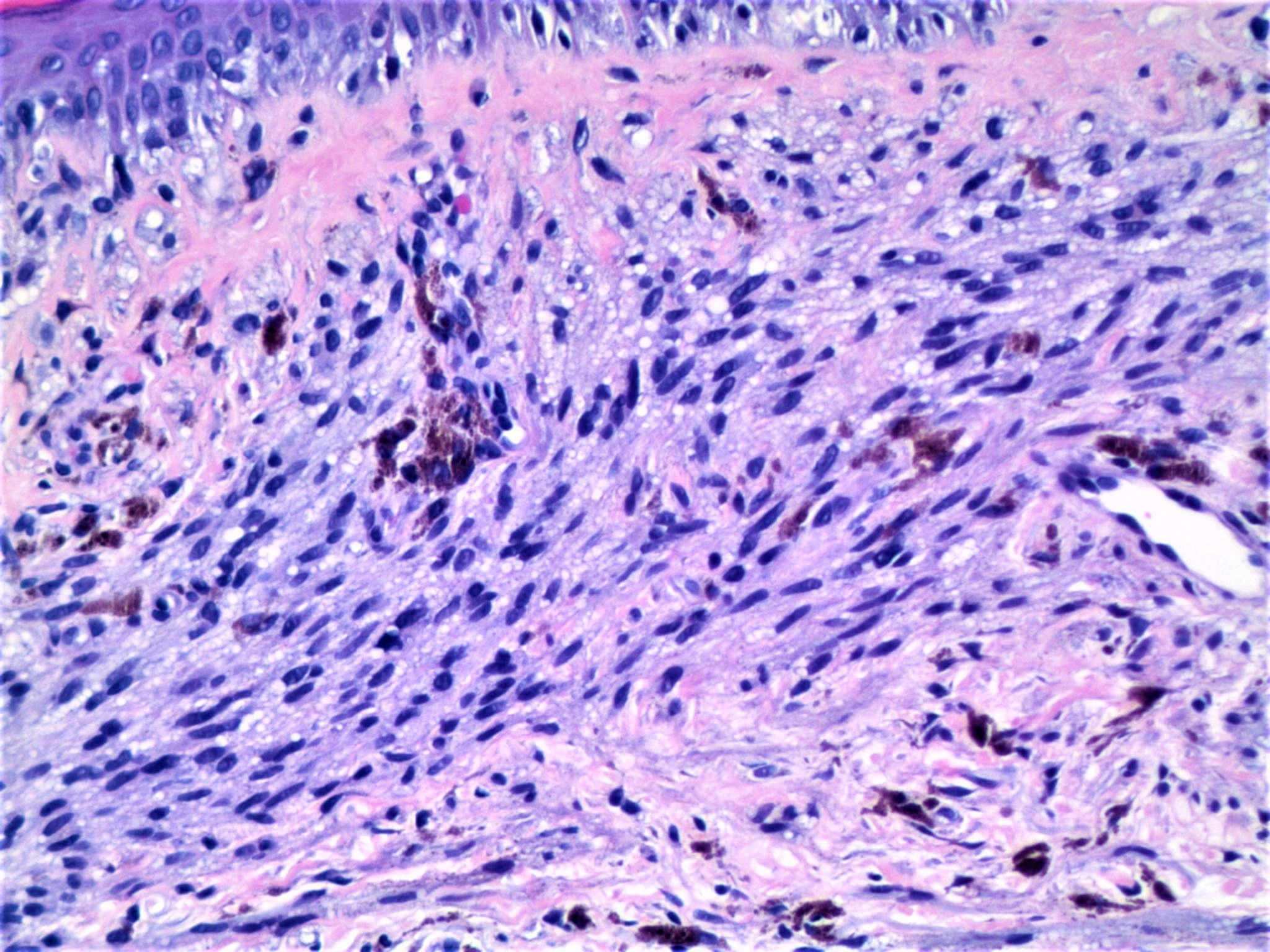
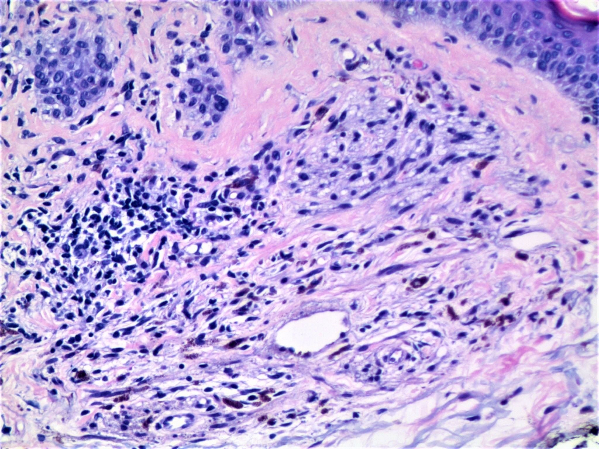
65 year old woman with lesion on forehead.


  Report Record

User Feedback


John Zhang

Posted

I am debating whether this should be classified as cellular blue nevus or just blue nevus. The upper cellular portion doesn't have dumbell shaped deep extensions. But maybe strictly speaking I should classify as cellular blue nevus. 

Share this comment


Link to comment
share_externally

Raul Perret

Posted

I am worried about this lesion. 65 year old, sun exposed skin, there is a continuous proliferation of melanocytes with some nuclear atypia. I would suggest complete excision, i am worried about melanoma

Share this comment


Link to comment
share_externally

Alice Roberts

Posted

I am also concerned.  We sometimes send borderline lesions for Myriad Mypath study to help us.  If this is a blue nevus, they tend to test far into the benign portion of the spectrum so might be helpful here.

Share this comment


Link to comment
share_externally

I have a hard time evaluating the epidermal component. So I would do Melan-A and SOX-10 to see whether there is an increase of melanocytes in the epidermis. If there is significant increase of melanocytes along the dermal-epidermal junction, I would call it malignant melanoma. Since I am not quite convinced there is an overlying melanoma in situ / lentigo maligna based on the available figures, I would consider the entity below:

Epithelioid and fusiform blue nevus of chronically sun-damaged skin, an entity distinct from the epithelioid blue nevus of the Carney complex.

Yazdan P, Haghighat Z, Guitart J, Gerami P.

Am J Surg Pathol. 2013 Jan;37(1):81-8


Share this comment


Link to comment
share_externally

My first thought: benign blue nevus in a sun damaged skin. Agree with Anh about Melan A utility to valuate the intraepidermal melanocytic growth.

Share this comment


Link to comment
share_externally

Uma Sundram

Posted

Great and extremely valuable comments. I too think this is a pre existing blue nevus on sun damaged skin. There is some atypia and given the age, I recommended excision. Mitf highlighted a small lentiginous junctional component.

Share this comment


Link to comment
share_externally


Join the conversation

You can post now and register later. If you have an account, sign in now to post with your account.

Guest
Add a comment...

×   Pasted as rich text.   Paste as plain text instead

  Only 75 emoji are allowed.

×   Your link has been automatically embedded.   Display as a link instead

×   Your previous content has been restored.   Clear editor

×   You cannot paste images directly. Upload or insert images from URL.

Loading...

×
×
  • Create New...