Jump to content
In this section we have spot diagnoses posted on a daily basis since June 2010, now over 4000! You can review the archived cases and read the suggested diagnoses by users and the final comment by the contributors.
Case are uploaded each week day by 10 am UK time with the correct diagnosis will generally be posted at 8 pm UK time. Why not view the most recent spot diagnosis and proffer a diagnosis?

Case Number : Case 2084 - 1 June 2018 Posted By: Dr. Richard Carr

Please read the clinical history and view the images by clicking on them before you proffer your diagnosis.
Submitted Date :
   (0 reviews)

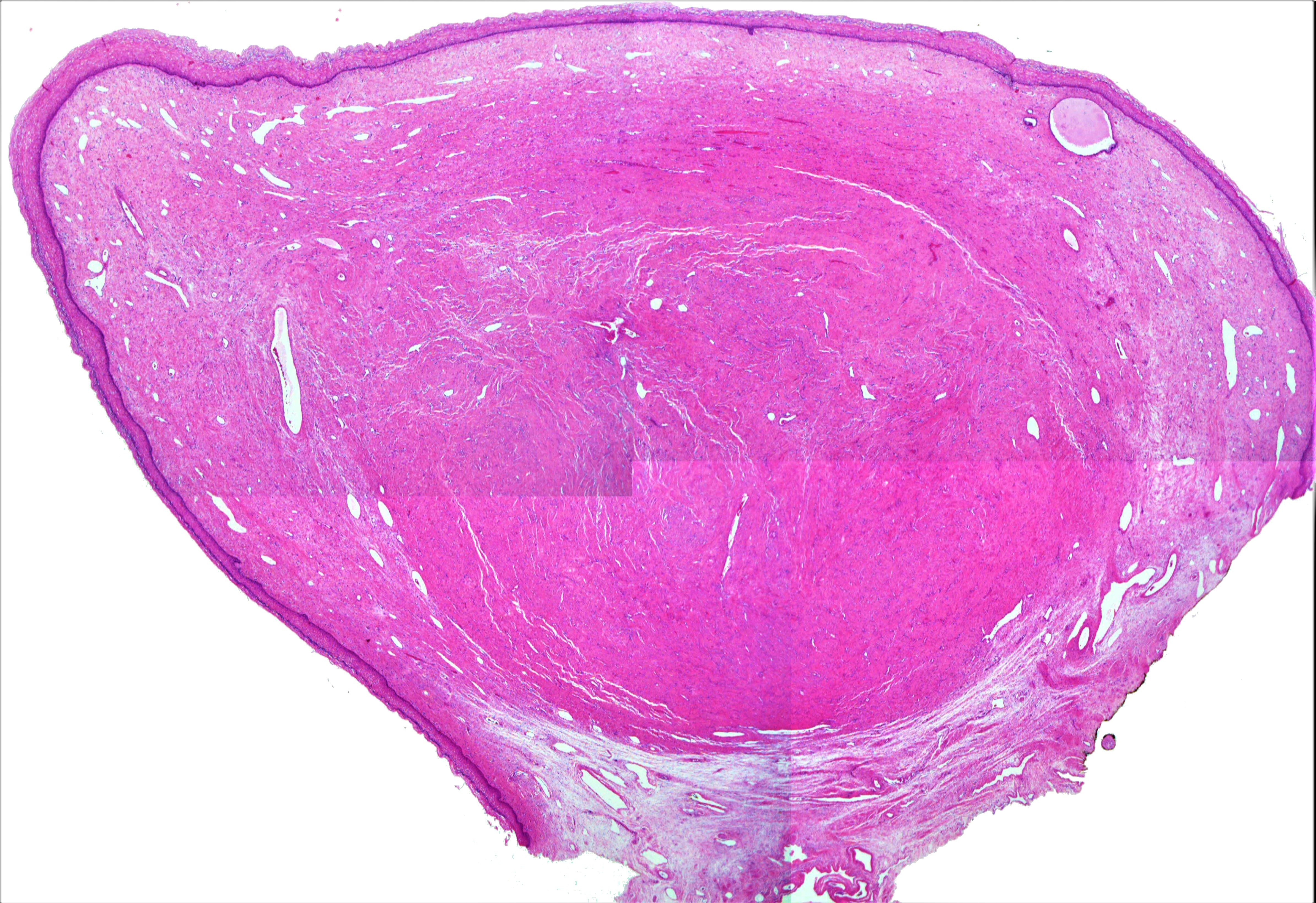
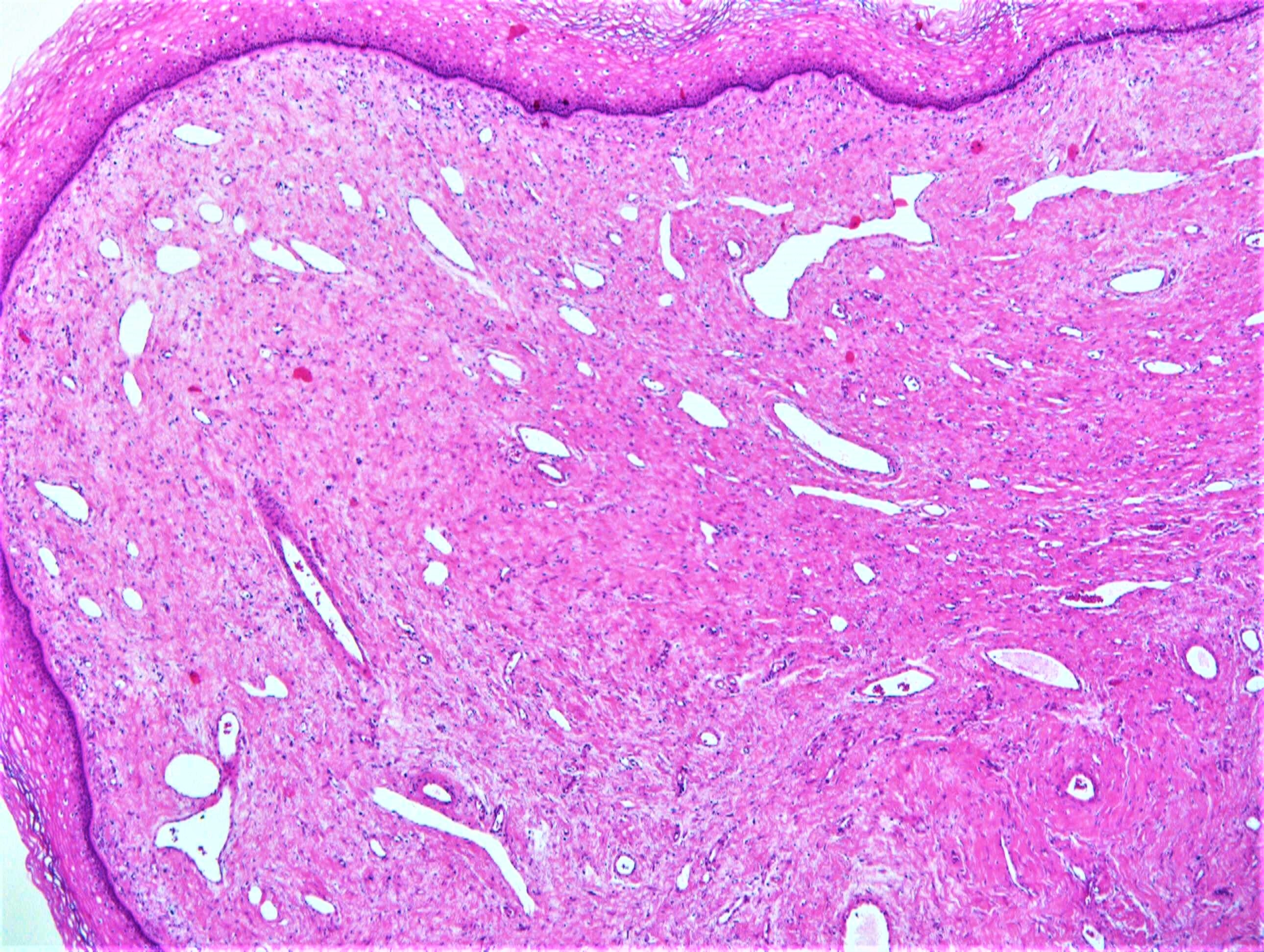
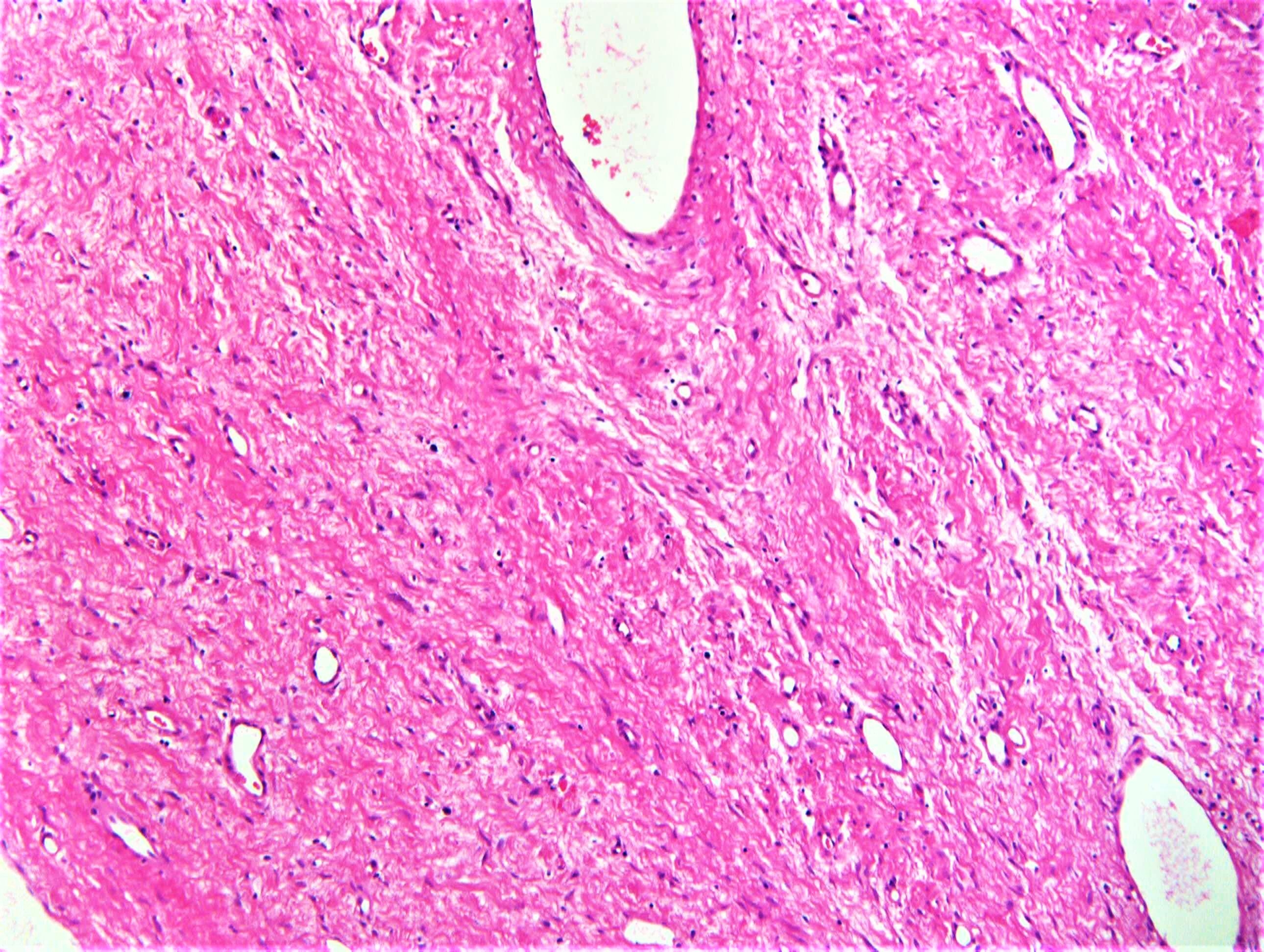
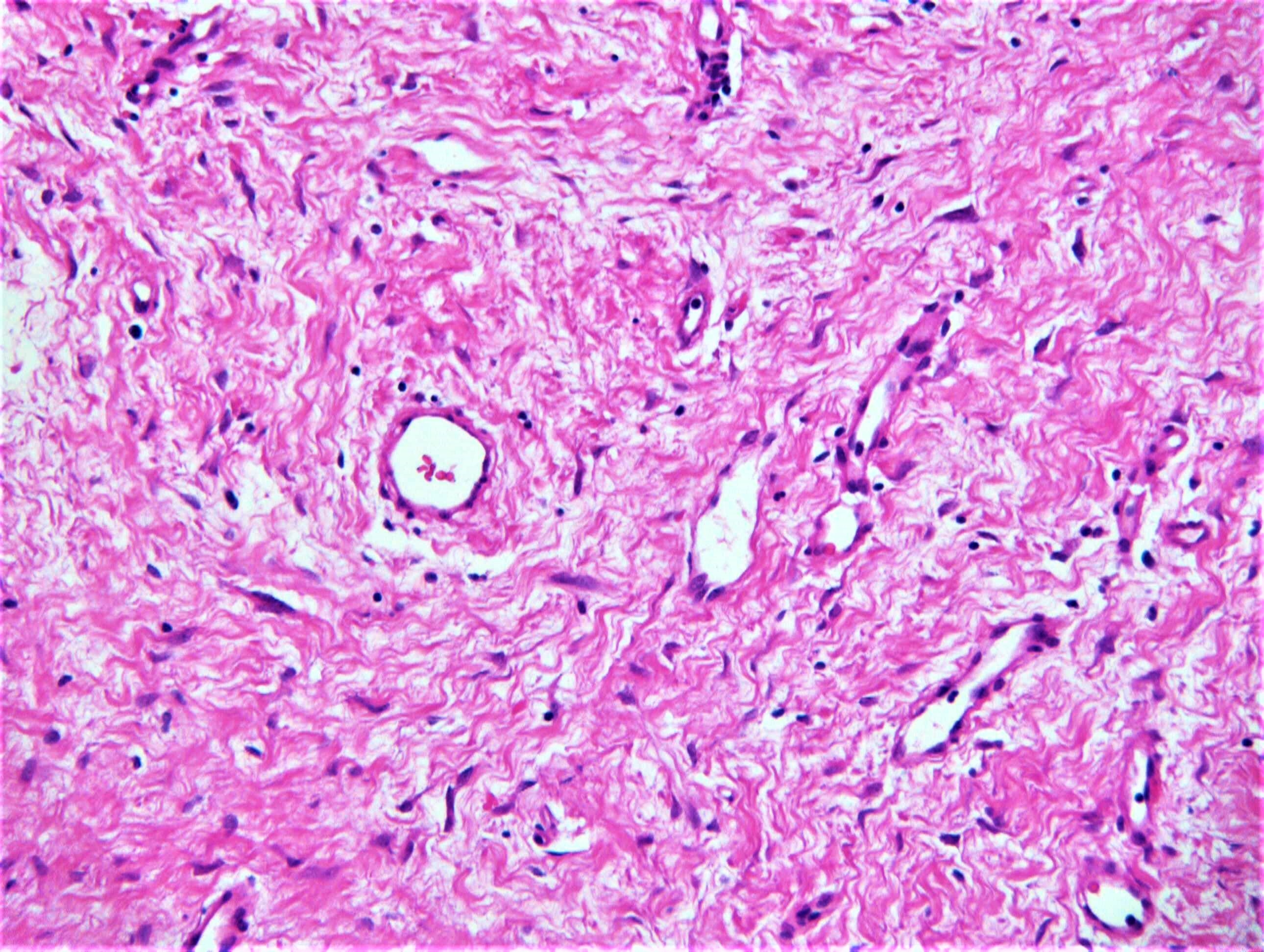
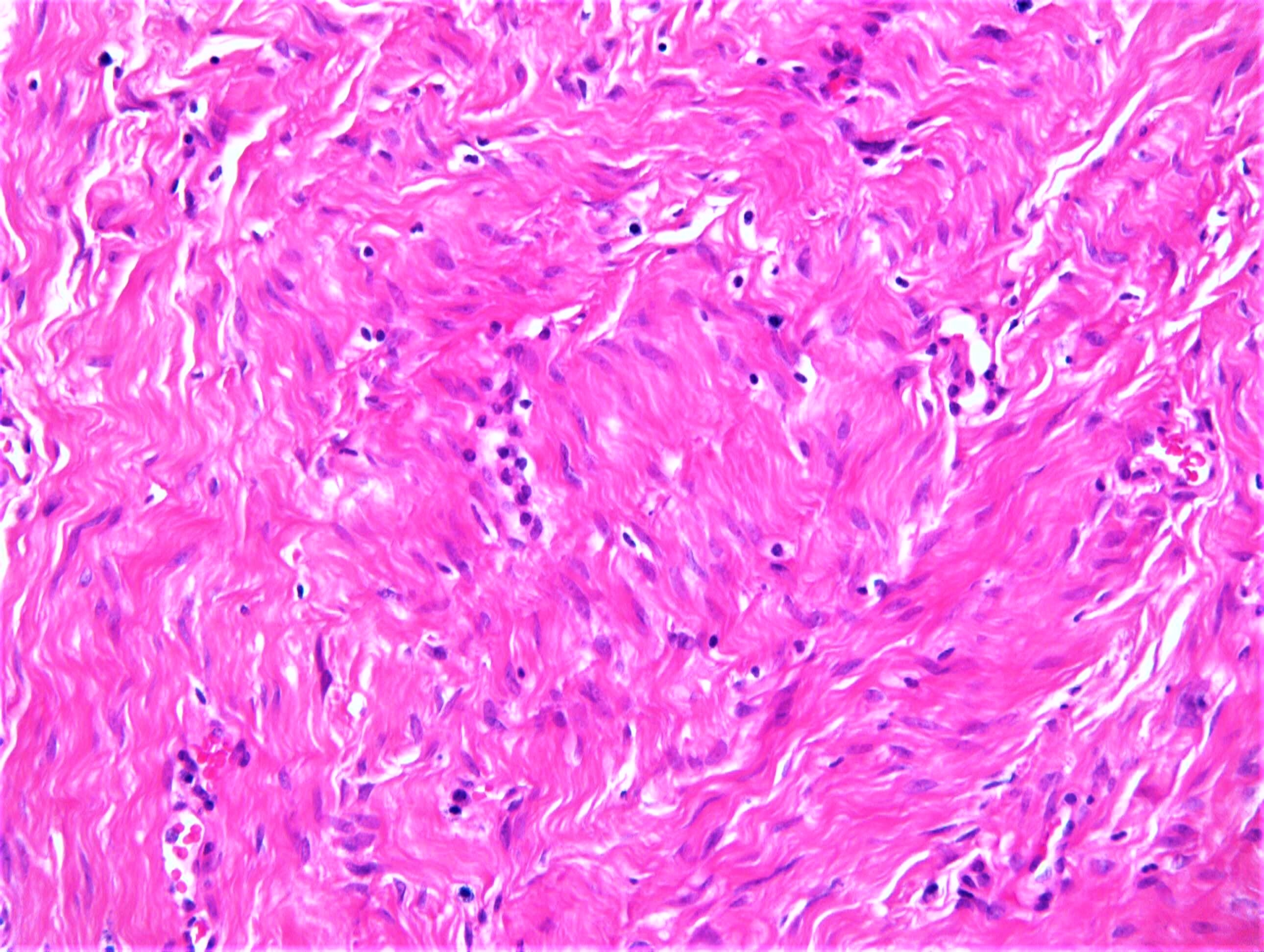
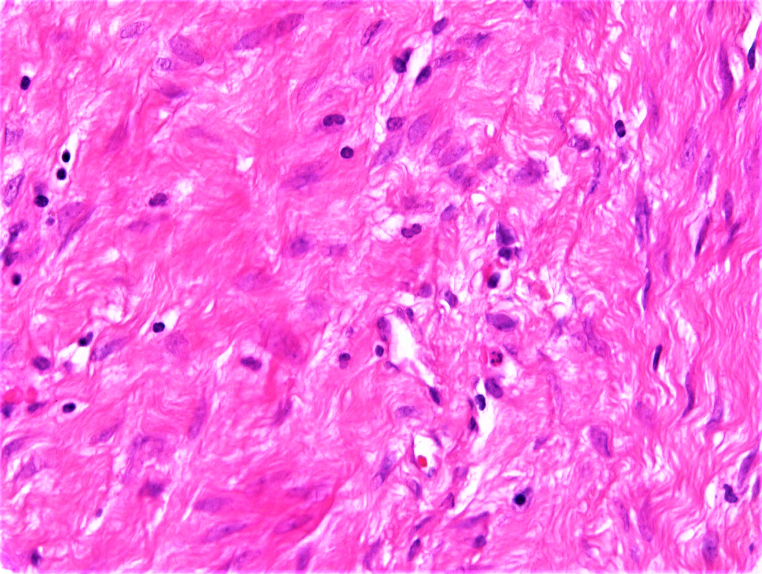
F70. Vaginal Cyst

Edited by Admin_Dermpath


  Report Record

User Feedback


msofopoulos

Posted

Looks like a Fibroepithelial (stromal) Polyp

Share this comment


Link to comment
share_externally

Agree. Just a little bit unusual because looks like there is a discrete hypocellular spindle cell nodule separated from the overlying mucosa by normal vaginal stroma.

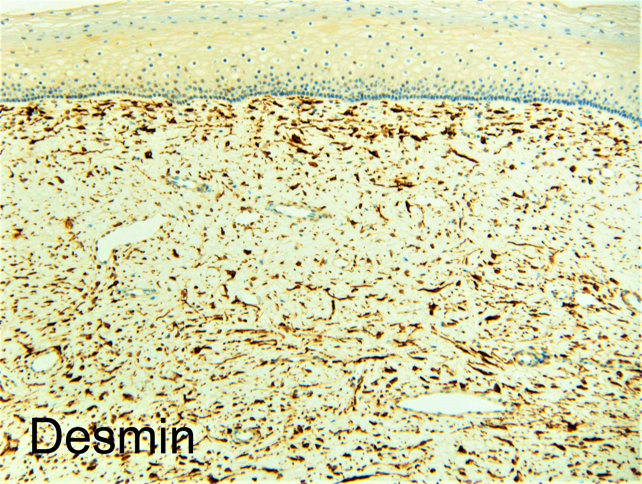
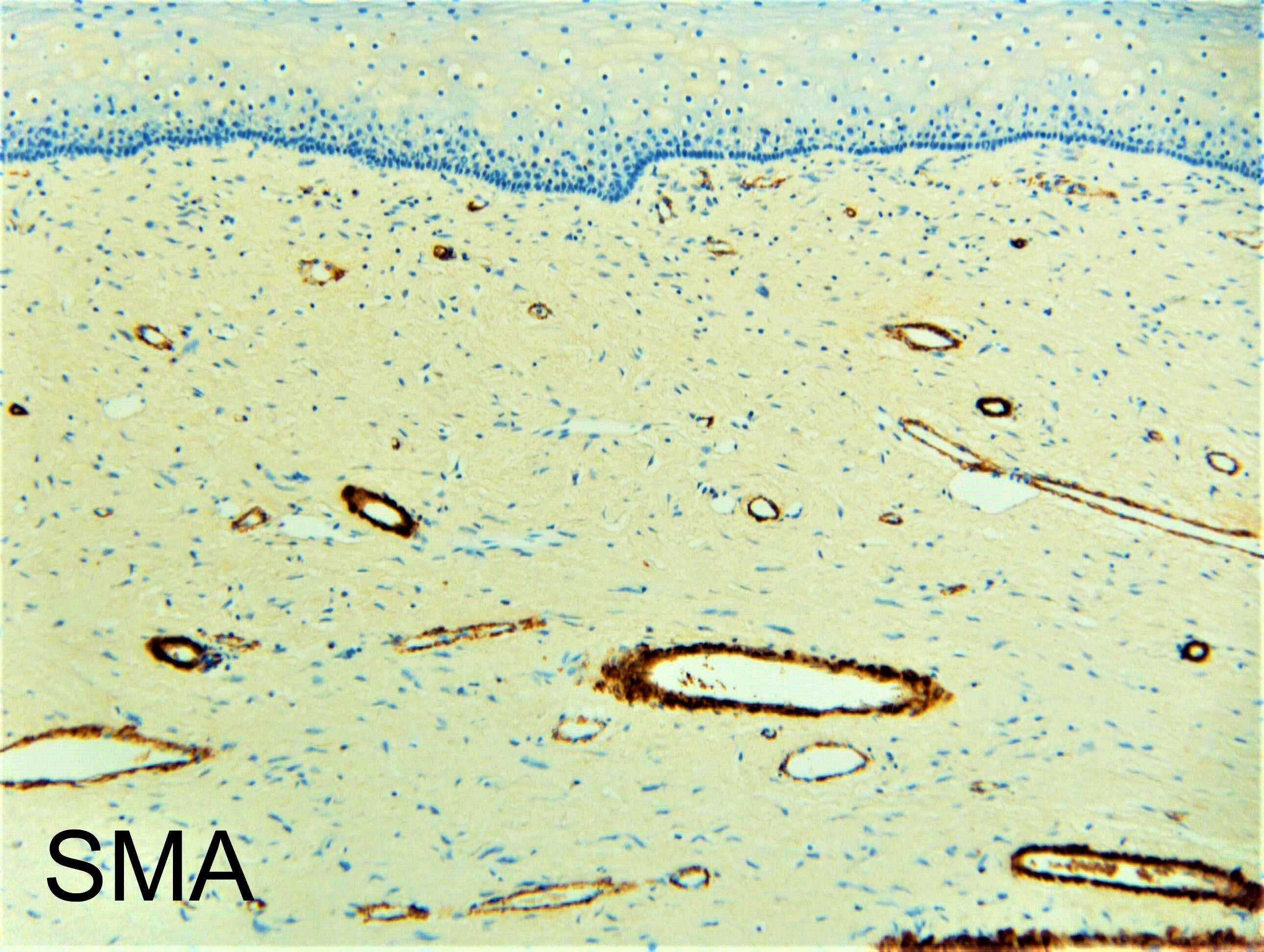
Just for fun, any Desmin, CD34, or ER stains?

Share this comment


Link to comment
share_externally

Anil Patki

Posted

Vulval vestibular papillomatosis. 

Share this comment


Link to comment
share_externally

Raul Perret

Posted

Fibroepithelial stromal polyp

Share this comment


Link to comment
share_externally

vincenzo polizzi

Posted

Angiofibroma, hypocellular variant. 

Share this comment


Link to comment
share_externally

Although the one that I have seen is usually more cellular, the immunopositivity for Desmin support the diagnosis of superficial cervicovaginal myofibroblastoma. The lesion is probably also positive for CD34 and ER. I personally think superficial cervicovaginal myofibroblastoma is probably a variant of fibropithelial stromal polyp, just somehow positive for Desmin.

Share this comment


Link to comment
share_externally

Superficial cervicovaginal myofibroblastoma: fourteen cases of a distinctive mesenchymal tumor arising from the specialized subepithelial stroma of the lower female genital tract.

Hum Pathol. 2001 Jul;32(7):715-25.

Abstract

The clinicopathologic features and immunohistochemical profiles of 14 cases of a distinctive mesenchymal tumor that arises in the superficial lamina propria of the cervix and vagina and is histologically distinguishable from mesodermal (fibroepithelial) stromal polyp, including the cellular (pseudosarcomatous) variant, angiomyofibroblastoma, aggressive angiomyxoma, and other well-recognized lesions that occur in this location, are described. The lesions presented as a polypoid (n = 10) or nodular (n = 4) mass in the vagina (n = 12) or cervix (n = 2) of women ranging in age from 40 to 74 years (median, 58 years). The tumors were subepithelial in location, were well circumscribed, and ranged in size from 1 to 6.5 cm. (mean, 2.7 cm). Microscopically, the process was moderately to highly cellular and composed of relatively bland spindled and stellate-shaped mesenchymal cells embedded in a finely collagenous stroma that was punctuated by myxoid and edematous foci in 9 cases. The lesions characteristically had a multipatterned architecture with tumor cells focally assuming a lacelike/sievelike growth pattern in the more stroma-rich areas of the tumor and a vague fascicular growth pattern in the more cellular foci. Mitotic activity was minimal, and no atypical mitotic figures were identified. The tumors were immunoreactive (in decreasing order of relative strength) for vimentin (5 of 5 cases), estrogen (10 of 10 cases), and progesterone (10 of 10 cases) receptors, desmin (13 of 13 cases), CD34 (11 of 13 cases), alpha-smooth muscle actin (5 of 11 cases), and muscle-specific actin (2 of 8 cases). The desmin and CD34 antibodies highlighted the interconnecting, dendritic processes associated with many of the tumor cells. No immunoreactivity was detected for S100 protein, epithelial membrane antigen, or keratins. Follow-up data for 11 patients (range, 1 to 20 years; median, 4 years) showed no recurrence or metastasis after local excision. The term "superficial cervicovaginal myofibroblastoma" is proposed because it reflects the distinguishing features of this benign, relatively site-specific mesenchymal tumor. The process probably arises as a neoplastic proliferation of hormonally responsive mesenchymal cells native to the unique subepithelial stromal layer normally found through the endocervix and vulva of adult women.


Because I think it's just a variant of fibroepithelial stromal polyp in the uterine cervix and vagina, I agreed with the diagnosis that most colleagues have suggested. I have seen only 2 or 3 cases so far (but I probably missed a lot because I did not want to do immunohistochemical studies for a benign lesion), and they just looked like a cellular  fibroepithelial stromal polyp.  If I have to do immunostaining, just for fun to bring attention to a not well known type of lesion in that area, I would just do Desmin (just to confuse the younger colleagues). CD34 is positive for a lot of spindle cell lesions, so I probably would not do, but according to the original article, they are positive (and I think the cases that I saw were positive too).

Share this comment


Link to comment
share_externally

Dr. Richard Carr

Posted

I was shown this case by a relatively experienced colleague who'd favoured leiomyoma and already done the makers I think. I also thought it fitted nicely with vulvo-vaginal fibroepithelial stromal polyp. Thanks for the great comments and reference. We did not do the ER or CD34.

Share this comment


Link to comment
share_externally


Join the conversation

You can post now and register later. If you have an account, sign in now to post with your account.

Guest
Add a comment...

×   Pasted as rich text.   Paste as plain text instead

  Only 75 emoji are allowed.

×   Your link has been automatically embedded.   Display as a link instead

×   Your previous content has been restored.   Clear editor

×   You cannot paste images directly. Upload or insert images from URL.

Loading...

×
×
  • Create New...