Jump to content
In this section we have spot diagnoses posted on a daily basis since June 2010, now over 4000! You can review the archived cases and read the suggested diagnoses by users and the final comment by the contributors.
Case are uploaded each week day by 10 am UK time with the correct diagnosis will generally be posted at 8 pm UK time. Why not view the most recent spot diagnosis and proffer a diagnosis?

Case Number : Case 2088 - 7 June 2018 Posted By: Raul Perret

Please read the clinical history and view the images by clicking on them before you proffer your diagnosis.
Submitted Date :
   (0 reviews)

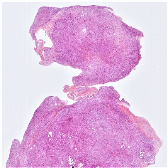
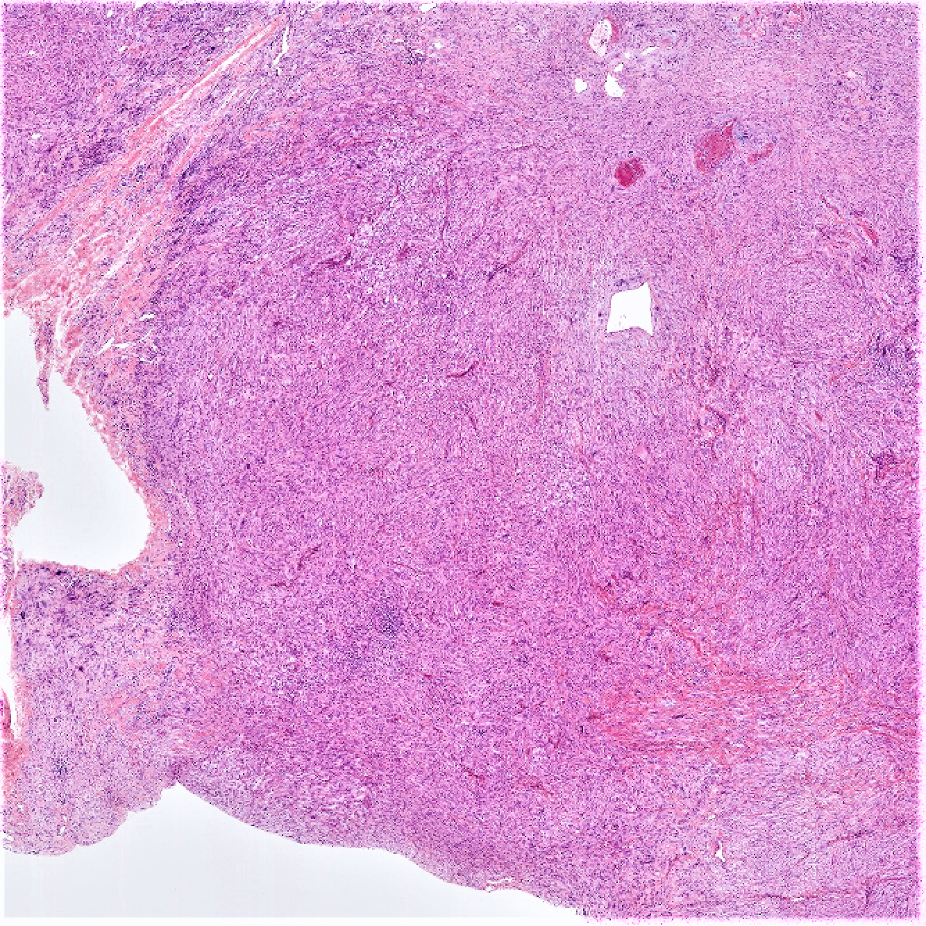
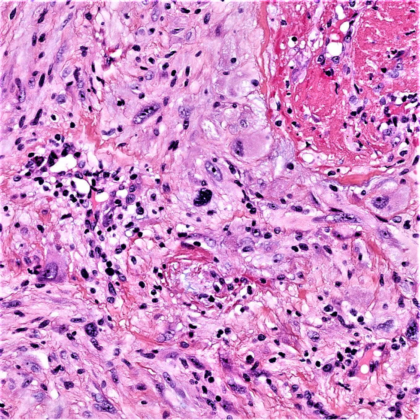
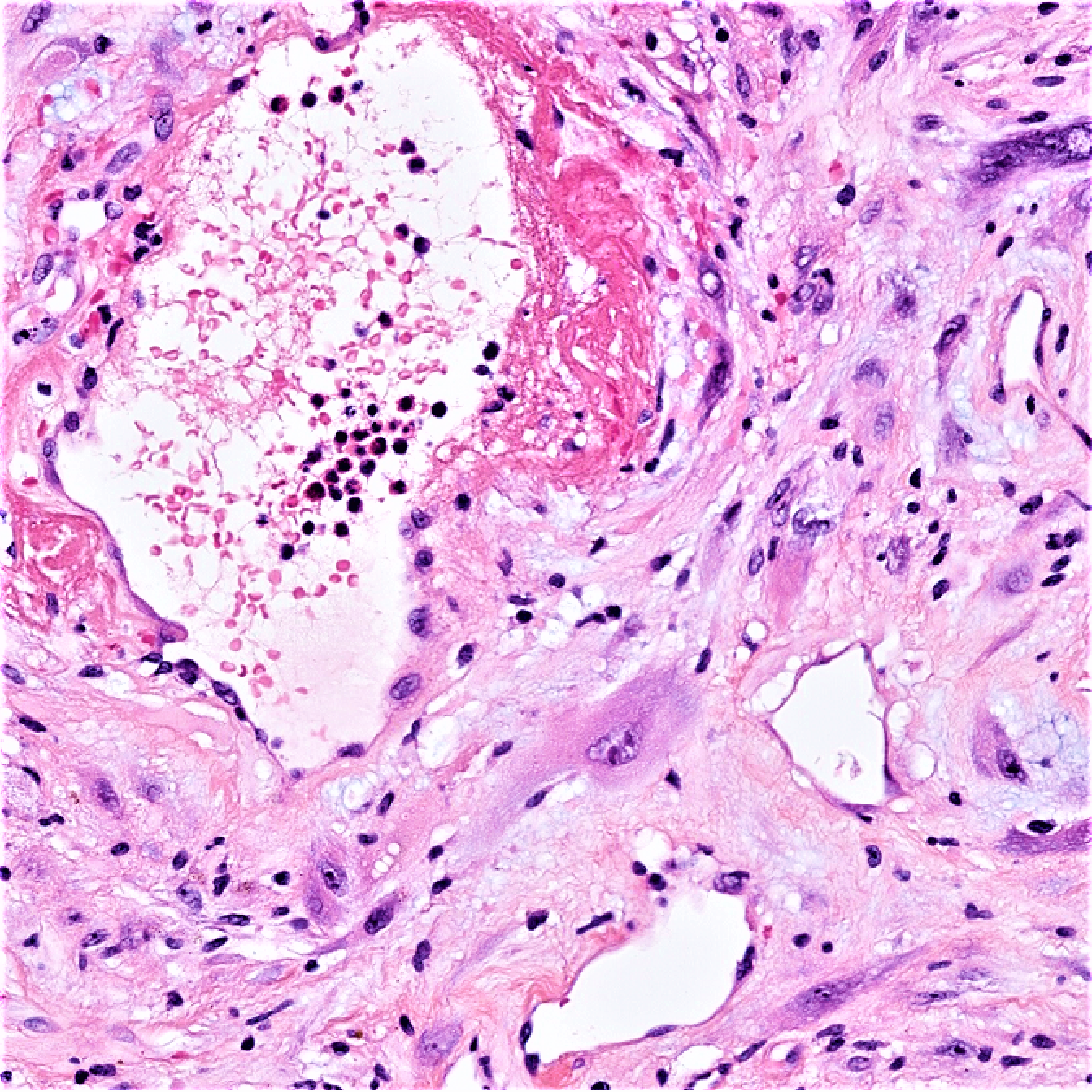
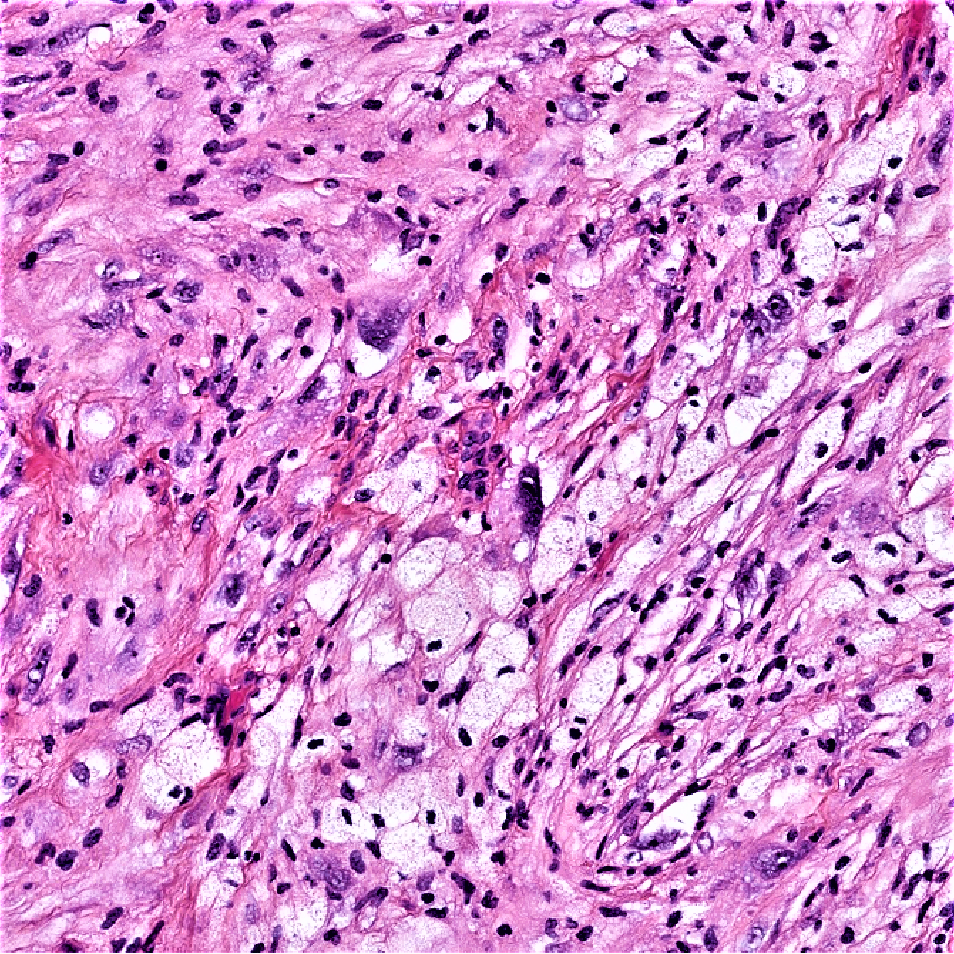
47 year old male with a 20 mm deep mass centered on the ankle.


  Report Record

User Feedback


John Zhang

Posted

If the lesion is negative for keratin, desmin and the GMS stain does not show organisms in the areas shown in the last photo, I intend to classify as pleomorphic undifferentiated sarcoma.

Share this comment


Link to comment
share_externally

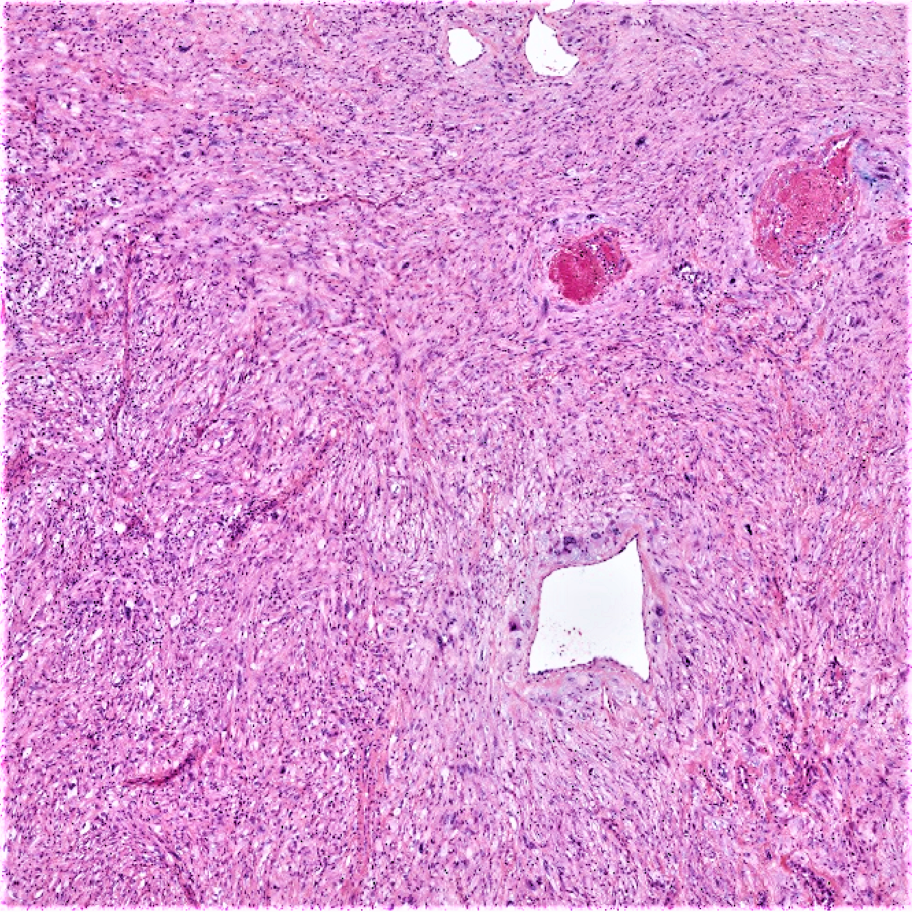
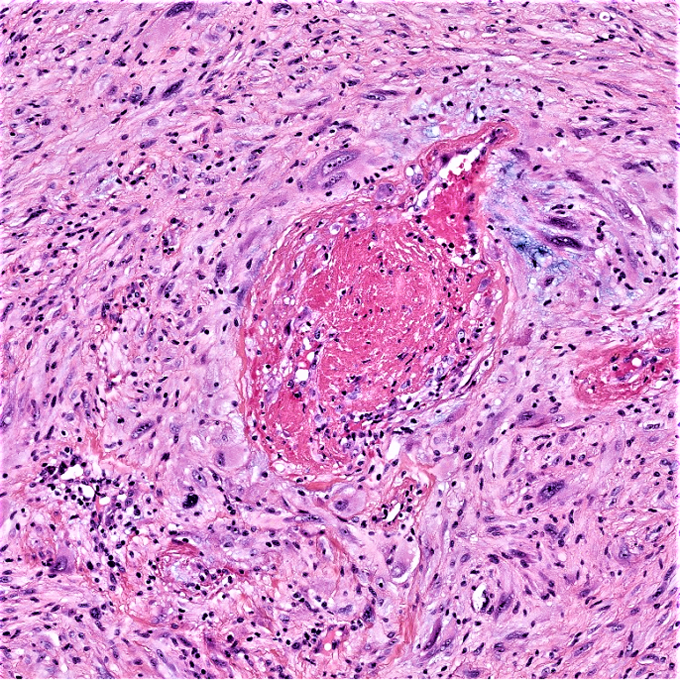
There is definitively cytologic atypia in the lesion, maybe even severe. But i do not see any mitoses or necrosis (at least in the figures). So there is a discrepancy between the degree of cytologic atypia and the mitotic activity. Can the cytologic atypia a degenerative phenomenom in nature? I also see foamy histiocytes and I usually back off a little bit from malignancy when I see that. There are also dilated blood vessels. If the lesion is positive for S_100 protein and SOX-10, I would consider anscient schwannoma.

Share this comment


Link to comment
share_externally

John Zhang

Posted

Anh has a good point. I would run S100, too.

Share this comment


Link to comment
share_externally

vincenzo polizzi

Posted

Which feature does win here?

the curvilinear vessels of Myxofibrosarcoma or the ganglion-like cells of myxoinflammatory fibroblastic sarcoma or the Angietactic sand pleomorphic but without hyalinization features of Pleomorphic hyalinizing angiectatic tumor?  The age and the site but also th microscopy make me favor PHAT. 

Share this comment


Link to comment
share_externally

vincenzo polizzi

Posted

I answered too soon. Here Anh wins. 

Share this comment


Link to comment
share_externally

Actually if S-100 is negative, I would start considering PHAT or maybe lipidized fibrous histiocytoma (because of the location) or atypical fibrous histiocytoma. So we definitely need immunos. That's the fun part of this activity. We all learn something new every day.

Share this comment


Link to comment
share_externally

Raul Perret

Posted

S100, desmin, h-caldesmon, mdm2, negatives. CK focally positive, CD34 diffusely positive. What do you guys think?

Share this comment


Link to comment
share_externally

msofopoulos

Posted

EMA? loss of INI?The immunohistochemistry leans towards epithelioid sarcoma but there are many histyocytes

Share this comment


Link to comment
share_externally

Raul Perret

Posted

2 minutes ago, msofopoulos said:

EMA? loss of INI?The immunohistochemistry leans towards epithelioid sarcoma but there are many histyocytes

INI1 positive (retained). EMA negative

Share this comment


Link to comment
share_externally

vincenzo polizzi

Posted

So why not a PHAT? Or the equivalent Hemosiderotic fibrohistiocytic lipomatous tumor?

Share this comment


Link to comment
share_externally

vincenzo polizzi

Posted

29 minutes ago, vincenzo polizzi said:


Share this comment


Link to comment
share_externally

Raul Perret

Posted

This was a case of Superficial CD34+ fibroblastic tumor. This entity was reported initially by Carter et al. in 2014 (https://www.nature.com/articles/modpathol2013139). It is characterized by a superficial location, diffuse expression of CD34, frequent focal co-expression of CK, presence of spindle cells with ample glassy eosinophilic cytoplasm (which can be quite focal), atypical bizarre nuclei, areas of inflammation, xanthomization and minimal mitotic activity.

Differential diagnosis to consider are (as Vincenzo initially suggested) PHAT and myxoinflammatory fibroblastic sarcoma which can share many morphological and immunophenotypic features (they are probable entities related with superficial CD34+ fibroblastic tumor). In order to rule out the latter differentials we performed a CGH to evaluate if there were amplifications of VGLL3 (https://www.ncbi.nlm.nih.gov/pmc/articles/PMC3361892/), the test demonstrated a flat profile.

This tumor seems to be of intermediate malignancy and will be probably better characterized in the future by performing NGS based techniques like RNAseq and whole exome sequencing. I also think they are probably underrecognized due to recent description and the presence of less than 50 cases reported in the literature. I am lucky enough to have already diagnosed 2 cases in resection specimens and will probably receive a third one soon (suspected on biopsy). Thanks for the great comments.

Share this comment


Link to comment
share_externally

vincenzo polizzi

Posted

Very great case Raul. Thanks. 

Share this comment


Link to comment
share_externally


Join the conversation

You can post now and register later. If you have an account, sign in now to post with your account.

Guest
Add a comment...

×   Pasted as rich text.   Paste as plain text instead

  Only 75 emoji are allowed.

×   Your link has been automatically embedded.   Display as a link instead

×   Your previous content has been restored.   Clear editor

×   You cannot paste images directly. Upload or insert images from URL.

Loading...

×
×
  • Create New...