Jump to content
In this section we have spot diagnoses posted on a daily basis since June 2010, now over 4000! You can review the archived cases and read the suggested diagnoses by users and the final comment by the contributors.
Case are uploaded each week day by 10 am UK time with the correct diagnosis will generally be posted at 8 pm UK time. Why not view the most recent spot diagnosis and proffer a diagnosis?

Case Number : Case 2090 - 11 June 2018 Posted By: Limin Yu

Please read the clinical history and view the images by clicking on them before you proffer your diagnosis.
Submitted Date :
   (0 reviews)

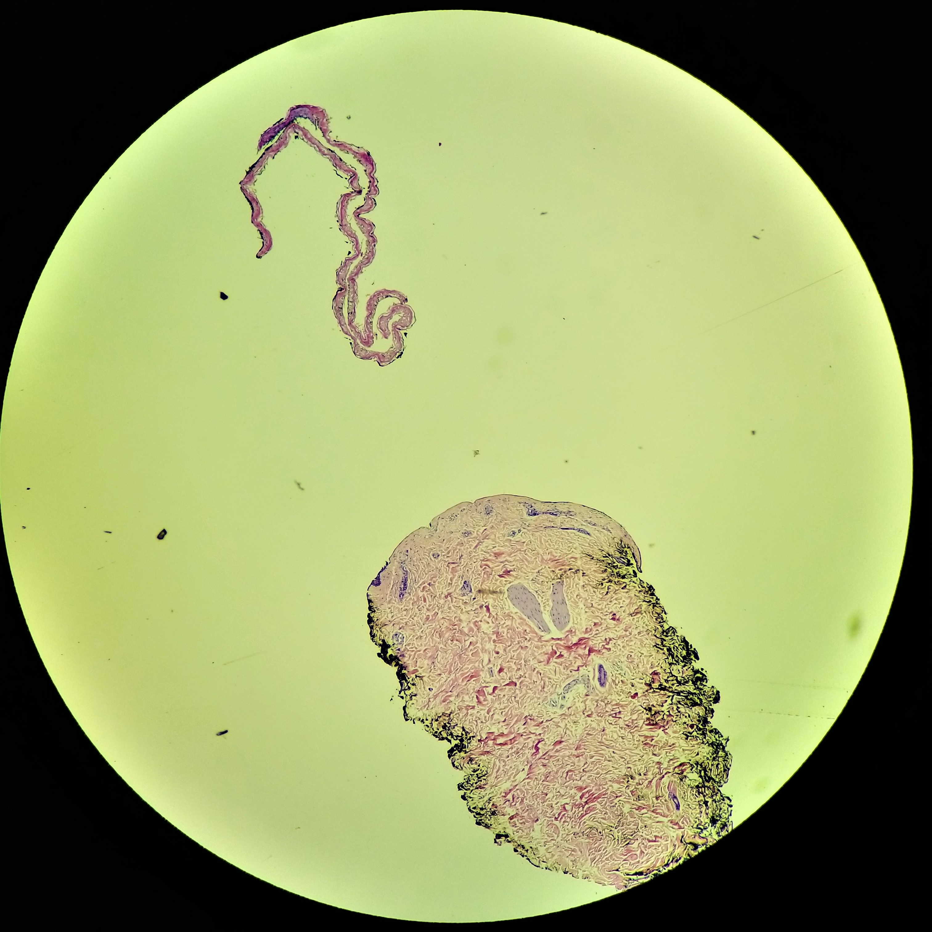
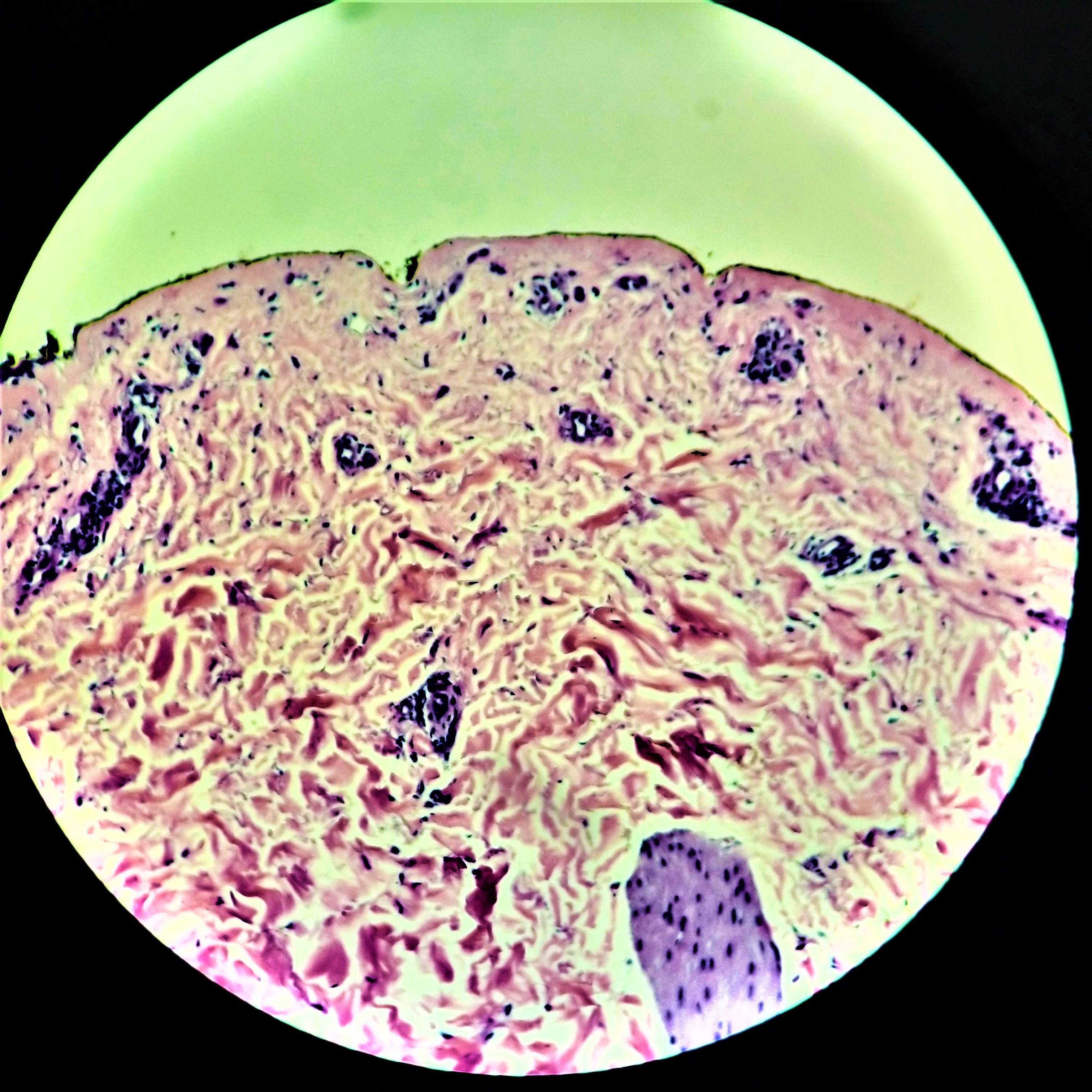
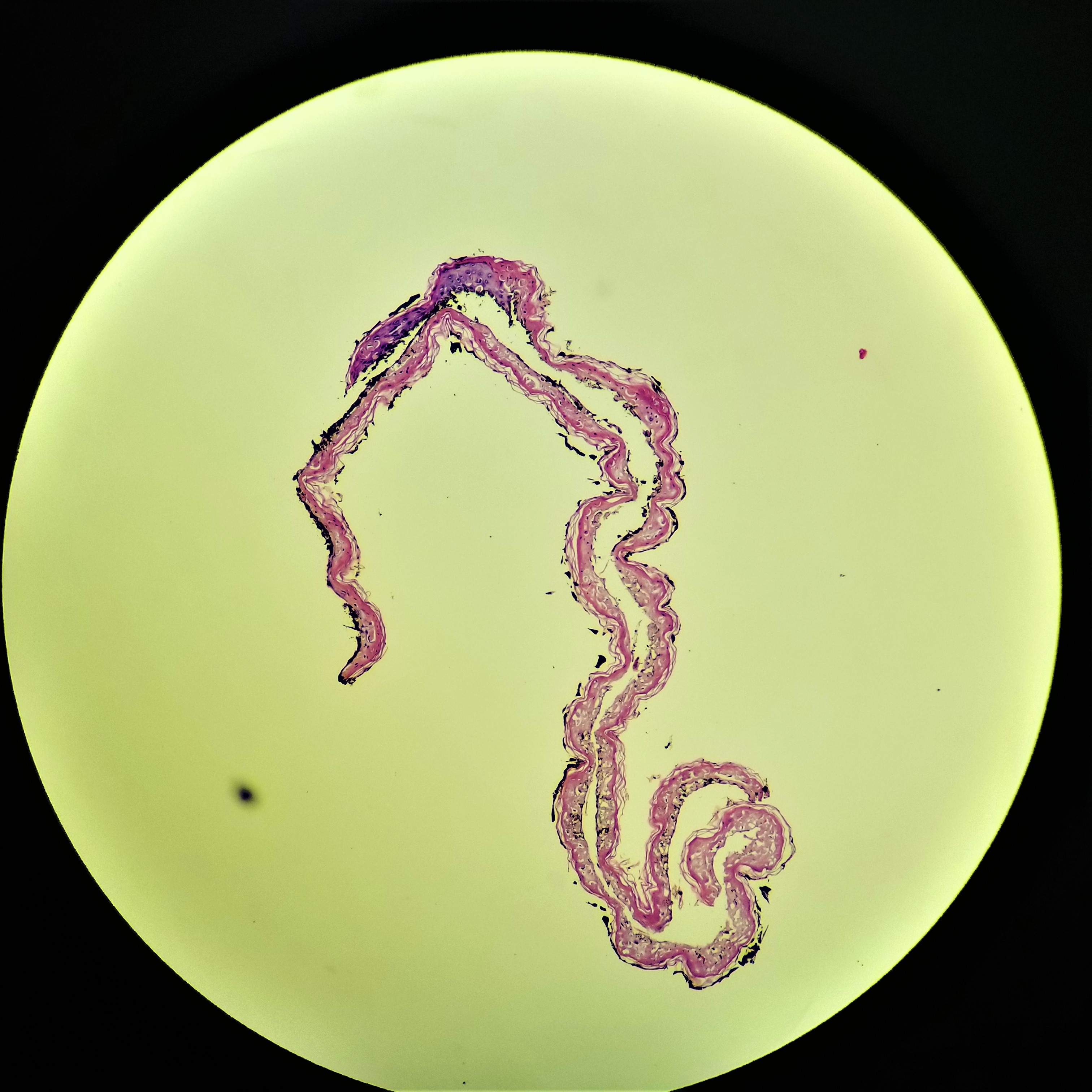
63 year old male with lesion on arm.

  Report Record

User Feedback


Anil Patki

Posted

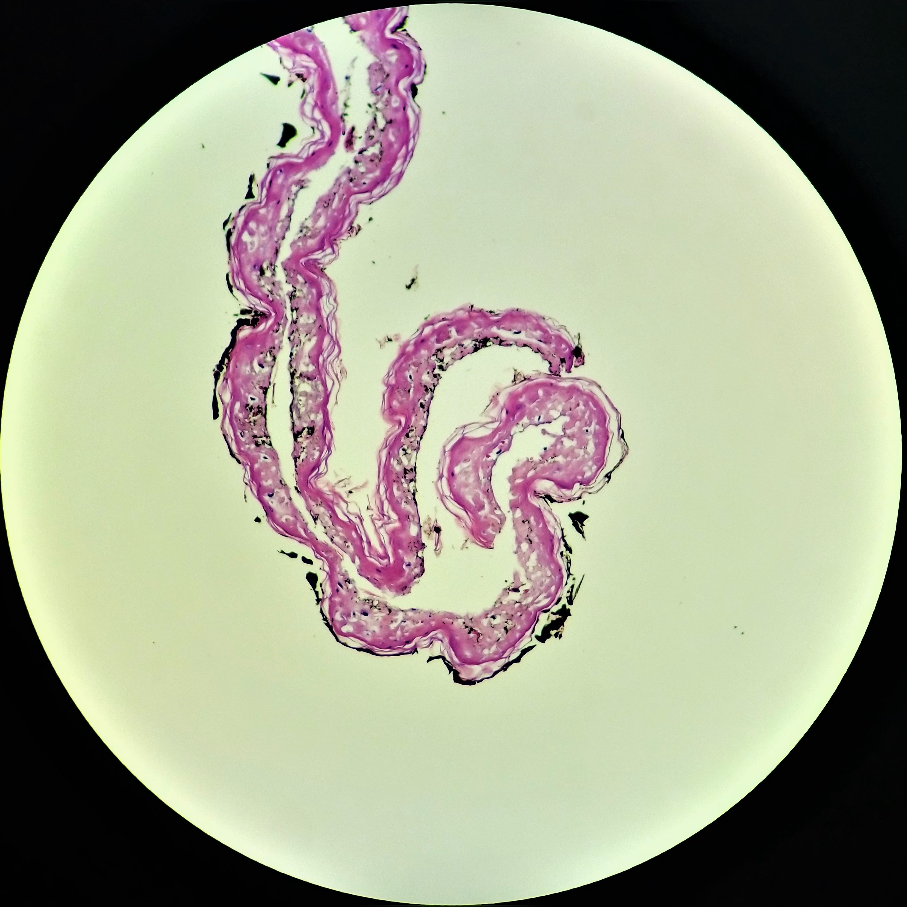
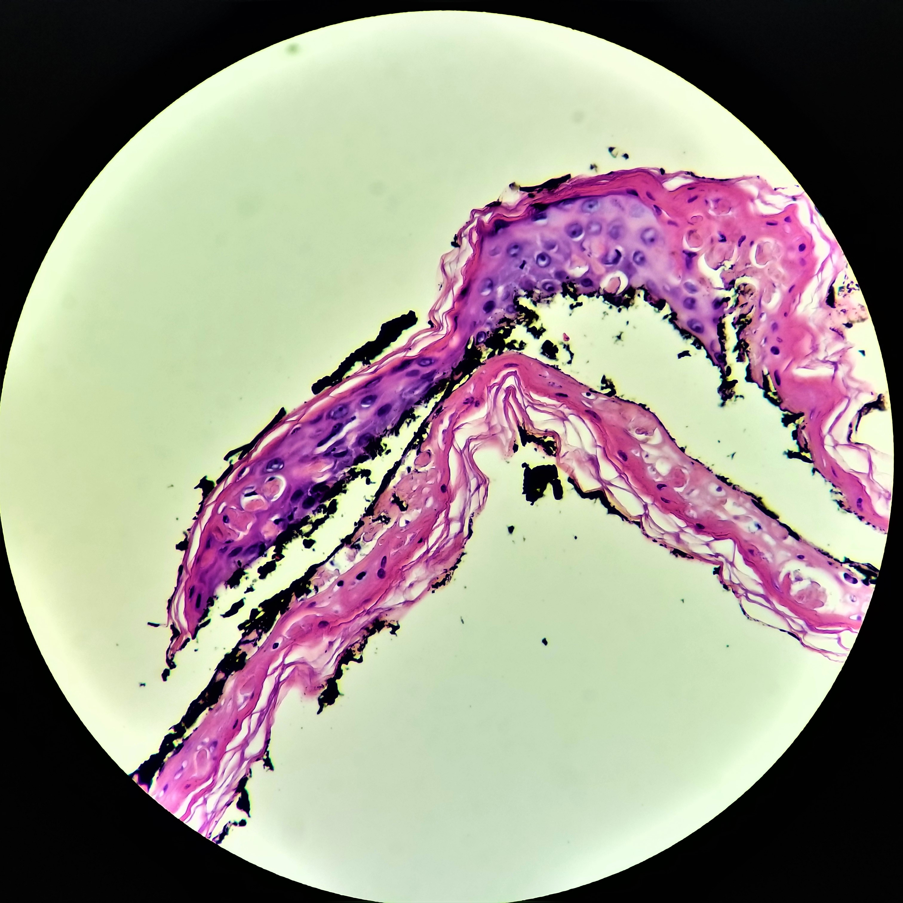
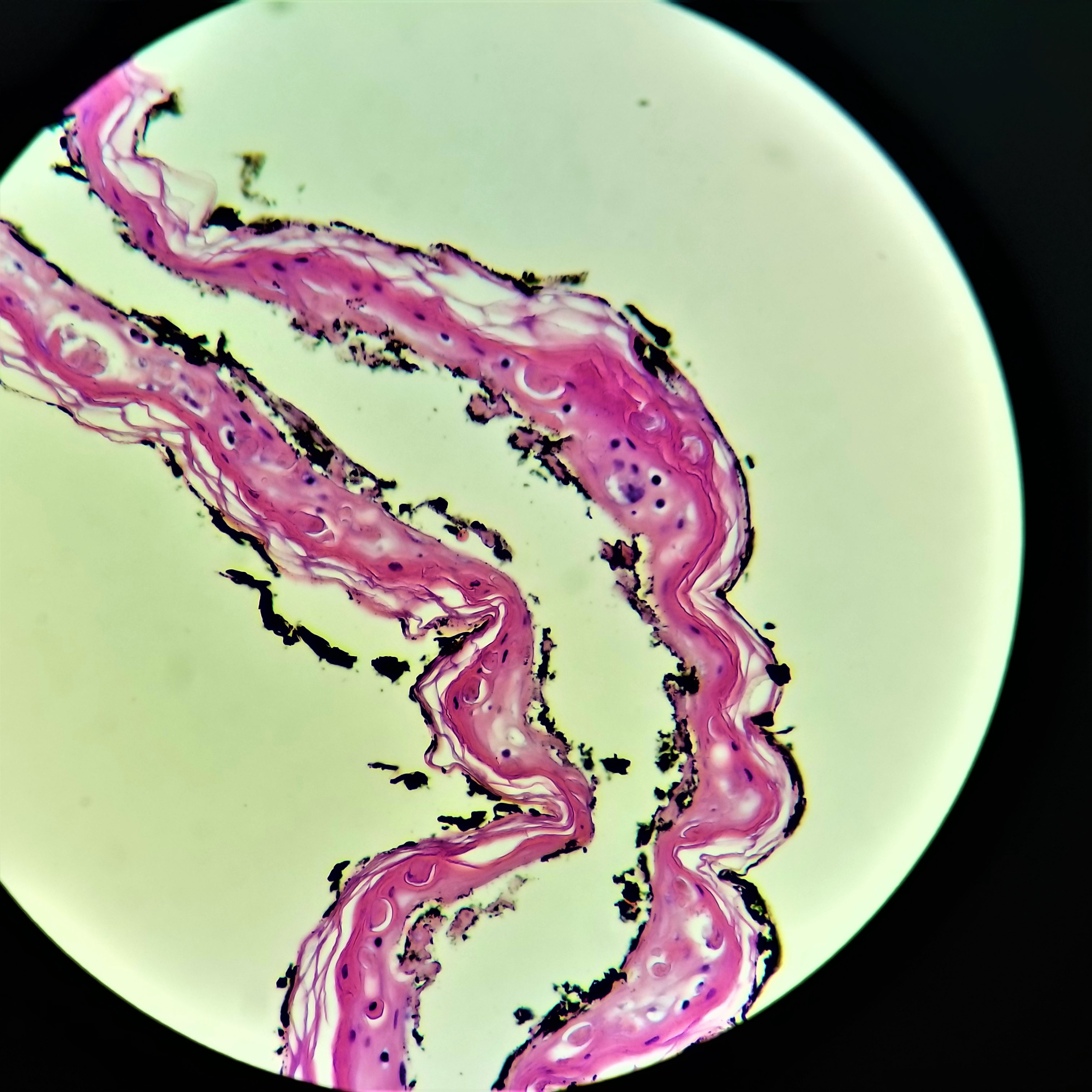
Subepidermal clevage with necrotic epidermis and dermal 'silence' . The only condition that comes to my mind is toxic epidermal necrolysis

Share this comment


Link to comment
share_externally

vincenzo polizzi

Posted

A single lesion of TEN?  Is it possible? Histologically full agree with Colleagues. 

Share this comment


Link to comment
share_externally

Limin Yu

Posted

The histologic changes are in the spectrum of EM/TEN/SJS. Clinically this lesion was more compatible with EM.

Share this comment


Link to comment
share_externally

John Zhang

Posted

My first thought was TEN. I am glad that we are all on the same page and Dr. Yu gave us the final answer which is consistent with what we thought. But I also want to comment that these histologic features are not pathognomonic for TEN. I believe that the epidermal necrosis can happen in any old blister. Therefore cell poor BP can show similar features like those in this case, and PCT cannot be excluded.

Share this comment


Link to comment
share_externally

Dr. Richard Carr

Posted

Actually here we see rounded pink bodies in the epidermis which is typical for individual cell necrosis (rather than coagulative) which is the clue for the diagnosis.

Share this comment


Link to comment
share_externally

Alice Roberts

Posted

I’d want to rule out pemphigoid and Clinical history should help. If any Clinical concern for pemphigoid, consider DIF.  I often see a rare necrotic keratinocytes in blister roof of pemphigoid and can be cell poor.  Old but interesting reference...

https://www.ncbi.nlm.nih.gov/pubmed/2409931

Share this comment


Link to comment
share_externally


Join the conversation

You can post now and register later. If you have an account, sign in now to post with your account.

Guest
Add a comment...

×   Pasted as rich text.   Paste as plain text instead

  Only 75 emoji are allowed.

×   Your link has been automatically embedded.   Display as a link instead

×   Your previous content has been restored.   Clear editor

×   You cannot paste images directly. Upload or insert images from URL.

Loading...

×
×
  • Create New...