Jump to content
In this section we have spot diagnoses posted on a daily basis since June 2010, now over 4000! You can review the archived cases and read the suggested diagnoses by users and the final comment by the contributors.
Case are uploaded each week day by 10 am UK time with the correct diagnosis will generally be posted at 8 pm UK time. Why not view the most recent spot diagnosis and proffer a diagnosis?

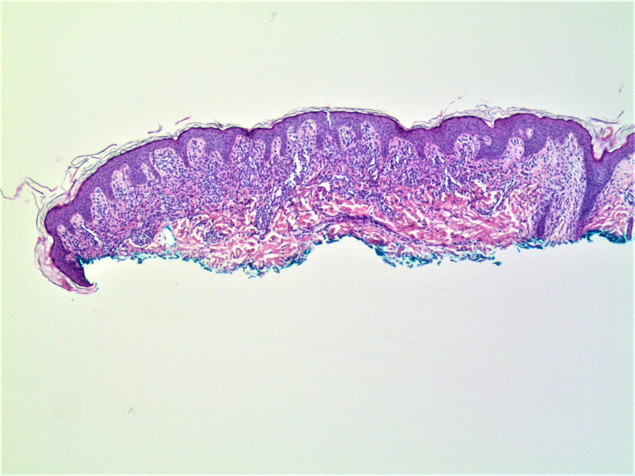
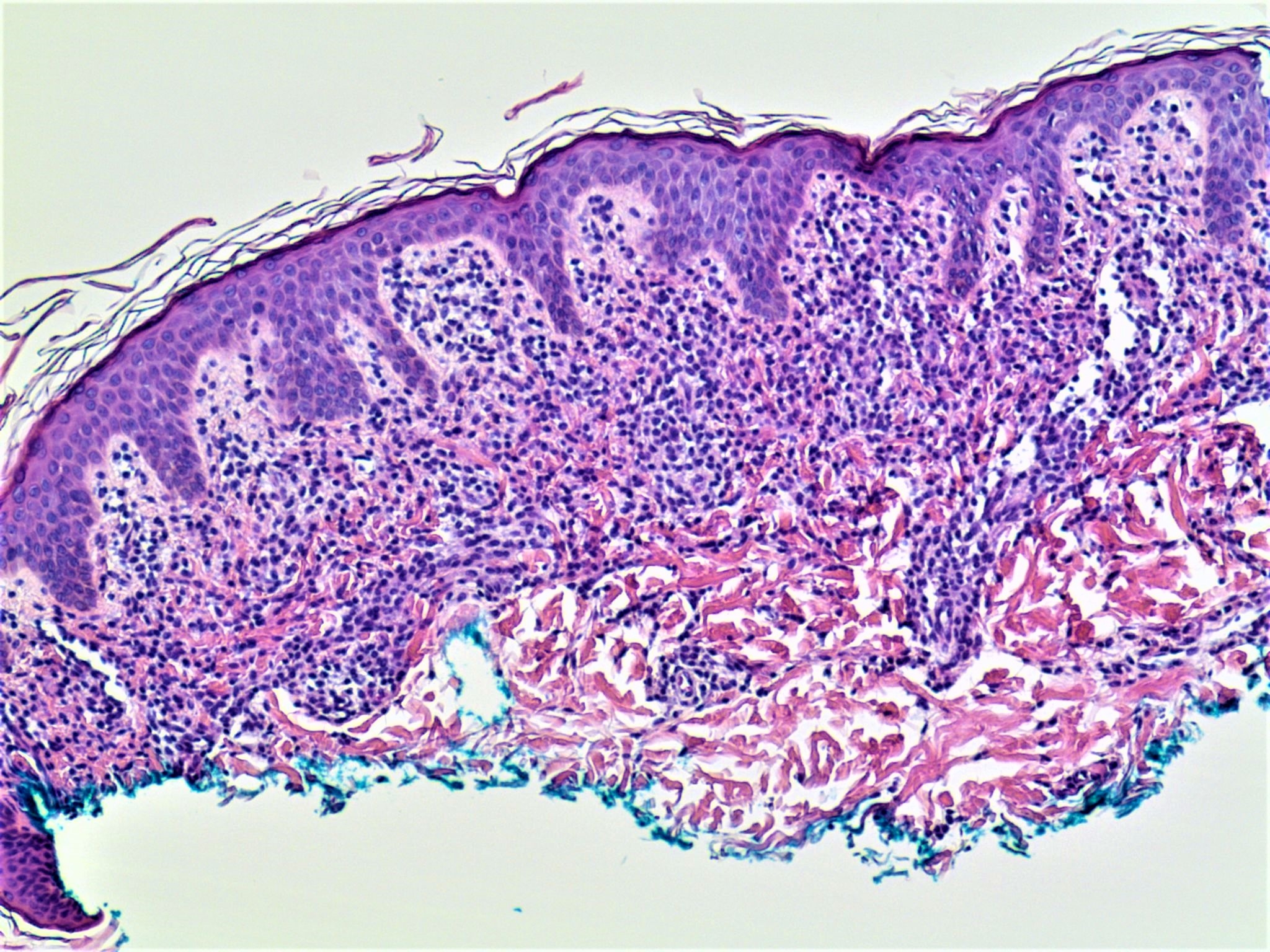
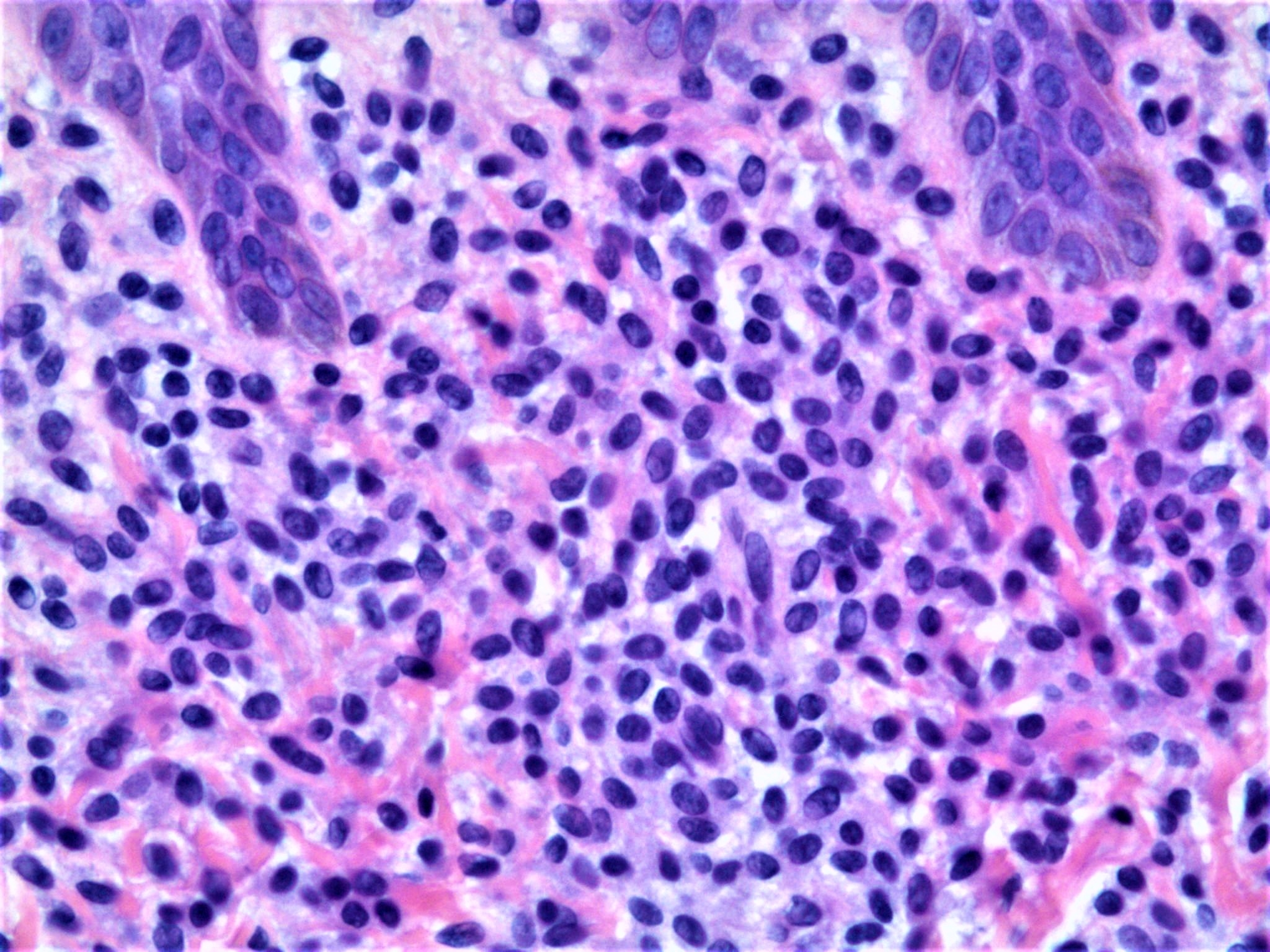
Case Number : Case 2101 - 26 June 2018 Posted By: Uma Sundram

Please read the clinical history and view the images by clicking on them before you proffer your diagnosis.
Submitted Date :
   (0 reviews)

8 year old female with solitary lesion on the back. The lesional cells are MITF positive.

Edited by Admin_Dermpath


  Report Record

User Feedback


I don't know about MITF1-positivity of mastocytosis, but for me it's a mastocytoma 

Share this comment


Link to comment
share_externally

vincenzo

Posted (edited)

Yes! Found this: 

Mast cell tryptase and microphthalmia transcription factor effectively discriminate cutaneous mast cell disease from myeloid leukemia cutis

First published: 09 October 2006
Cited by: 7

Abstract

Background:  Cutaneous mast cell disorders are uncommon, but a subset, especially mastocytoma and mast cell leukemia, can histologically mimic myeloid leukemia cutis. Our objective was to employ a panel of cytochemical and immunohistochemical markers to determine which ones would be most useful in separating these two entities.

Methods:  We stained 17 cases of cutaneous mast cell disease and 20 cases of myeloid leukemia cutis with Giemsa, toluidine blue, or pinacyanol erythrosinate (PE), as well as with antibodies against mast cell tryptase, microphthalmia transcription factor (MiTF), CD117 (c‐kit), myeloperoxidase, CD43, CD25, CD2, and CD68.

Results:  Mast cell tryptase and MiTF emerged as highly sensitive and specific markers for mast cell disease in this context, as both antibodies stained all cases of mast cell diseases but none of myeloid leukemia cutis. Although CD117 stained all cases of mast cell disease, it also stained 2 of 18 cases of myeloid leukemia cutis. PE appeared to be specific for mast cell disease, as 11 of 12 cases stained with this marker, compared with 0 of 18 cases of myeloid leukemia cutis.

Conclusions:  Our results show that mast cell tryptase and MiTF are equally effective in distinguishing mast cell disease from myeloid leukemia cutis.

 
I'm learning a lot in this site.
 
 
Edited by vincenzo

Share this comment


Link to comment
share_externally

Raul Perret

Posted

Agree, those are mast cells. Thanks for the paper

Share this comment


Link to comment
share_externally

Uma Sundram

Posted

Yes, mastocytoma. Did not mean to plug myself :)

But it's important to know, especially in this context, that MITF positivity does not mean that this is a melanocytic proliferation.

Share this comment


Link to comment
share_externally


Join the conversation

You can post now and register later. If you have an account, sign in now to post with your account.

Guest
Add a comment...

×   Pasted as rich text.   Paste as plain text instead

  Only 75 emoji are allowed.

×   Your link has been automatically embedded.   Display as a link instead

×   Your previous content has been restored.   Clear editor

×   You cannot paste images directly. Upload or insert images from URL.

Loading...

×
×
  • Create New...