Jump to content
In this section we have spot diagnoses posted on a daily basis since June 2010, now over 4000! You can review the archived cases and read the suggested diagnoses by users and the final comment by the contributors.
Case are uploaded each week day by 10 am UK time with the correct diagnosis will generally be posted at 8 pm UK time. Why not view the most recent spot diagnosis and proffer a diagnosis?

Case Number : Case 2063 - 3 May 2018 Posted By: Raul Perret

Please read the clinical history and view the images by clicking on them before you proffer your diagnosis.
Submitted Date :
   (0 reviews)

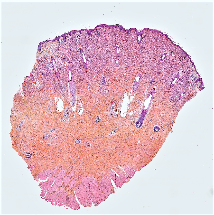
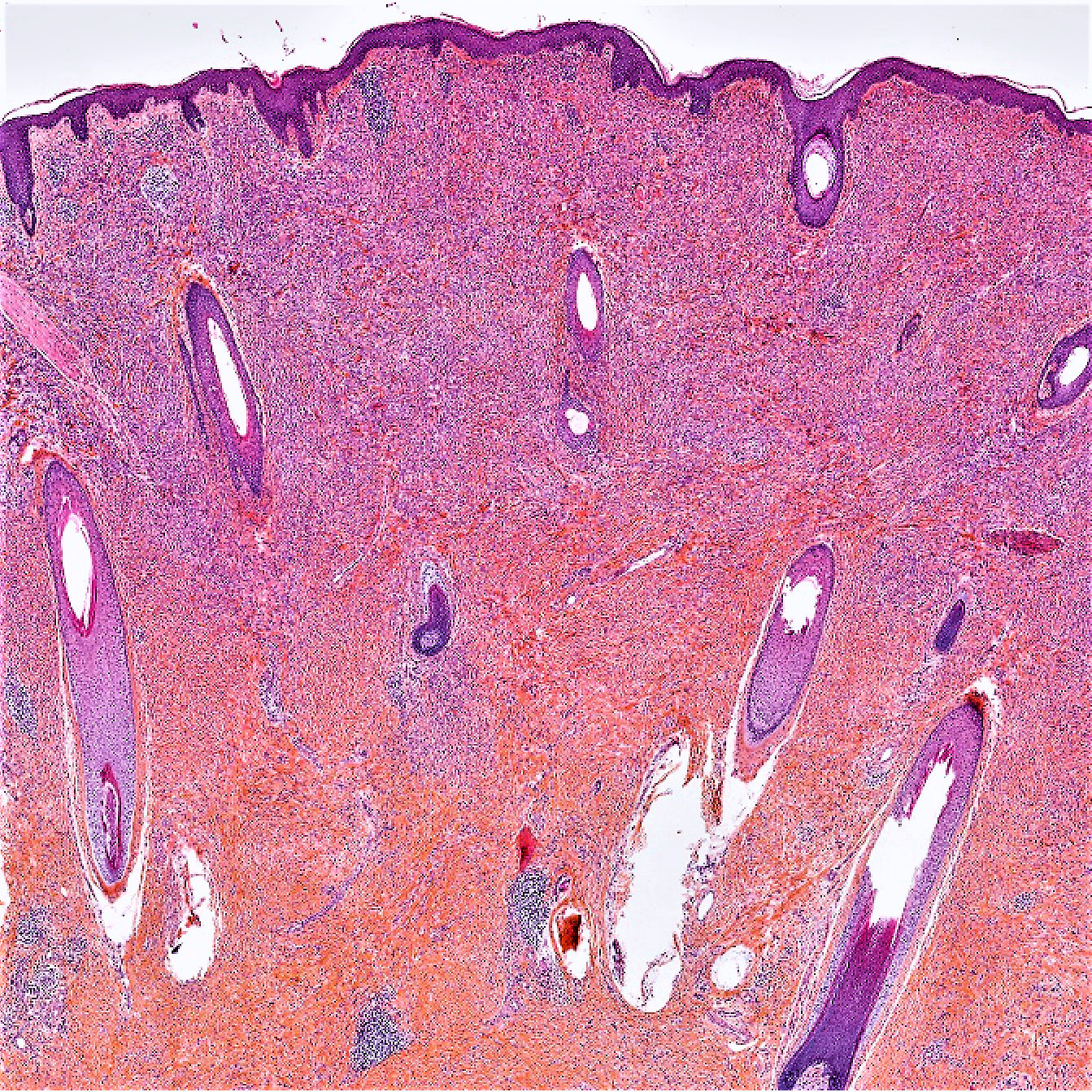
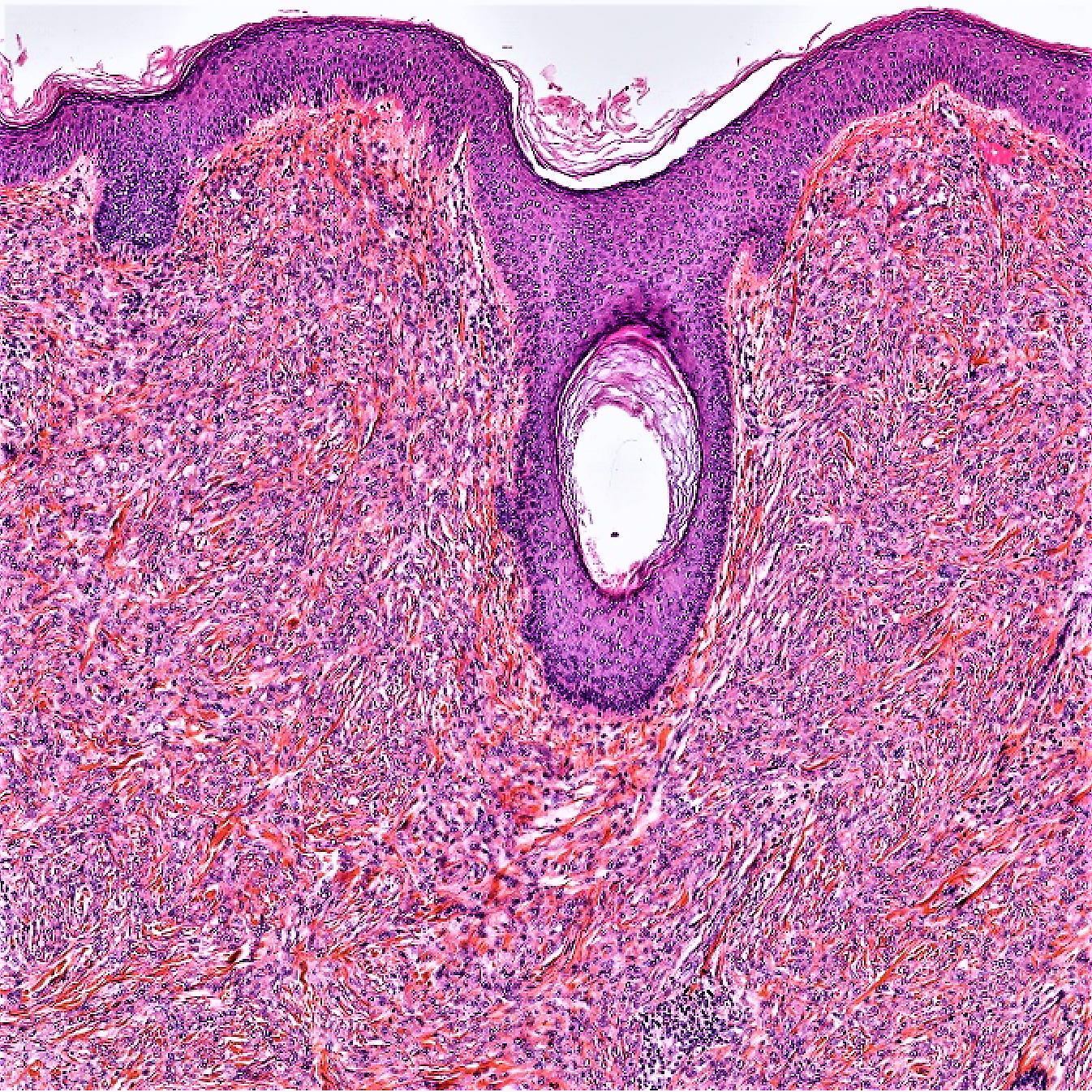
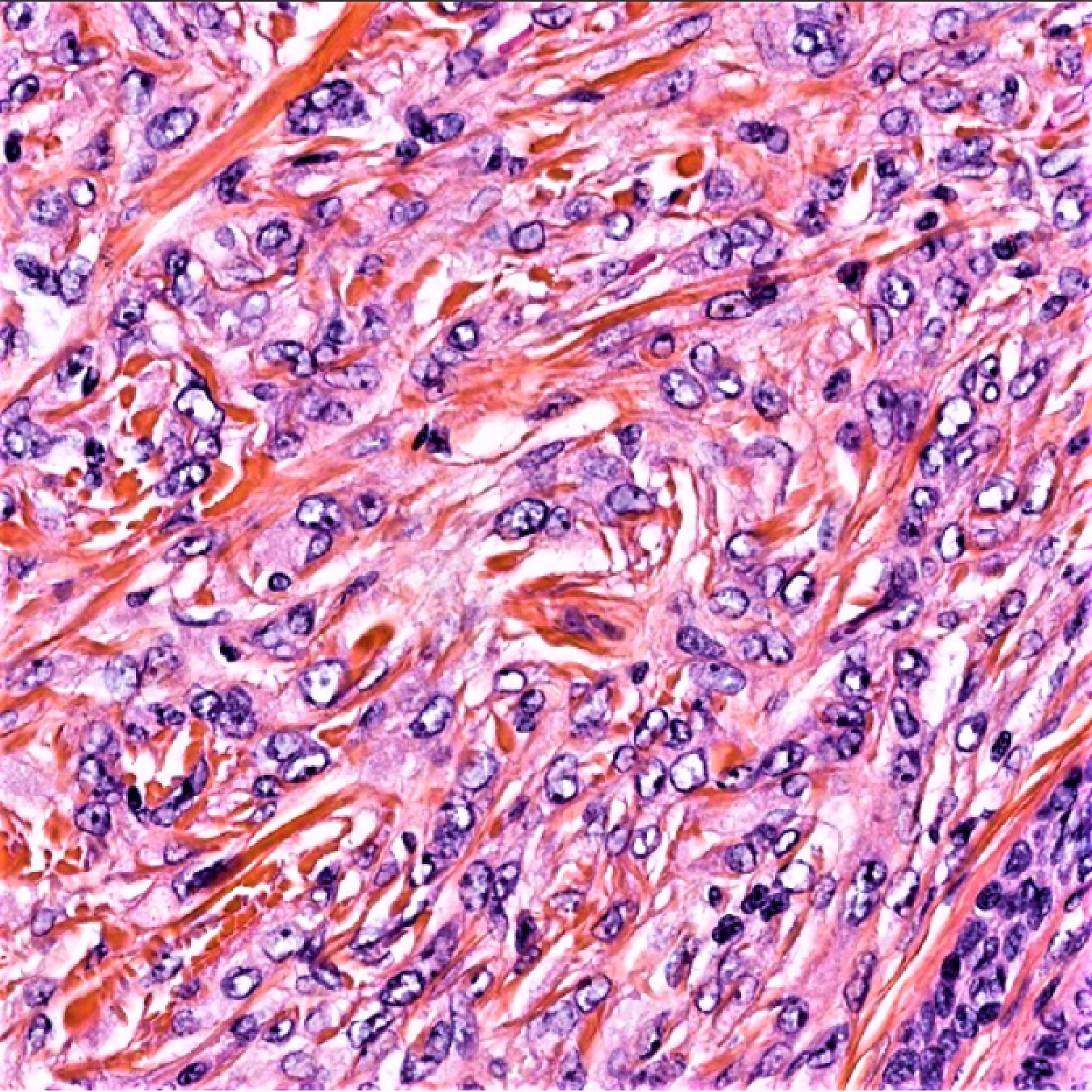
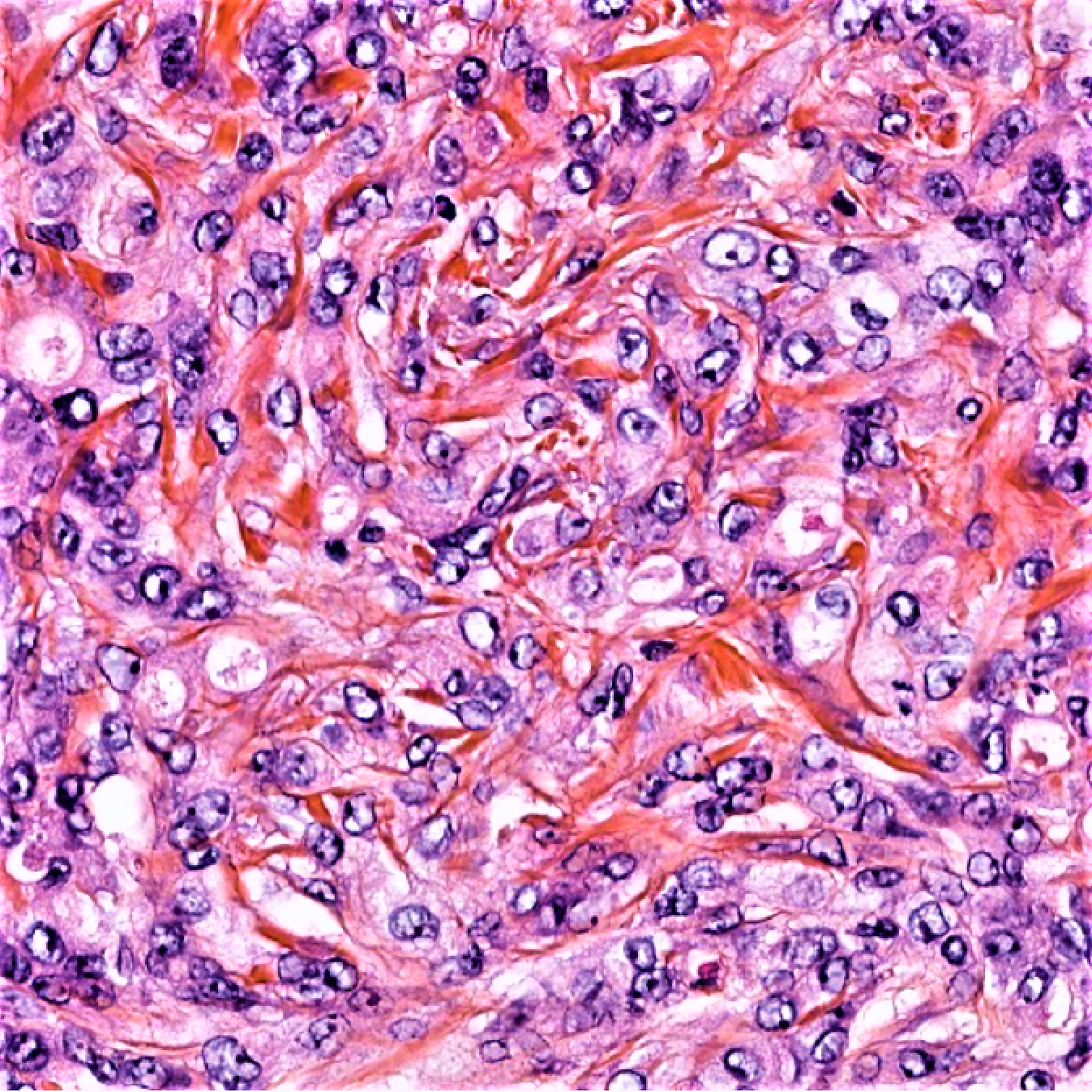
Case 54: 49 y old male with a recently developed lesion of the lip.


  Report Record

User Feedback


vincenzo polizzi

Posted

I'm struggling with this case... but no features enough convincing for a comfortable spot diagnosis..there are two an entity more convincing for me: EED-like Granuloma Faciale, late stage...and amelanotic cellular blue nevus...

Share this comment


Link to comment
share_externally

Anil Patki

Posted

Was just wondering about the hair follicles in a lip biopsy. Fibrous histiocytoma ?

Share this comment


Link to comment
share_externally

Dr. Richard Carr

Posted

Apologies but I have now finally posted my last two Friday cases.  I had problems logging in during my hols.

Share this comment


Link to comment
share_externally

Dr. Richard Carr

Posted

Challenging indeed. Couple of quick thoughts. Cellular neurothekeoma an epithelioid angiomatous nodule or a haemangioendothelioma variant. Likely to be some soft tissue teaser I suspect. I'd throw in PECOMA. Even histioid leprosy! Brown stains needed.

Share this comment


Link to comment
share_externally

I like epithelioid angiomatous nodule

Share this comment


Link to comment
share_externally

vincenzo polizzi

Posted

Yeahh. Histioid Leprosy is a good spot. I was completely off road. 

Share this comment


Link to comment
share_externally

Raul Perret

Posted (edited)

This is a case of a poorly differentiated adenocarcinoma, of probable metastatic origin. CKAE1-AE3 was diffusely positivite and the tumor cells also coexpressed CK7, CK20 and CDX2. I suggested ruling out a primitive from the GI tract (particularly from the stomach) pancreatic or eventually a cholangiocarcinoma. P63, GATA3, Glypican 3 were all negative.

I thought this case was particularly challenging due to the overall architecture of the lesion (simulating a dermatofibroma) and the moderate atypia of the cells. The giveaway were the isolated intracytoplasmic mucin vacuoles (hard to see on HE but beautifully demonstrated by alcian blue).

The case was sent to our institute as a probable "atypical dermatofibroma". As a rule I always perform an initial small pannel including a pan-keratin, a couple of muscle markers, CD34 and s-100/sox-10 in cutaneous tumors sent as atypical variants of DF.

Thanks for the comments and enjoy your sunday!

Edited by Raul Perret

Share this comment


Link to comment
share_externally

Arash Daryakar

Posted

Nice case dear Raul.

Actually i thought of epithelioid angiomatous nodule and Epithelioid histiocytoma.

Although,in this case IHC study solved our dilemma.

and nice image quality!

Share this comment


Link to comment
share_externally

Raul Perret

Posted

2 hours ago, Arash Daryakar said:

Nice case dear Raul.

Actually i thought of epithelioid angiomatous nodule and Epithelioid histiocytoma.

Although,in this case IHC study solved our dilemma.

and nice image quality!

Thank you Arash, nice to see you posting at the site again

Share this comment


Link to comment
share_externally


Join the conversation

You can post now and register later. If you have an account, sign in now to post with your account.

Guest
Add a comment...

×   Pasted as rich text.   Paste as plain text instead

  Only 75 emoji are allowed.

×   Your link has been automatically embedded.   Display as a link instead

×   Your previous content has been restored.   Clear editor

×   You cannot paste images directly. Upload or insert images from URL.

Loading...

×
×
  • Create New...