Jump to content
In this section we have spot diagnoses posted on a daily basis since June 2010, now over 4000! You can review the archived cases and read the suggested diagnoses by users and the final comment by the contributors.
Case are uploaded each week day by 10 am UK time with the correct diagnosis will generally be posted at 8 pm UK time. Why not view the most recent spot diagnosis and proffer a diagnosis?

Case Number : Case 2071 - 15 May 2018 Posted By: Uma Sundram

Please read the clinical history and view the images by clicking on them before you proffer your diagnosis.
Submitted Date :
   (0 reviews)

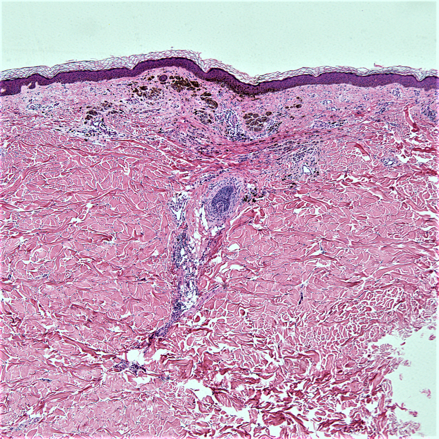
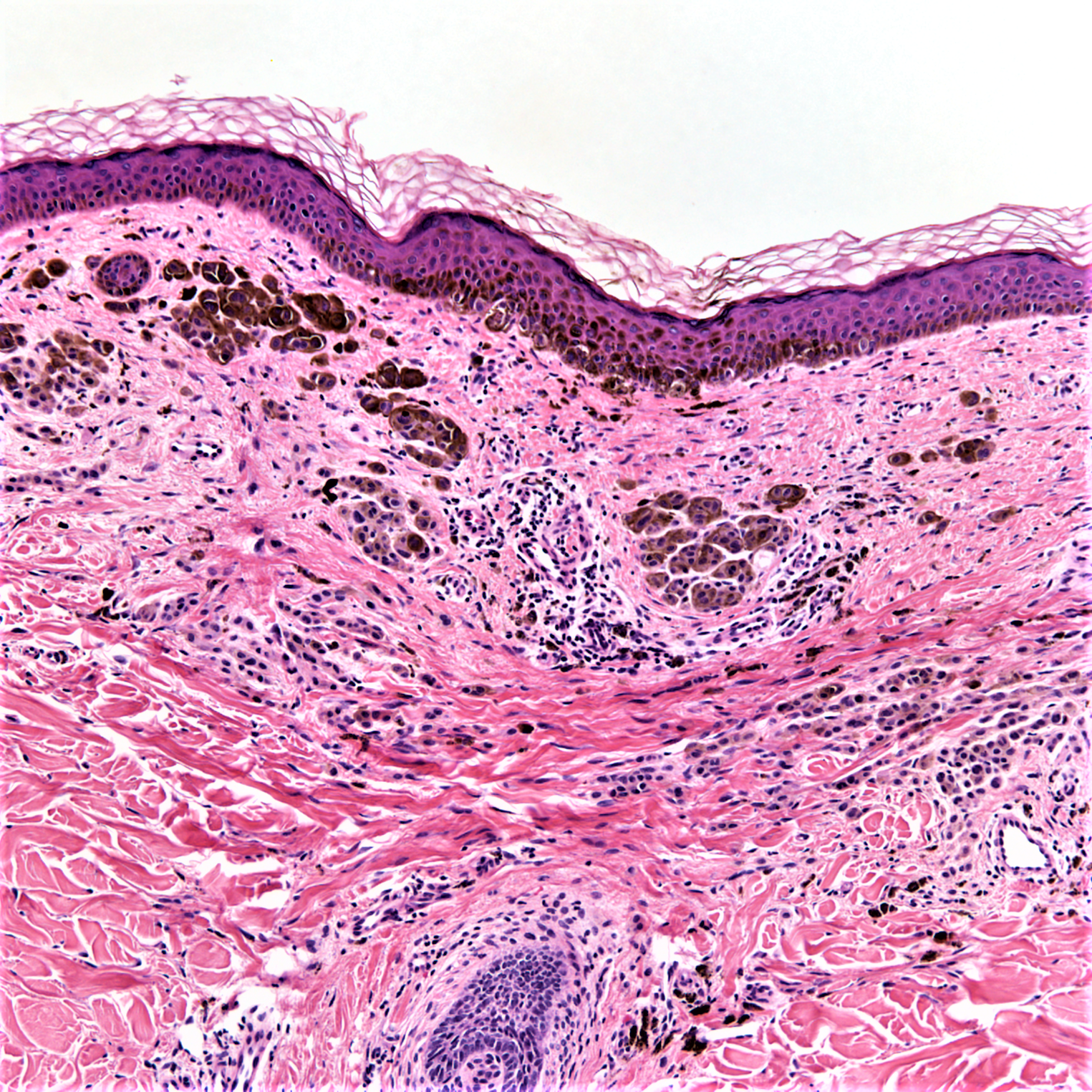
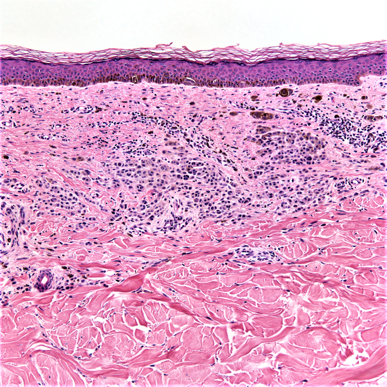
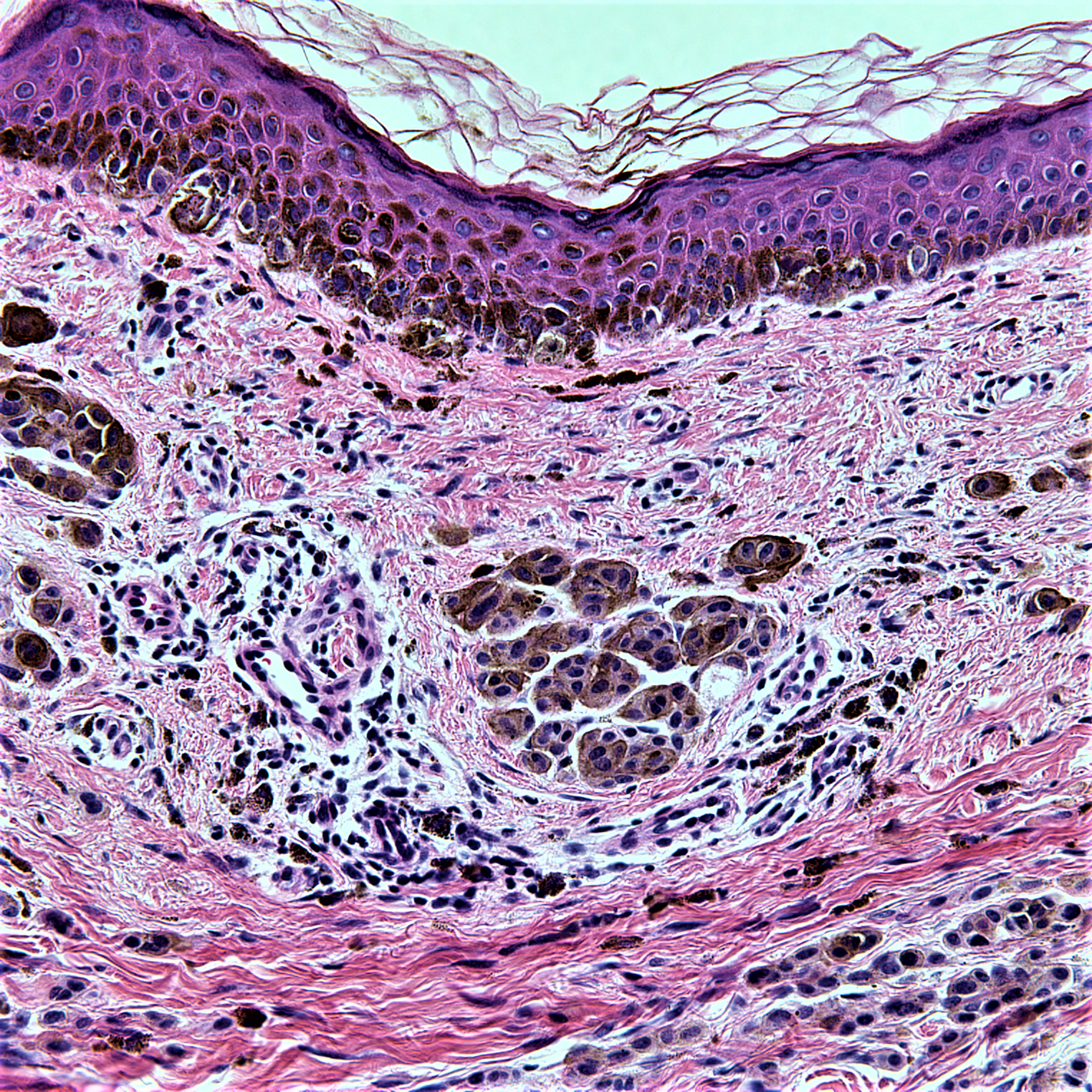
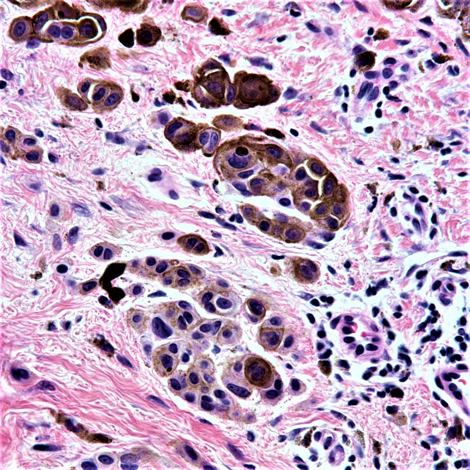
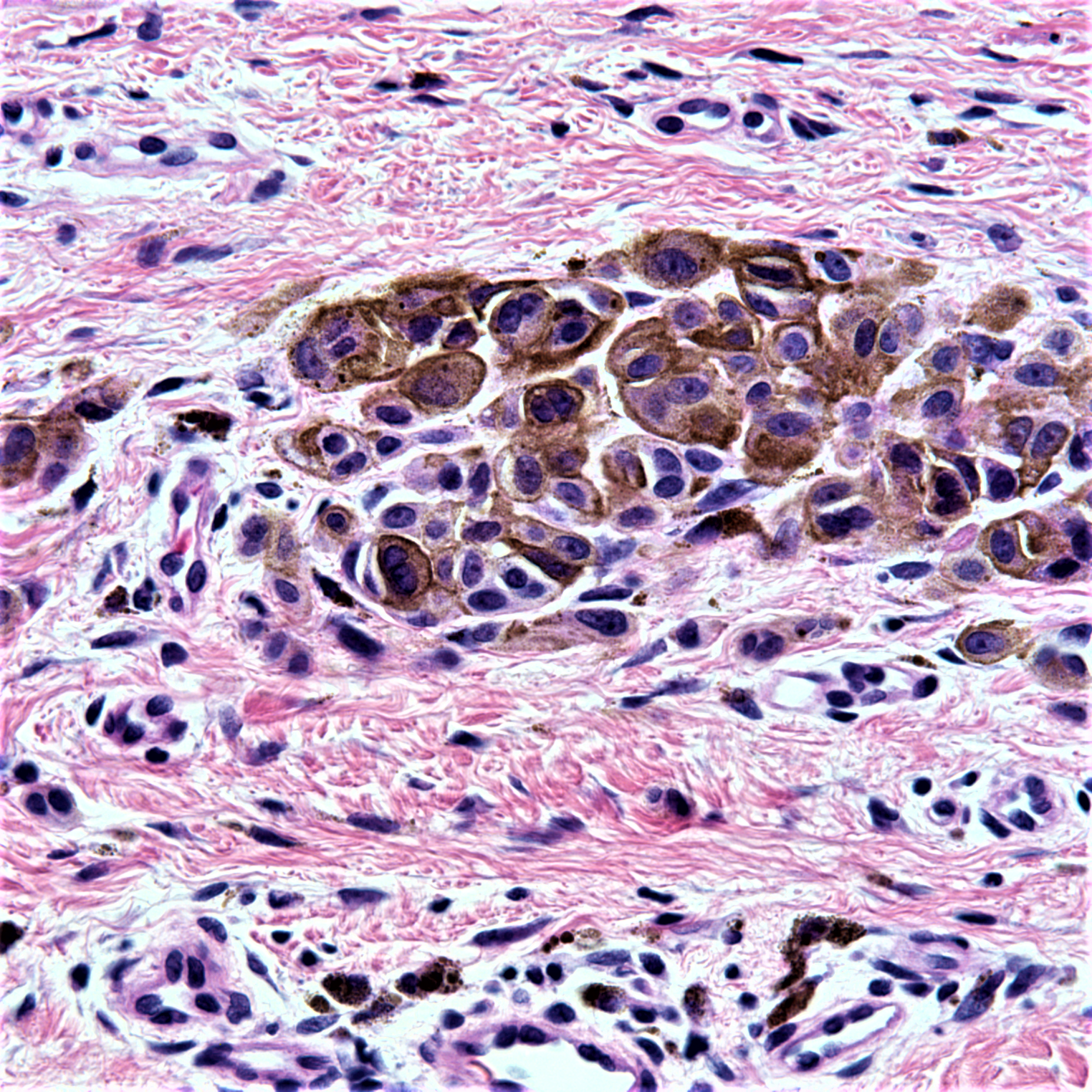
77 year old male with a few month history of a pigmented papule of left shoulder.


  Report Record

User Feedback


Anil Patki

Posted

Age of the patient and big hyperchromatic nuclei of melanocytes make me think of melanoma

Share this comment


Link to comment
share_externally

dermpath1

Posted

Pigmented epithelioid melanocytoma.

Share this comment


Link to comment
share_externally

vincenzo polizzi

Posted

What about a partially regressed dermal nevus?  

Share this comment


Link to comment
share_externally

Anil Patki

Posted

It's really a maze - pigmented epithelioid melanocytoma, animal type melanoma, Epithelioid blue nevus, pigment producing melanoma etc etc... Waiting for enlightenment from the experts. 

Share this comment


Link to comment
share_externally

Raul Perret

Posted

Considering the age I would be worried about partially regressed melanoma. Would add a note in the report and talk with the dermatologist

Share this comment


Link to comment
share_externally

Uma Sundram

Posted

My apologies for the late comment. I spent most of Monday in a plane to London but am here now for the London Dermatopathology Symposium. A welcome to all those who are here, and a plug for the LDS for those who've never been - it's a very relaxed way to learn about dermatopathology from the experts with tons of new material and review of old material. Good times!

Regarding the posted case, a difficult one indeed. It's a regressing lesion; difficult to say if it's a regressing melanoma or a regressing nevus. I favored nevus because the remaining cells appear relatively bland, with mild cytologic atypia. Nevertheless, given the age of the patient, I recommended excision of any remaining lesion. While PEM and animal type melanoma are good thoughts, those tend to have larger, more epithelioid cells, whereas here, the cells really seem 'nevoid'. 

Share this comment


Link to comment
share_externally


Join the conversation

You can post now and register later. If you have an account, sign in now to post with your account.

Guest
Add a comment...

×   Pasted as rich text.   Paste as plain text instead

  Only 75 emoji are allowed.

×   Your link has been automatically embedded.   Display as a link instead

×   Your previous content has been restored.   Clear editor

×   You cannot paste images directly. Upload or insert images from URL.

Loading...

×
×
  • Create New...