Jump to content
In this section we have spot diagnoses posted on a daily basis since June 2010, now over 4000! You can review the archived cases and read the suggested diagnoses by users and the final comment by the contributors.
Case are uploaded each week day by 10 am UK time with the correct diagnosis will generally be posted at 8 pm UK time. Why not view the most recent spot diagnosis and proffer a diagnosis?

Case Number : Case 2078 - 24 May 2018 Posted By: Iskander H. Chaudhry

Please read the clinical history and view the images by clicking on them before you proffer your diagnosis.
Submitted Date :
   (0 reviews)

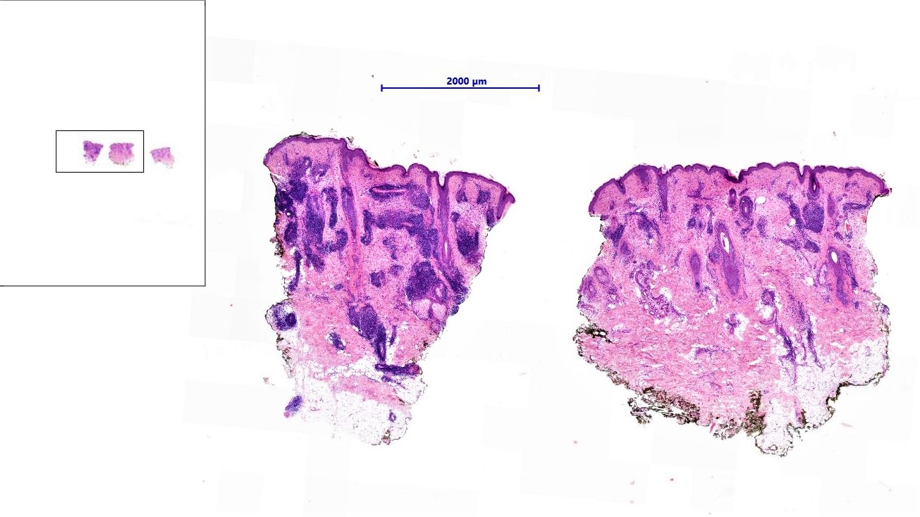
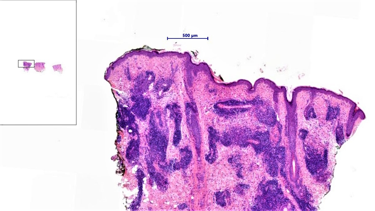
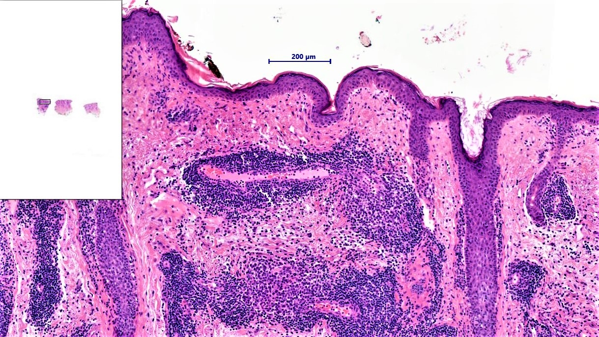
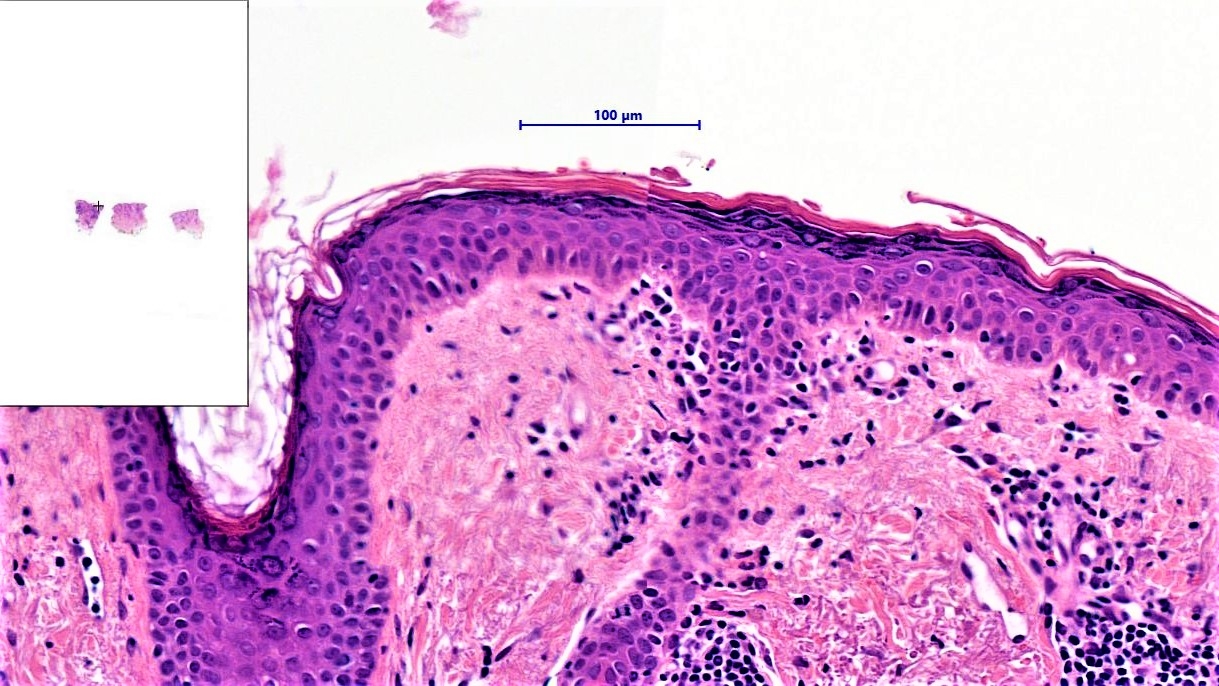
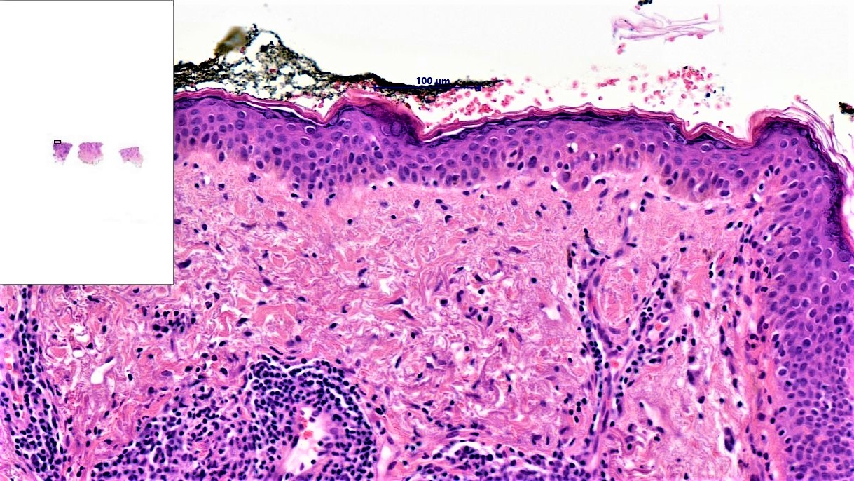
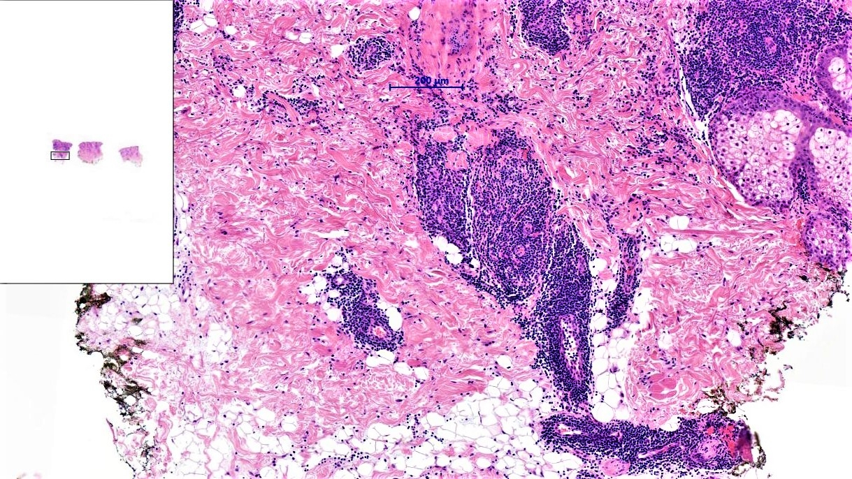
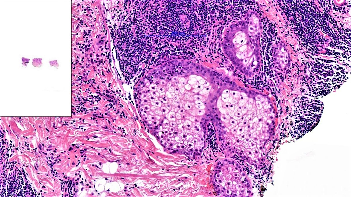
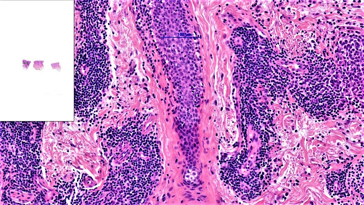
Left cheek incision biopsy.

Edited by Admin_Dermpath


  Report Record

User Feedback


Chillblain or lymphoid hyperplasia

Share this comment


Link to comment
share_externally

vincenzo polizzi

Posted

CD31?HHV8?  Angiosarcoma is my first thought. 

Share this comment


Link to comment
share_externally

Anil Patki

Posted

Superficial and deep dense perivascular and periadnexal infiltrate of lymphocytes and ?  mucin in upper dermis. I think this is lupus tumidus. Cutaneous lymphoid hyperplasia is the second possibility which can be ruled out by clinical correlation.

Share this comment


Link to comment
share_externally

vincenzo polizzi

Posted

I'm better watching this case on my computer, and agree with Anil comment. 

Share this comment


Link to comment
share_externally

Dr. Richard Carr

Posted

I would favour Jessner's which some regards as a variant of lupus. I am assuming the infiltrate is mainly T-cells of course (if not would have to re-consider).

Share this comment


Link to comment
share_externally

Admin_Dermpath

Posted

Dear all,

Some good guesses here so I've included full observations/diagnosis below:

In the dermis there is a superficial and deep, light perivascular and to a lesser extent periadnexal chronic inflammatory cell infiltrate. Basement membrane thickening and mucin deposition are not appreciable (DPAS negative). There are no fungal hyphae on special stains. The main histological differentials are Jessner's lymphocytic infiltration of skin, tumid lupus erythematosus, lymphocytoma cutis, and polymorphous light eruption.
The features are those of a superifical and deep perivsacular dermatitis; and in the right clinical context are in keeping with Jessner's lymphocytic infiltration of skin.

Dr Iskander Chaudhry


Share this comment


Link to comment
share_externally


Join the conversation

You can post now and register later. If you have an account, sign in now to post with your account.

Guest
Add a comment...

×   Pasted as rich text.   Paste as plain text instead

  Only 75 emoji are allowed.

×   Your link has been automatically embedded.   Display as a link instead

×   Your previous content has been restored.   Clear editor

×   You cannot paste images directly. Upload or insert images from URL.

Loading...

×
×
  • Create New...