Jump to content
In this section we have spot diagnoses posted on a daily basis since June 2010, now over 4000! You can review the archived cases and read the suggested diagnoses by users and the final comment by the contributors.
Case are uploaded each week day by 10 am UK time with the correct diagnosis will generally be posted at 8 pm UK time. Why not view the most recent spot diagnosis and proffer a diagnosis?

Case Number : Case 2079 - 25 May 2018 Posted By: Dr. Richard Carr

Please read the clinical history and view the images by clicking on them before you proffer your diagnosis.
Submitted Date :
   (0 reviews)

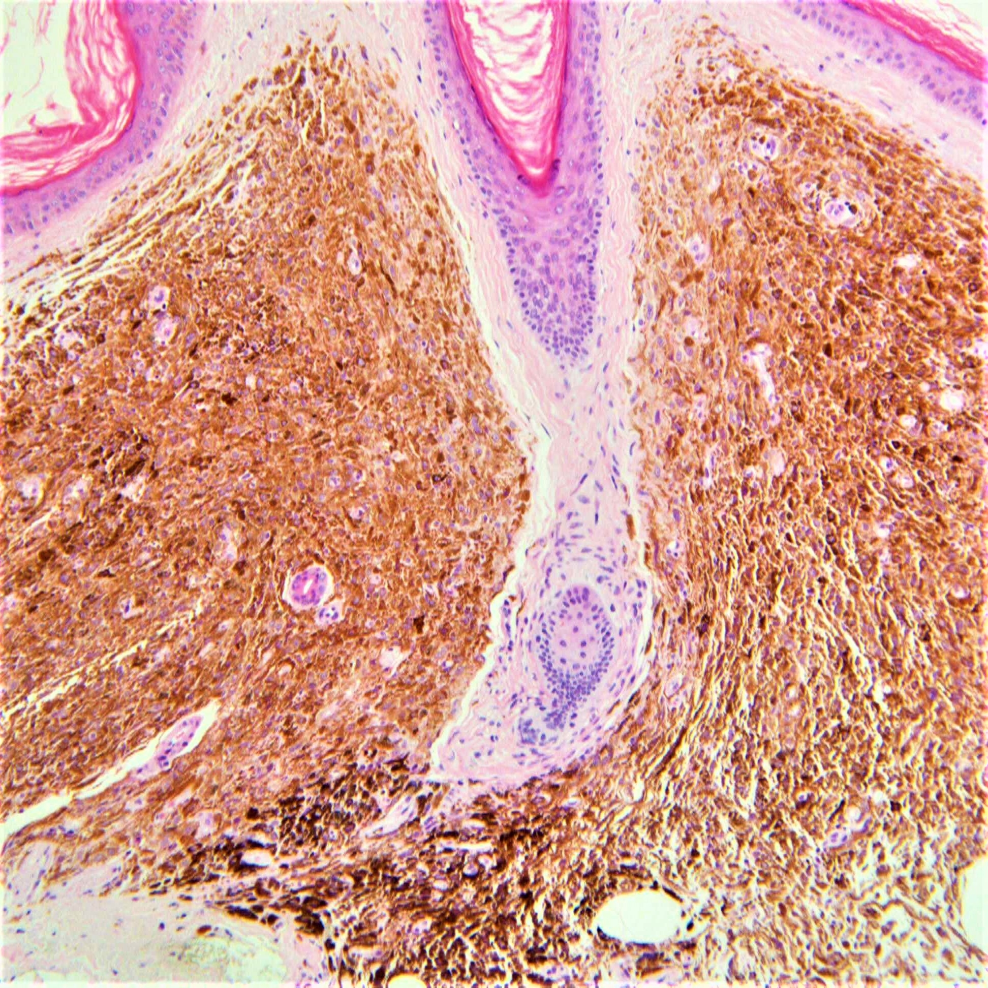
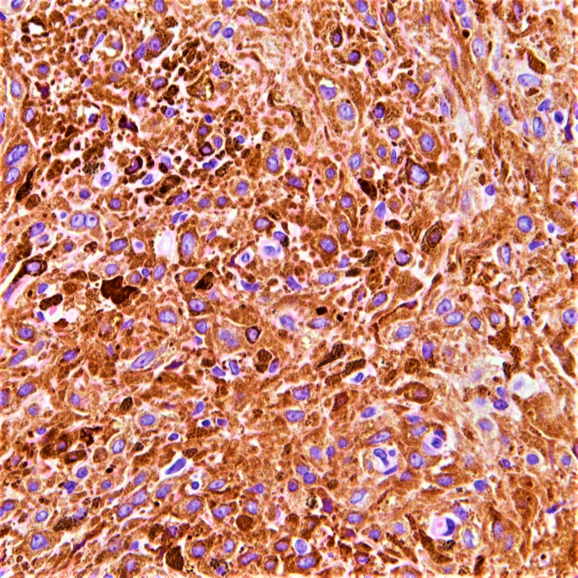
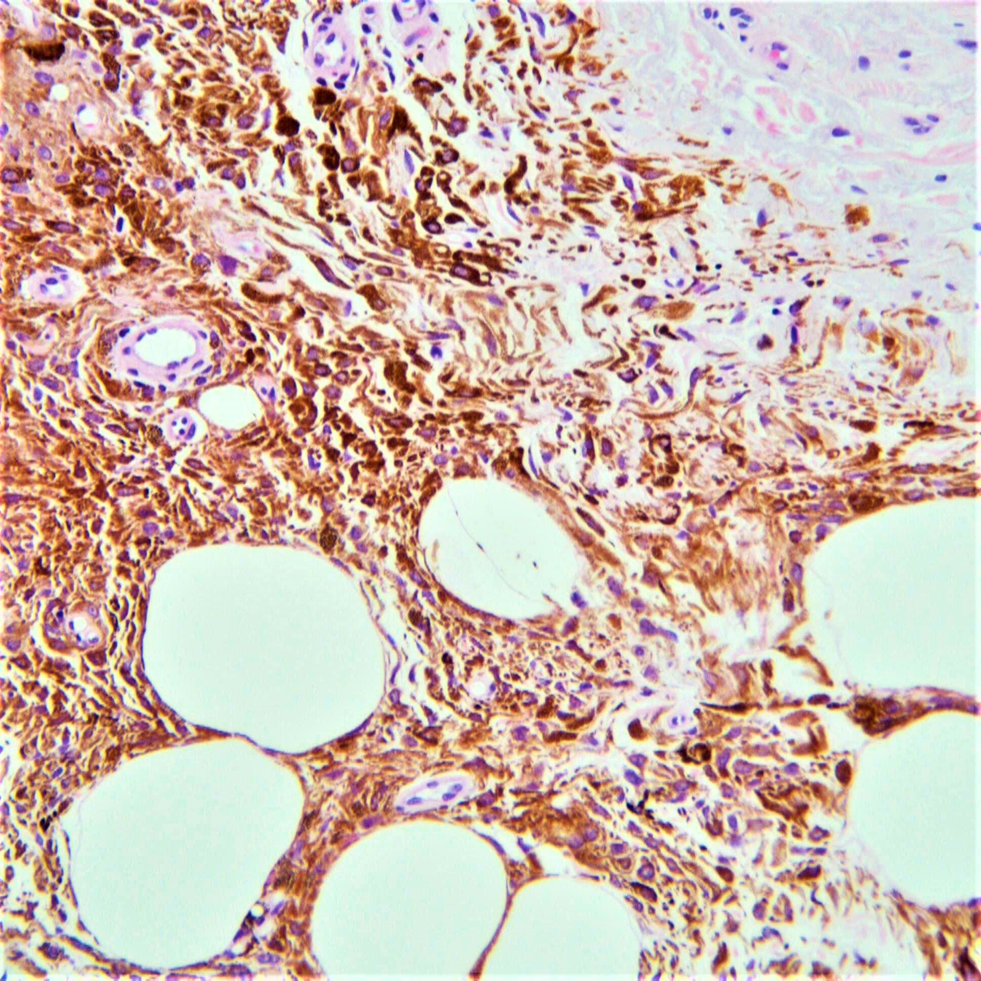
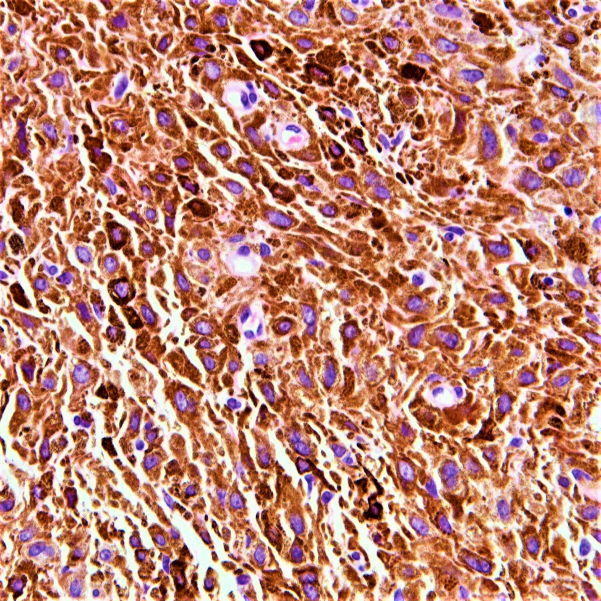
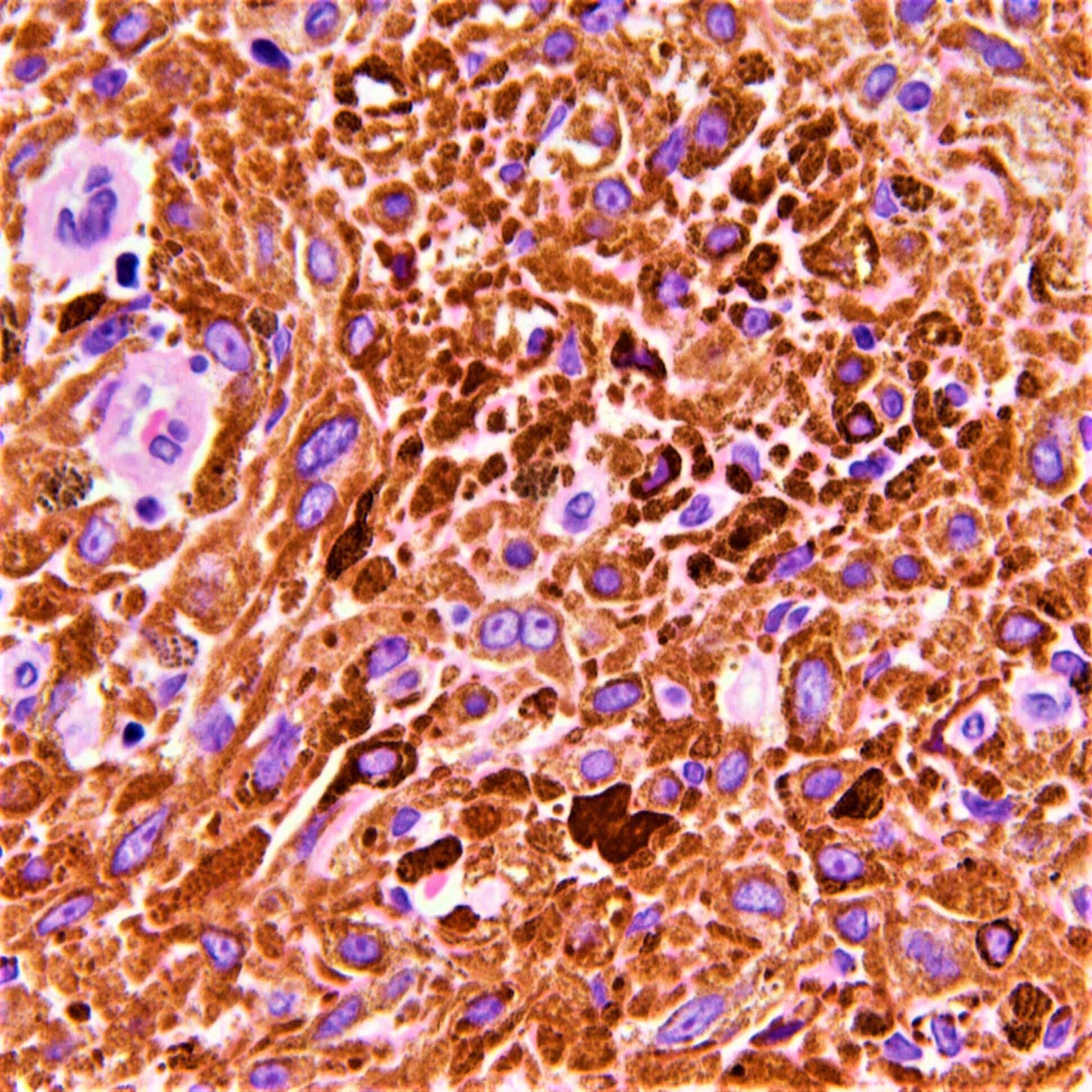
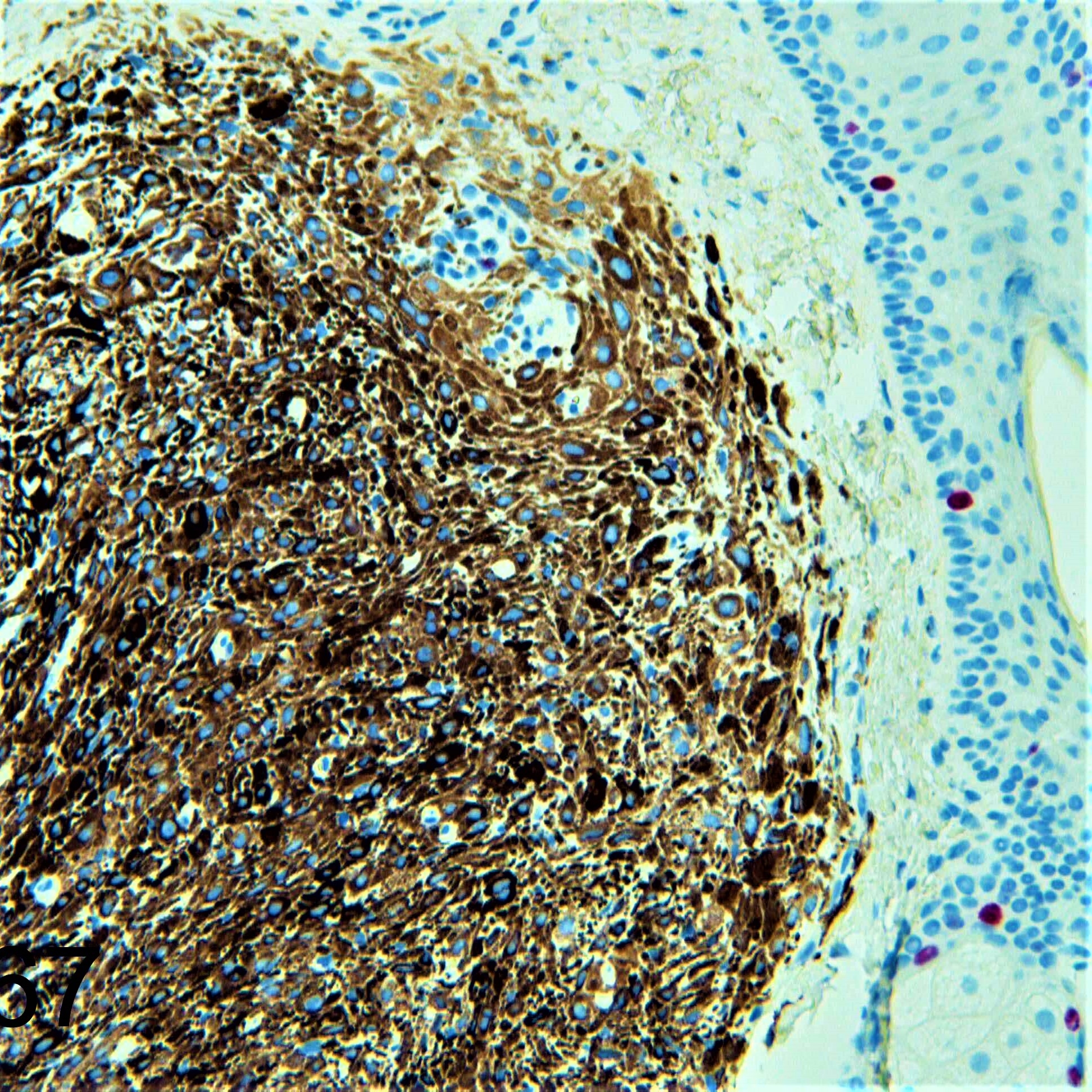
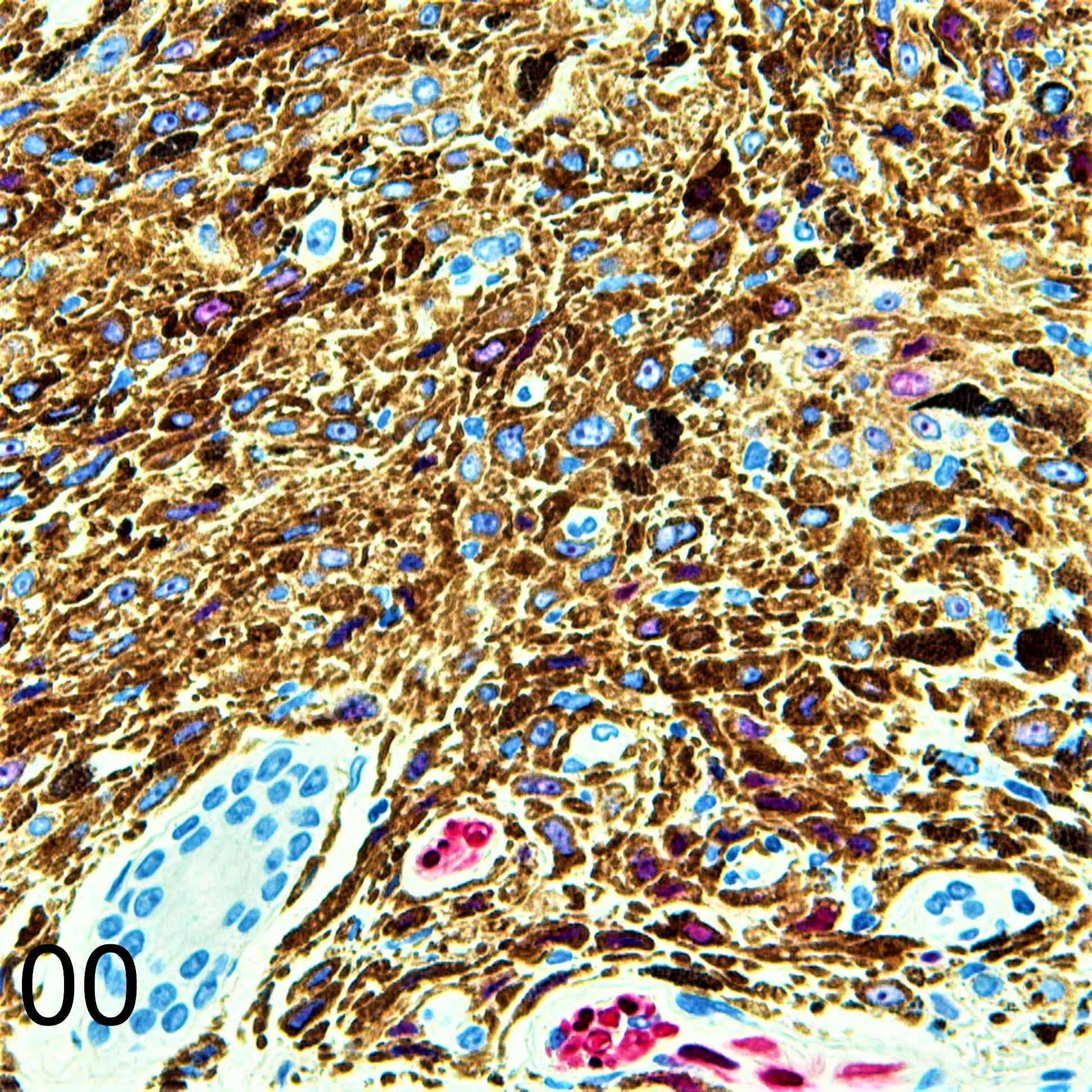
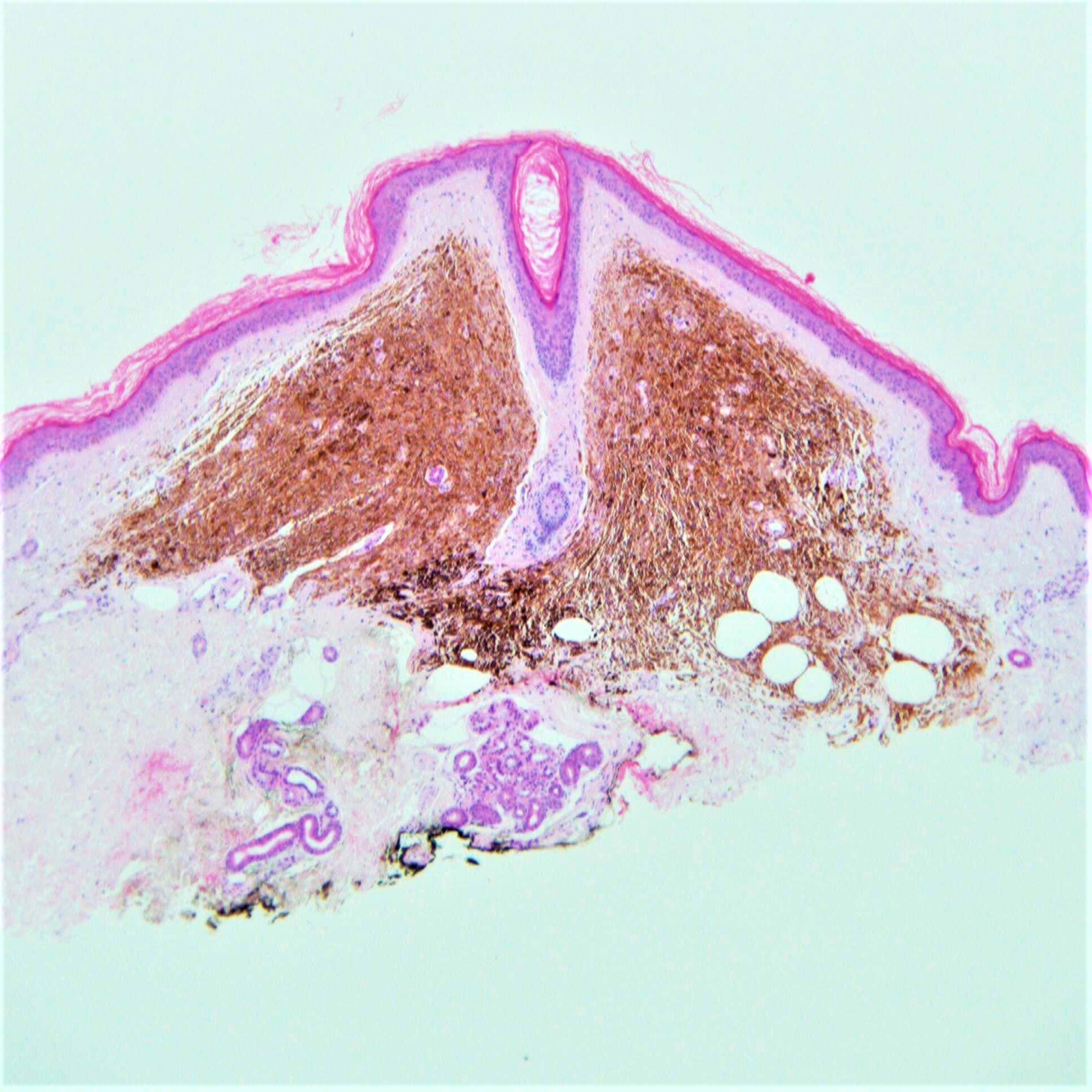
Clinical Details: F75. Right forearm. 2 years, well demarcated, oval, darkly pigmented lesion 6 x 4mm. c/o Dr George Powell

Edited by Admin_Dermpath


  Report Record

User Feedback


anh

Posted (edited)

Pigmented epithelioid melanocytoma? Epithelioid blue nevus?

Edited by anh

Share this comment


Link to comment
share_externally

Dr. Richard Carr

Posted

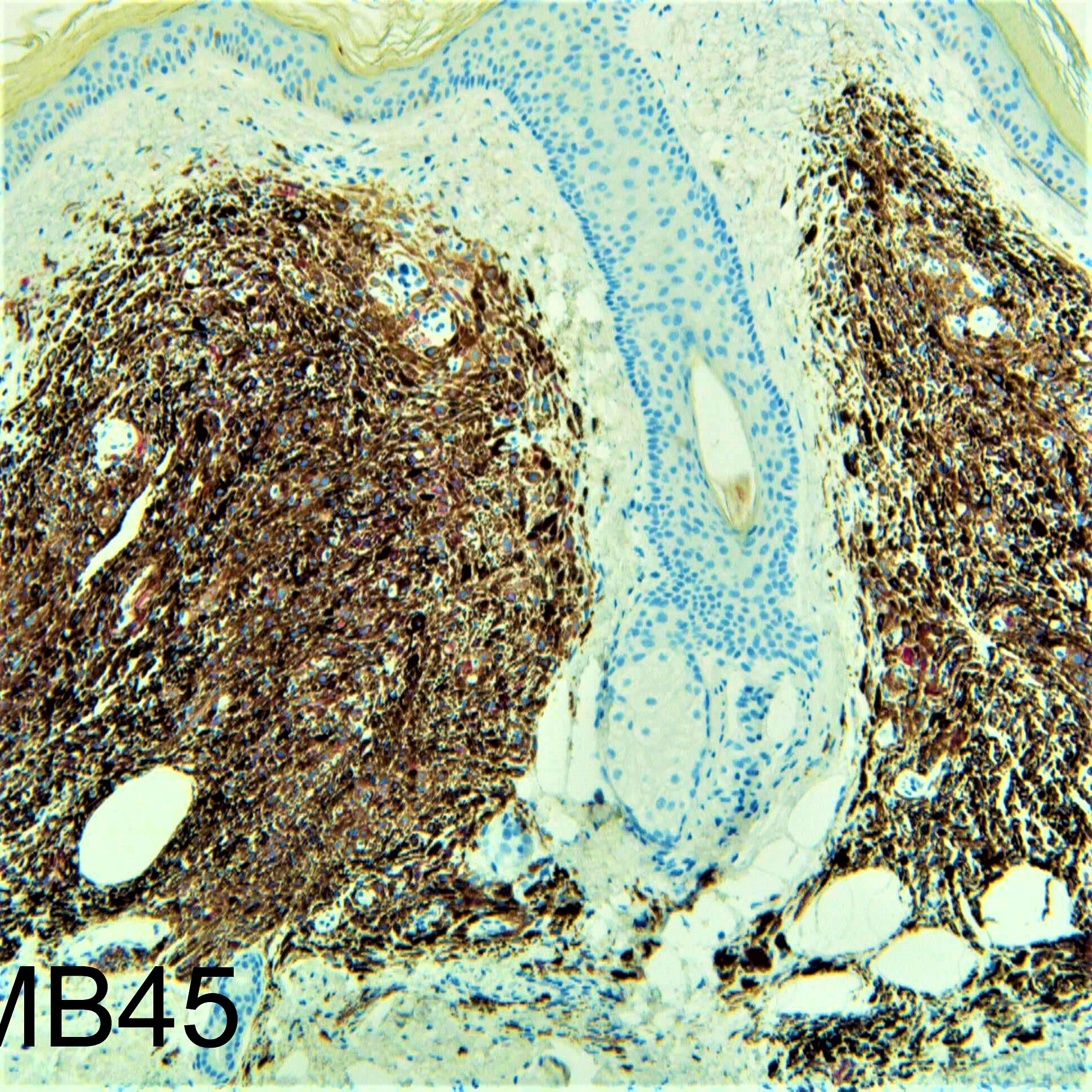
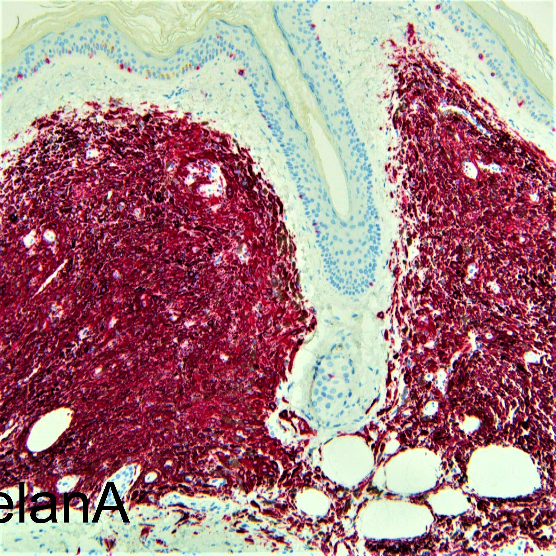
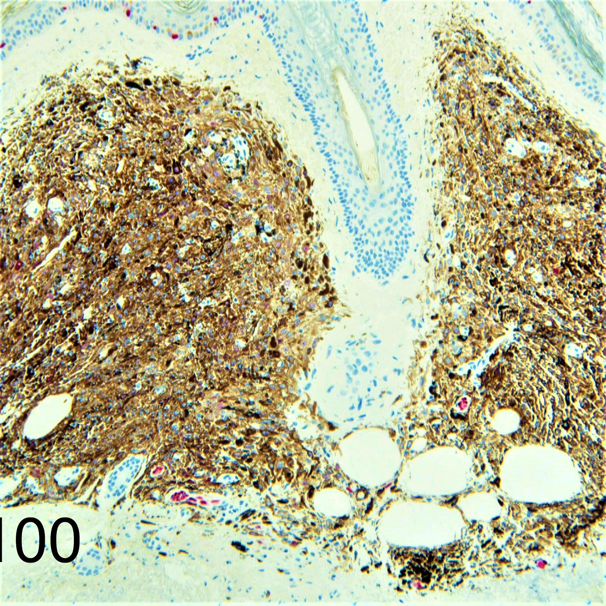
I favoured this was an example of a blue naevus with epithelioid cells rather than a PEM (aka epithelioid blue naevus when seen in the setting of Carney's). Interesting that the S100 is largely negative following our findings in most other blue naevi. Data for PEM is rather sparse but seems most cases were S100 positive.

Reference:

Yazdan P1, Haghighat Z, Guitart J, Gerami P. Epithelioid and fusiform blue nevus of chronically sun-damaged skin, an entity distinct from the epithelioid blue nevus of the Carney complex. Am J Surg Pathol. 2013 Jan;37(1):81-8.

Abstract

Epithelioid blue nevus (EBN) was first described in patients with Carney complex (CNC) and subsequently shown to also occur sporadically. Over 50% of patients with CNC harbor mutations in the gene PRKAR1A, which codes for protein kinase A regulatory subunit 1α (R1α) involved in the signaling pathway regulating melanogenesis and melanocytic proliferation. Immunohistochemical expression of R1α has been shown to be absent in the majority of pigmented epithelioid melanocytomas and all CNC-associated EBNs but present in melanomas and other melanocytic nevi. We have observed several examples of EBN occurring in chronically sun-damaged (CSD) skin with a predominance of epithelioid morphology but also containing a component of fusiform and conventional blue nevus cells, which we have termed epithelioid and fusiform blue nevus of CSD skin. Several of these cases demonstrated notable pleomorphism and nuclear atypia with rare mitotic activity raising concern for the possibility of melanoma; however, the clinical outcomes, detailed histologic review, and molecular results were most consistent with a benign melanocytic neoplasm. We report our clinical, histopathologic, immunohistochemistry, and fluorescence in situ hybridization experience with this distinct entity of epithelioid and fusiform blue nevus and demonstrate that it is a unique subtype of blue nevus occurring on CSD skin with a higher frequency of an associated conventional blue nevus component compared with EBN and without association with CNC or loss of R1α expression typically found in pigmented epithelioid melanocytoma and CNC-associated EBN. We also postulate that the epithelioid pattern may represent a subclone of the conventional blue nevus component induced by chronic UV damage.

Share this comment


Link to comment
share_externally


Join the conversation

You can post now and register later. If you have an account, sign in now to post with your account.

Guest
Add a comment...

×   Pasted as rich text.   Paste as plain text instead

  Only 75 emoji are allowed.

×   Your link has been automatically embedded.   Display as a link instead

×   Your previous content has been restored.   Clear editor

×   You cannot paste images directly. Upload or insert images from URL.

Loading...

×
×
  • Create New...