Jump to content
In this section we have spot diagnoses posted on a daily basis since June 2010, now over 4000! You can review the archived cases and read the suggested diagnoses by users and the final comment by the contributors.
Case are uploaded each week day by 10 am UK time with the correct diagnosis will generally be posted at 8 pm UK time. Why not view the most recent spot diagnosis and proffer a diagnosis?

Case Number : Case 2192 - 2 November 2018 Posted By: Dr. Richard Carr

Please read the clinical history and view the images by clicking on them before you proffer your diagnosis.
Submitted Date :
   (0 reviews)

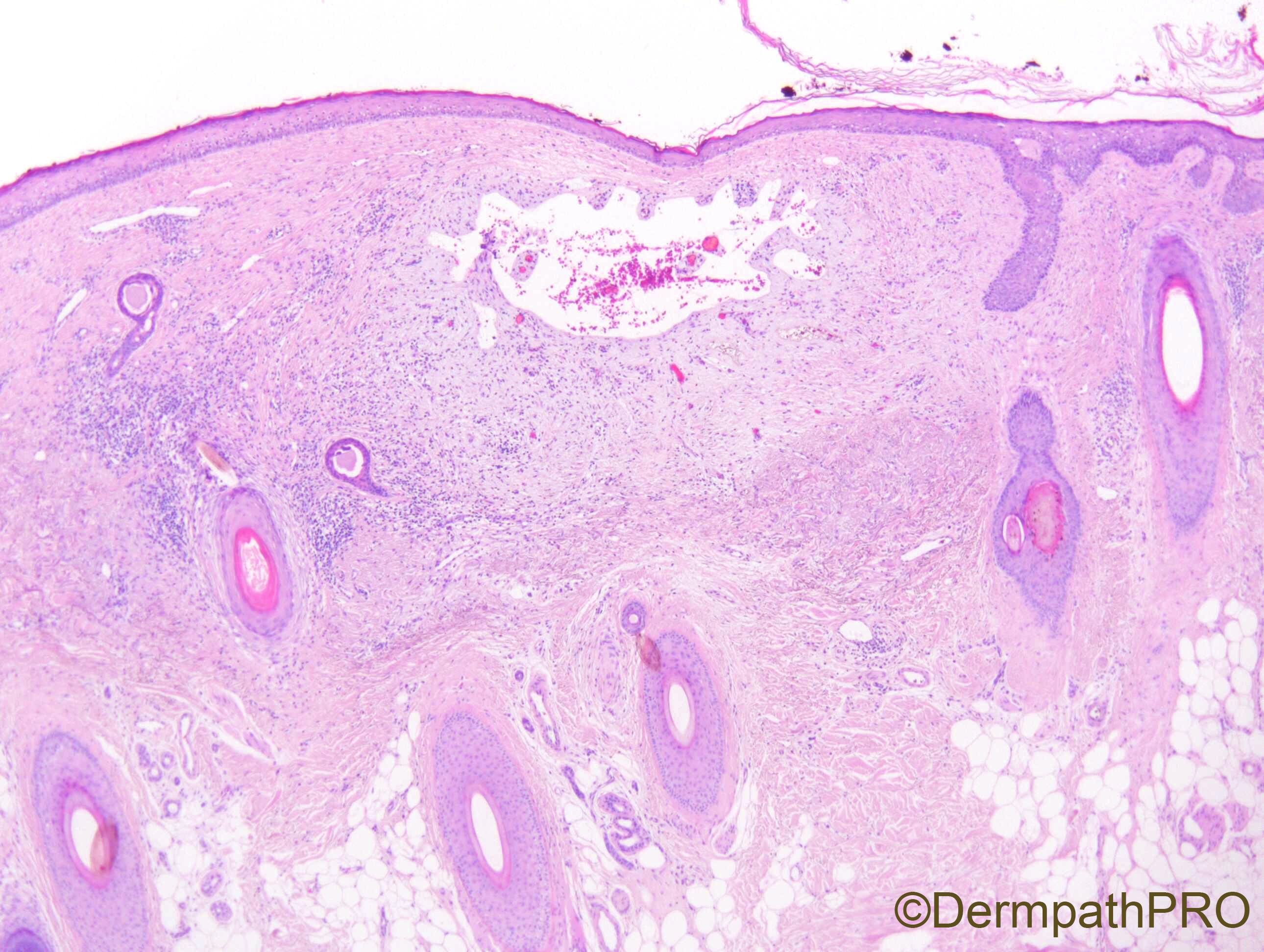
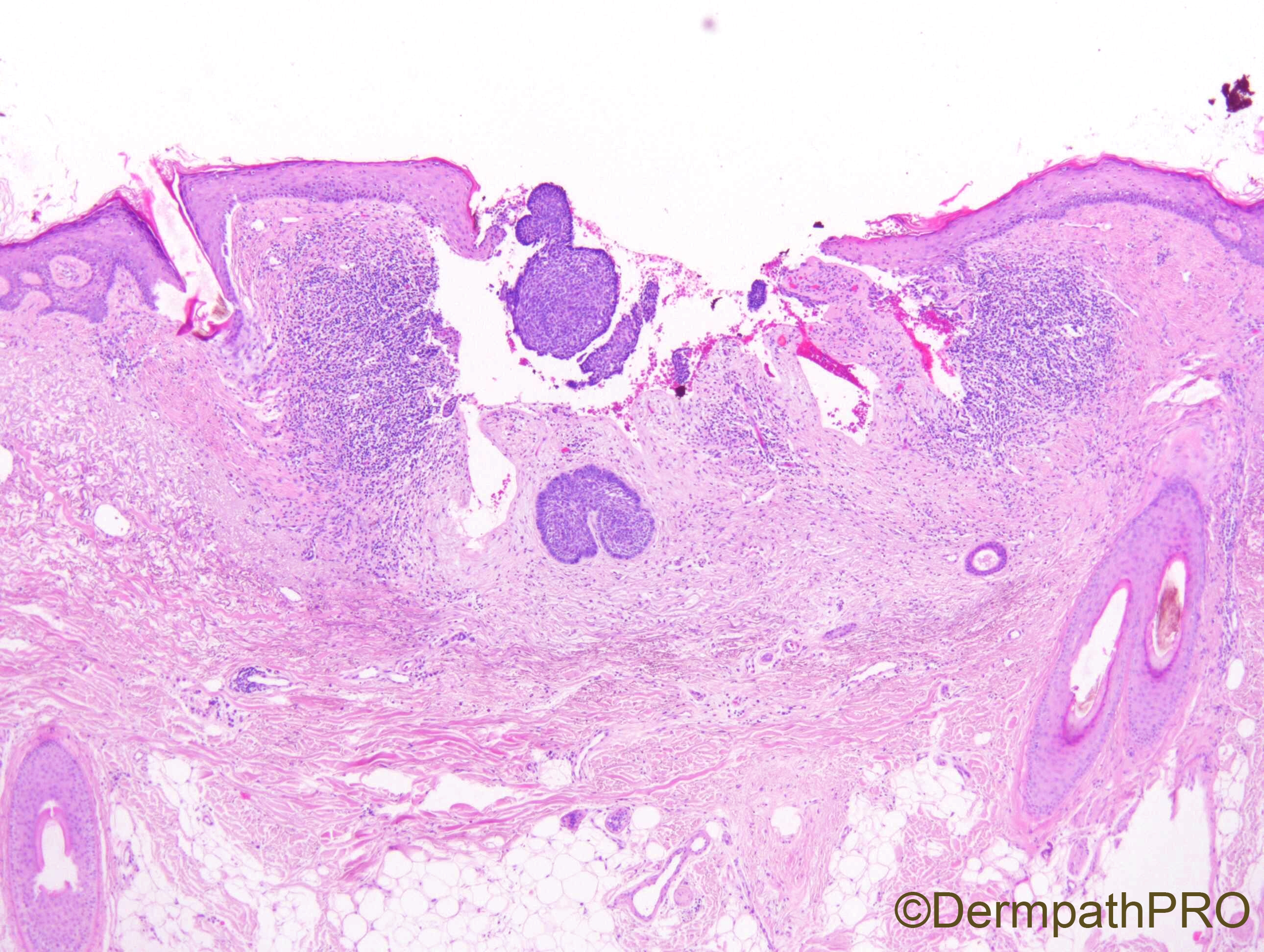
F63. Vertex of scalp.

Edited by Admin_Dermpath


  Report Record

User Feedback


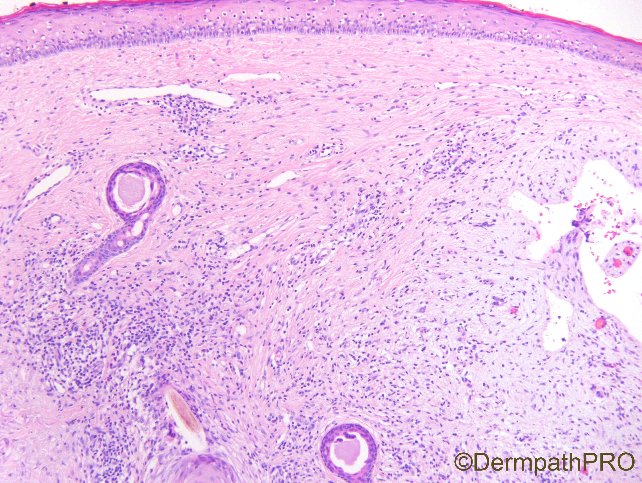
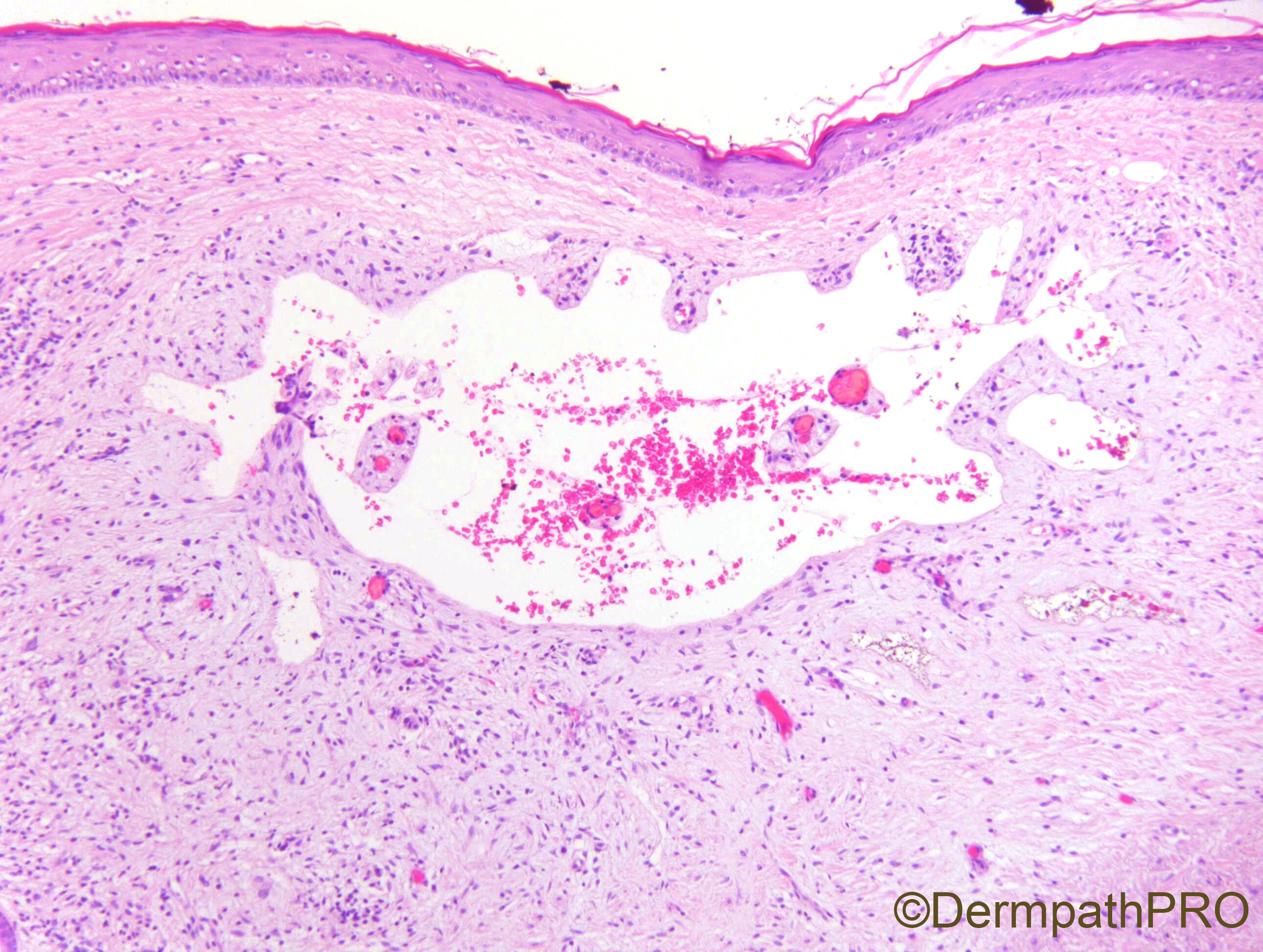
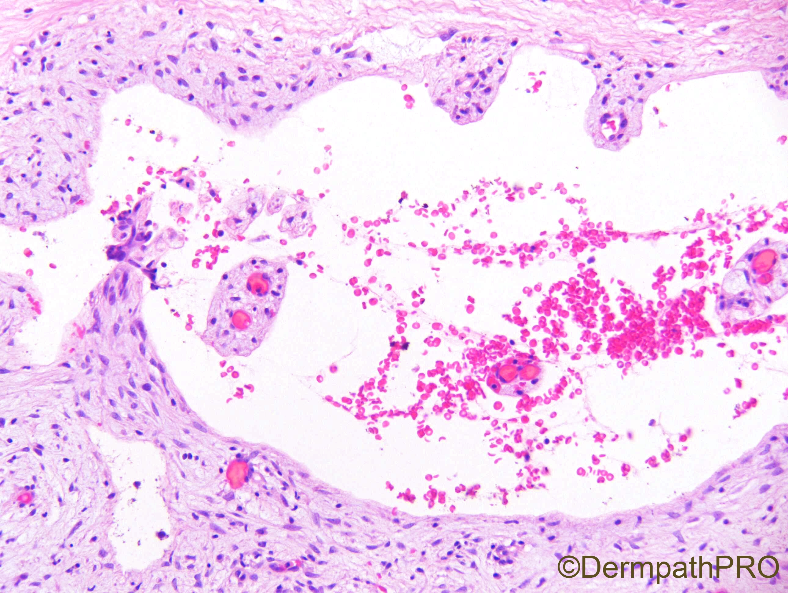
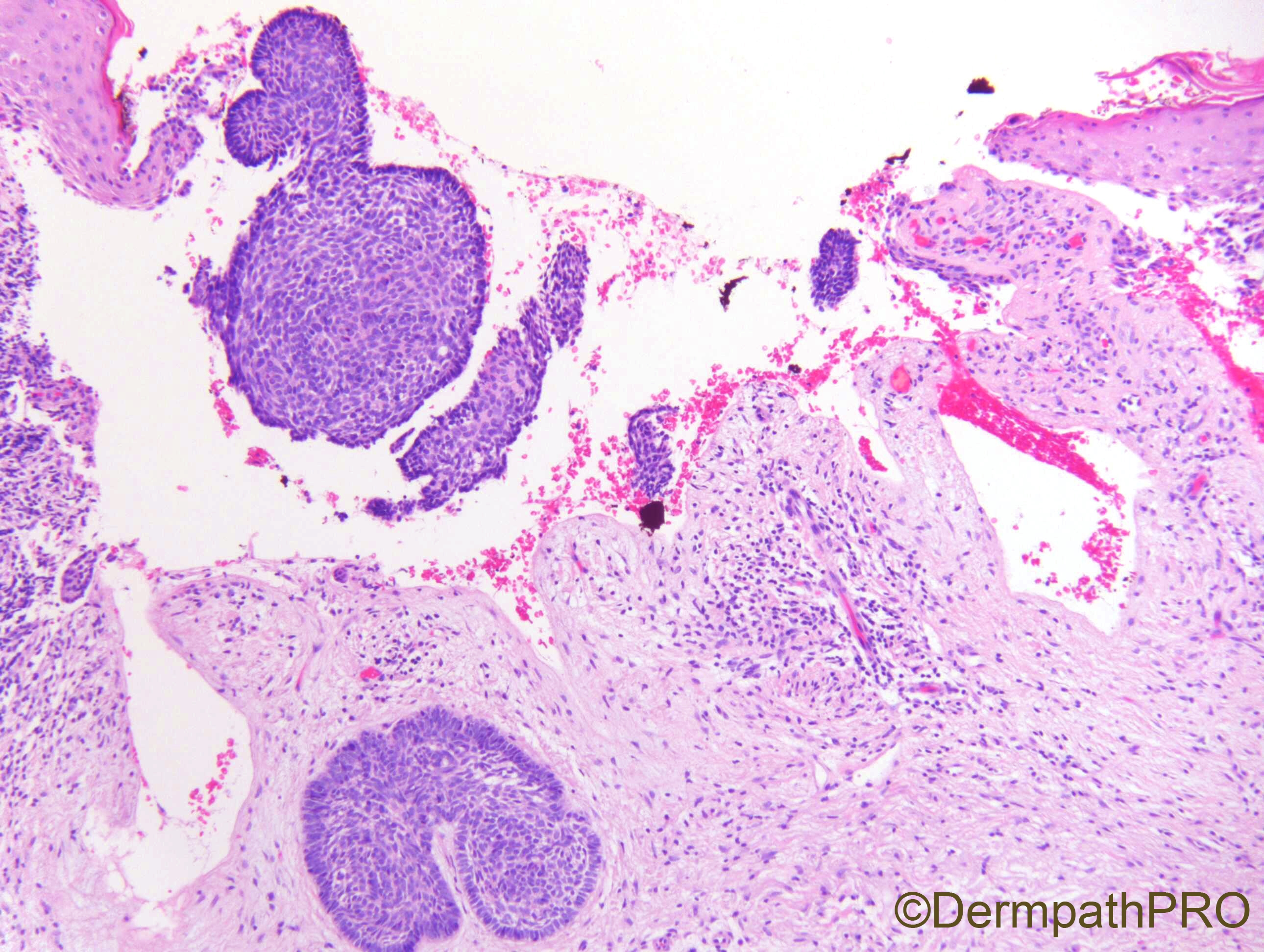
Difficult to interpret!! I can see some dermal fibrosis and an empty space in the superficial dermis. Everytime I see some empty space in the dermis I think about basal cell carcinoma. The external border of this space is corrugated wich is odd. Wating for more comments.

Share this comment


Link to comment
share_externally

Krishnakumar subramanian

Posted

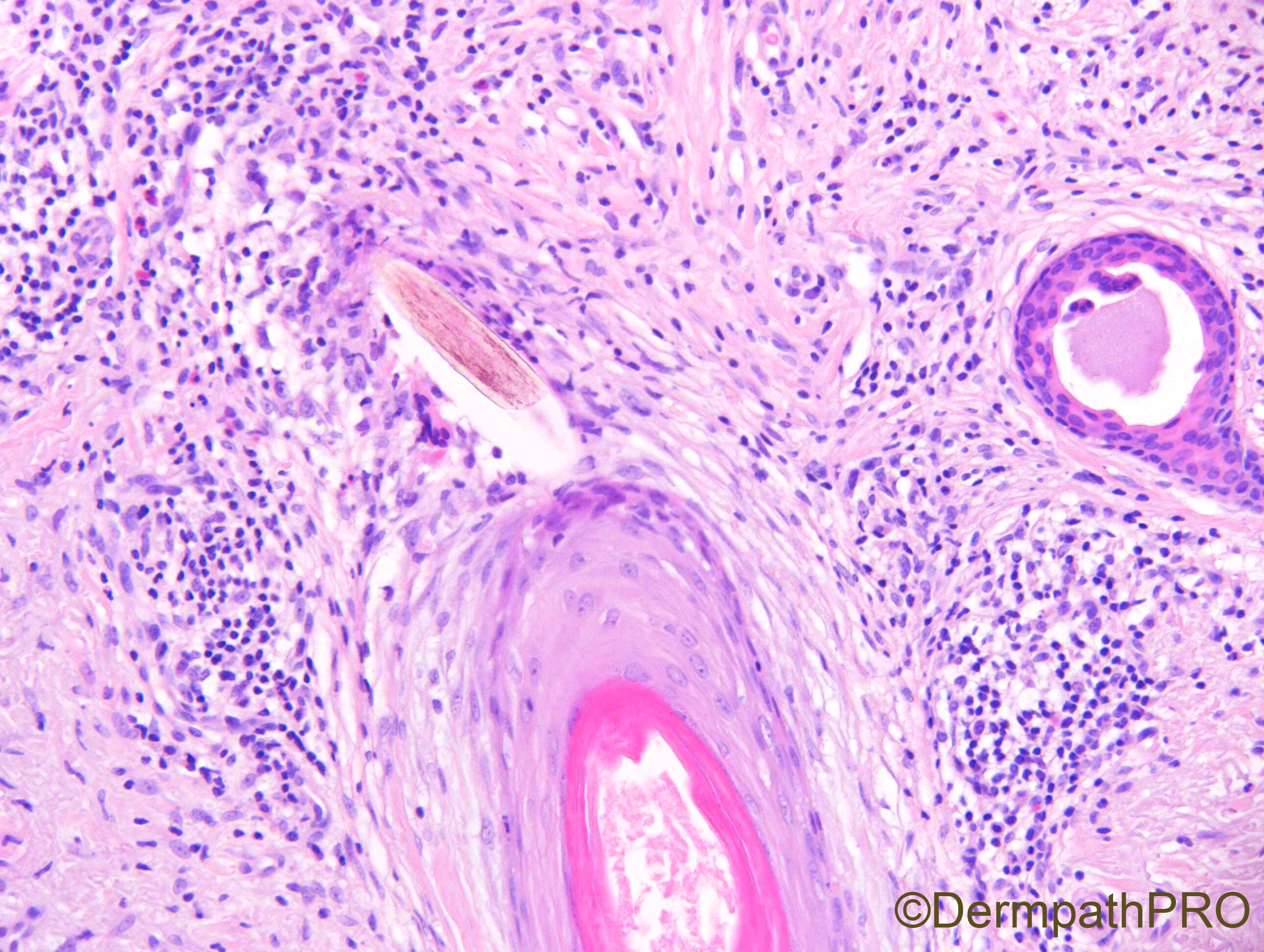
dermal myxoid and fibrosis.perifollicular fibrosis and mixed inflammation and eosinophils seen. the empty space has villi like projections, may be some parasitic 


Share this comment


Link to comment
share_externally

The empty space indicates that something must have popped off during processing. Eosinophils in the mixed infiltrate suggest that it could be a foreign body,  may be an artificial hair fiber used for treating alopecia? 

Share this comment


Link to comment
share_externally

Alex-Ventura-Leon

Posted

Agree that its difficult. But I dont think in BCC because i dont see any signs of solar damage.

I favor some foreign body material or some kind of parasit infection.

Share this comment


Link to comment
share_externally

vincenzo

Posted (edited)

Dilated ectatic vessels superficially and compressed vessels with some hemosiderin in deep dermis--->why not a Hobnail Hemangioma/alias hemosiderotic targetoiod hemangioma?

Edited by vincenzo

Share this comment


Link to comment
share_externally

Igor SC was right, a BCC that has popped off!


Share this comment


Link to comment
share_externally

So I was completely off side!!! Very interesting case! I often saw these empty spaces in the superficial dermis, as marker of popped off BCC, but this time I did not really think about it. Congrats to Igor!!!

Share this comment


Link to comment
share_externally

Dr. Richard Carr

Posted

Yes well done IgorSC. I remember well a similar case from many years ago that came to review at a skin meeting. Amazingly if was from the dorsum of the foot (not common for a BCC). The pathologist had described a cyst and mentioned a "carry-over" of basal cell carcinoma. The problem was the "cyst" had no lining cells and the "carry-over" was the same shape as the hole! The "cyst" wall often looks a little loose and myxoid as here. The first batch of images don't show it but there is quite a lot of solar elastosis in this case (extra images - 3rd down on the right).

Share this comment


Link to comment
share_externally


Join the conversation

You can post now and register later. If you have an account, sign in now to post with your account.

Guest
Add a comment...

×   Pasted as rich text.   Paste as plain text instead

  Only 75 emoji are allowed.

×   Your link has been automatically embedded.   Display as a link instead

×   Your previous content has been restored.   Clear editor

×   You cannot paste images directly. Upload or insert images from URL.

Loading...

×
×
  • Create New...